Implications
The present study demonstrates that acute cold exposure induces porcine white adipose tissue (WAT) browning, whereas chronic cold exposure impairs the browning process. Thus, browning of porcine WAT represents a novel strategy that could protect piglets against cold stress and thereby reduce piglet mortality. During the browning process, WAT could be burned to produce heat. However, the mechanisms by which this occurs in porcine WAT browning remain largely unknown. The browning of WAT could promote thermogenesis and increase the body temperature of piglets that tend to be characteristically cold intolerant.
Introduction
Distinct types of adipose tissues exist with different functions; WAT stores energy, whereas brown adipose tissue (BAT) contains abundant mitochondria with activated uncoupling protein 1 (UCP1), to dissipate chemical energy in the form of heat (Seale and Lazar, Reference Seale and Lazar2009; Fedorenko et al., Reference Fedorenko, Lishko and Kirichok2012). Subcutaneous WAT (sWAT) is capable of switching to the beige phenotype, thereby promoting metabolic heat production (Shabalina et al., Reference Shabalina, Petrovic, de Jong, Kalinovich, Cannon and Nedergaard2013), in a process referred to as ‘browning’ (Wu et al., Reference Wu, Bostrom, Sparks, Ye, Choi, Giang, Khandekar, Virtanen, Nuutila, Schaart, Huang, Tu, van Marken, Hoeks, Enerback, Schrauwen and Spiegelman2012; Kajimura et al., Reference Kajimura, Spiegelman and Seale2015). Browning of WAT and activation of BAT have become efficient therapeutic approaches to increase basal metabolism to combat the worldwide obesity epidemic (Harms and Seale, Reference Harms and Seale2013; Yoneshiro et al., Reference Yoneshiro, Aita, Matsushita, Kayahara, Kameya, Kawai, Iwanaga and Saito2013). Previous studies have used mice to mimic the potential effects of browning on human WAT, and considerable research has been devoted to the browning of mice adipose tissue (Bi et al., Reference Bi, Shan, Liu, Yue, Yang, Liang, Wang, Li, Carlesso, Liu and Kuang2014; Dodd et al., Reference Dodd, Decherf, Loh, Simonds, Wiede, Balland, Merry, Munzberg, Zhang, Kahn, Neel, Bence, Andrews, Cowley and Tiganis2015). The latest research finding suggested that pigs lacked classic BAT, but had beige adipocytes (Lin et al., Reference Lin, Cao, Tao, Ye, Dong, Zheng, Wang, Jiang, Qin, Yan, Li, Speakman, Wang, Jin and Zhao2017). However, browning of WAT in pigs, which are similar to humans in anatomy and physiology, more closely reflects human metabolism than mice do in energy metabolism, remain largely unknown (Bellinger et al., Reference Bellinger, Merricks and Nichols2006; Spurlock and Gabler, Reference Spurlock and Gabler2008). Therefore, browning of porcine WAT following cold exposure was explored in present study.
Cold exposure is a potent trigger for the recruitment of beige adipocytes (van der Lans et al., Reference van der Lans, Hoeks, Brans, Vijgen, Visser, Vosselman, Hansen, Jorgensen, Wu, Mottaghy, Schrauwen and van Marken2013; Yoneshiro et al., Reference Yoneshiro, Aita, Matsushita, Kayahara, Kameya, Kawai, Iwanaga and Saito2013). Both acute and chronic cold exposure could induce browning in humans and mice. Mechanistically, the sympathetic nervous system could be activated by exposure to cold conditions, which is followed by the production of noradrenaline and its subsequent binding to β3 adrenergic receptors that thereby increases levels of intercellular cyclic adenosine monophosphate. In turn, this stimulates protein kinase A and p38 mitogen-activated protein kinase (MAPK), thus promoting lipolysis and expression of UCP1 (Lowell and Spiegelman, Reference Lowell and Spiegelman2000; Collins, Reference Collins2011; Cypess et al., Reference Cypess, Weiner, Roberts-Toler, Franquet, Kessler, Kahn, English, Chatman, Trauger, Doria and Kolodny2015). Moreover, chronic cold exposure or β3 adrenergic stimuli could aggravate the degree to which browning occurs in humans (van der Lans et al., Reference van der Lans, Hoeks, Brans, Vijgen, Visser, Vosselman, Hansen, Jorgensen, Wu, Mottaghy, Schrauwen and van Marken2013; Sidossis et al., Reference Sidossis, Porter, Saraf, Borsheim, Radhakrishnan, Chao, Ali, Chondronikola, Mlcak, Finnerty, Hawkins, Toliver-Kinsky and Herndon2015) and mice (Rosenwald et al., Reference Rosenwald, Perdikari, Rulicke and Wolfrum2013).
Besides the UCP1-dependent pathway discussed above, UCP1-independent pathways also modulate the process of browning (Kajimura et al., Reference Kajimura, Spiegelman and Seale2015), including the creatine and peptidase M20 domain containing 1 (PM20D1) pathways that are involved in mitochondrial thermogenesis. The creatine-driven substrate cycle could compensate for a lack of UCP1 thermogenesis, through creatine metabolism marked by the expression of creatine kinase mitochondrial 1 (CKMT1) and creatine kinase mitochondrial 2 (CKMT2) (Kazak et al., Reference Kazak, Chouchani, Jedrychowski, Erickson, Shinoda, Cohen, Vetrivelan, Lu, Laznik-Bogoslavski, Hasenfuss, Kajimura, Gygi and Spiegelman2015). Moreover, N-acyl amino acids have been reported to activate mitochondrial thermogenesis under the regulation of PM20D1 during UCP1-independent respiration (Long et al., Reference Long, Svensson, Bateman, Lin, Kamenecka, Lokurkar, Lou, Rao, Chang, Jedrychowski, Paulo, Gygi, Griffin, Nomura and Spiegelman2016). Thus, the UCP1-independent thermogenic pathways were analysed in the present study.
Here, the existence of porcine WAT browning and whether browning could be a novel strategy to increase the body temperature of piglets were explored (Balsbaugh et al., Reference Balsbaugh, Curtis, Meyer and Norton1986).
Material and methods
Animals and treatments
Piglets handling protocols were conducted following the guidelines and regulations approved by the Animal Ethics Committee of Northwest A&F University (Yangling, China). Five-day-old piglets were provided by the experimental farm of Northwest A&F University. For the acute cold exposure experiment, four piglets of one group were housed at 4°C, and four in the control group were housed at room temperature of 25°C for 10 h. Five piglets subjected to chronic cold exposure were housed at 8°C for 15 days, and five in the control group were housed at room temperature of 25°C for 15 days. Piglets were fed ad libitum, and then sacrificed. The sWAT at the back of the neck was isolated, photographed (Sony α6000,Tokyo, Japan) and picture processed using the crop tool of Photoshop software (Adobe, CA, USA), collected, quickly fixed in 4% paraformaldehyde for frozen sections, or frozen in liquid nitrogen and stored at −80°C for subsequent analysis.
Quantitative real-time polymerase chain reaction analysis
Adipose tissues were ground using a Tissue Lyser (Scientz-48; Huakeda, Wuhan, China). Total RNA was extracted from the sWAT using the TRIzol reagent (TaKaRa, Otsu, Japan) following standard procedures. The total RNA was processed into single-stranded cDNA using the High Capacity Reverse Transcription Kit (TaKaRa). The qRT-PCR was performed using the One-Step SYBR PrimeScript RT-PCR Kit (TaKaRa) on the Bio-iQ5 Real-Time PCR System (Bio-Rad, Berkeley, CA, USA). The β-actin gene was used as a housekeeping gene to normalise the expression of other genes. The
 $${\rm 2}^{{{\minus}\Delta \Delta C_{t} }} $$
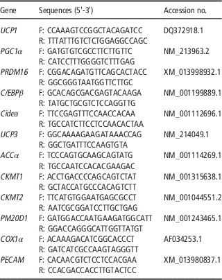
algorithm was employed to estimate the relative expression level of each gene. The sequences of the primers used are presented in Table 1.
Primers for quantitative real-time polymerase chain reaction or the calculation of mitochondrial DNA copy number in porcine white adipose tissue

Western blot analysis
Total proteins of adipose tissue were extracted using radioimmunoprecipitation buffer (Applygen, Beijing, China) supplemented with protease inhibitor (Roche, Basel, Switzerland). Protein concentration was measured using the bicinchoninic acid (BCA) Assay (Cwbiotech, Beijing, China). Total protein was electrophoresed in SDS-polyacrylamide gel (Heart, Xian, China) and transferred to polyvinylidene difluoride membranes (Bio-Rad). After blocking in 5% skim milk for 2 h, the membranes were incubated overnight at 4°C in primary antibodies, diluted with antibody diluent (Jiancheng, Nanjing, China) at 1 : 1000: PGC1α (ab54481; Abcam, Cambridge, UK); PGC1β (ab176328; Abcam); Leptin (ab3583; Abcam); FABP4 (SC150; Santa Cruz, CA, USA); CyclinE (sc-247; Santa Cruz); proliferating cell nuclear antigen (PCNA) (sc-56; Santa Cruz); β-Tubulin (KM9003; Sungene Biotech, Tianjin, China); p38-P (9211; CST, Danvers, MA, USA); and p38-T (9212; CST). Immuno-labelling was detected using the enhanced chemiluminescence reagent (Millipore, Boston, MA, USA).
Histology
Isolated adipose tissue was fixed in 4% paraformaldehyde in phosphate-buffered saline for 12 h, before dehydration in 30% sucrose solution for 24 h. After being sliced with a freezing microtome (CM1950; Leica, Wetzlar, Germany), sections were stained with haematoxylin and eosin (H&E) following the standard protocol. Then, sections were examined by light microscopy. Photomicrographs were captured using a Nikon TE2000 microscope (Nikon, Tokyo, Japan), and adipocyte diameter statistics by Image-pro plus 6.0.
Mitochondrial DNA copy number
Adipose tissues were ground using a Tissue Lyser (Scientz-48, Huakeda, Wuhan, China) and digested with proteinase K for 10 h. Genomic DNA of adipose tissue was isolated by standard procedure for quantitative PCR. The mitochondrial DNA (mtDNA) copy number was calculated from the ratio of DNA abundance of the mitochondrial gene COX1α to that of the nuclear gene platelet endothelial cell adhesion molecule (PECAM). The sequences of COX1α and PECAM primers are presented in Table 1.
Statistical analysis
All data are presented as mean+SEM and were analysed using the Prism software (GraphPad). Statistical significance was determined using Mann Whitney U-test by SPSS. The differences are indicated as follows: *0.01<P⩽0.05, **P⩽0.01.
Results
Acute cold exposure induces browning in porcine subcutaneous WAT
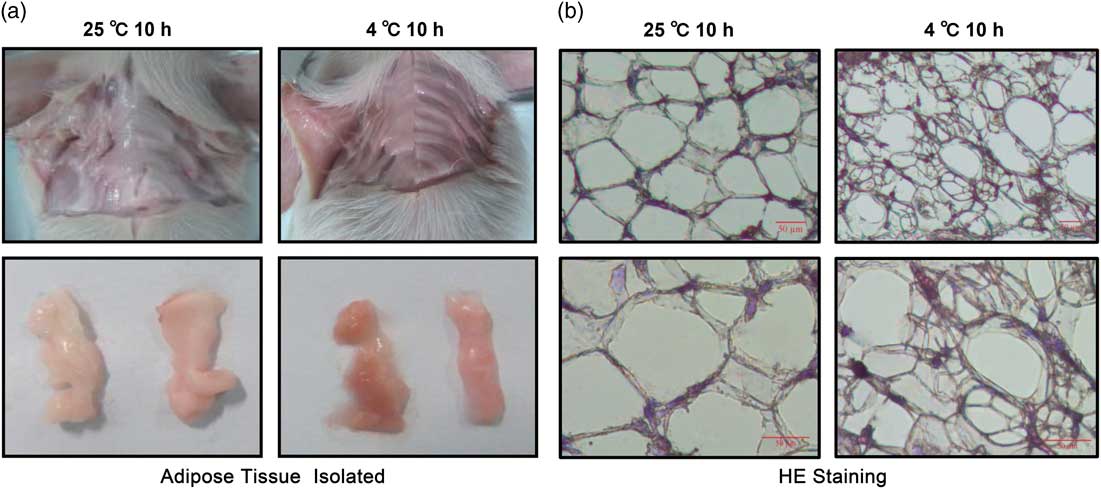
Piglets under acute cold exposure showed more reddish porcine sWAT than exposed to room temperature (Figure 1a). Moreover, on the cellular level, sWAT sections using H&E staining in the acute cold exposure group showed shrinking of the adipocytes compared with that of control (Figure 1b and Supplementary Material Figure S1).
Acute cold exposure induces morphological characteristics of browning in porcine subcutaneous white adipose tissue (sWAT). (a) Differences in the sWAT of piglets between acute cold exposure (4°C, 10 h, right) and room temperature (25°C, 10 h, left). (b) Haematoxylin and eosin (H&E) staining of frozen sWAT sections; thickness of all sections is 8 µm. Scale bar=50 µm.

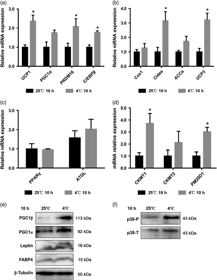
Because of the browning tendency of porcine sWAT observed following acute cold exposure, the treated tissues were further analysed to detect the expression of UCP1-dependent and -independent thermogenesis, mitochondrial biogenesis, lipogenic and lipolytic genes. The results showed that messenger RNA (mRNA) expression of browning marker genes, including UCP1, PRD1-BF1-RIZ1 homologous domain containing 16 (PRDM16)and CCAAT/enhancer binding protein beta (C/EBPβ), were significantly upregulated following acute cold exposure, whereas mRNA level of peroxisome proliferative activated receptor gamma co-activator 1 alpha (PGC1α) showed no change (Figure 2a). Furthermore, the expression of mitochondrial thermogenic genes, such as the cell death activator CIDE-A (Cidea) and uncoupling protein 3 (UCP3), were increased. However, no differences were found in the mRNA levels ofcytochrome oxidase subunit 1 alpha (Cox1α) and acetyl-CoA carboxylase alpha (ACCα) (Figure 2b), nor in the lipogenic gene peroxisome proliferator-activated receptor gamma (PPARγ) and lipolytic gene adipose triglyceride lipase (ATGL) (Figure 2c). Notably, UCP1-independent mitochondrial thermogenesis genes, including CKMT1 and PM20D1, showed significantly higher levels following acute cold exposure (Figure 2d). In addition, consistent with the mRNA expression, protein expression levels of PGC1α, peroxisome proliferative activated receptor gamma co-activator 1 beta (PGC1β) and leptin in the group subjected to cold exposure were markedly increased. However, no differences were noted in the expression of fatty acid binding protein 4 (FABP4) (Figure 2e and Supplementary Material Figure S2). Moreover, the level of phosphorylated p38 MAPK was also highly expressed following acute cold exposure (Figure 2f and Supplementary Material Figure S3). Overall, these data suggest that browning of porcine sWAT was induced following acute cold exposure.
Browning of porcine subcutaneous white adipose tissue (sWAT) is activated upon acute cold exposure. (a) Relative mRNA expression of browning marker genes (UCP1, PGC1α, PRDM16 and C/EBPβ); (b) mitochondrialthermogenic genes (Cidea, UCP3, ACCα and COX1α); (C) lipogenic (PPARγ) and lipolytic (ATGL) genes; and (d) UCP1-independent thermogenesis related genes (CKMT1, CKMT2 and PM20D1) upon acute cold exposure. (e) Protein expression of PGC1α, PGC1β, leptin and FABP4; β-Tubulin was used as a reference gene. (f) Phosphorylated changes of p38 mitogen-activated protein kinase upon acute cold exposure (n=4, values shown are mean+SEM, *P<0.05).

Chronic cold stress does not induce morphological characteristics of browning
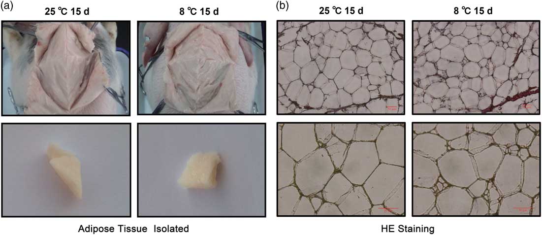
No significant colour differences of adipose tissue were observed between piglets housed at room temperature and those subjected to chronic cold exposure (Figure 3a). Haematoxylin and eosin staining showed that there were no morphological changes in the adipocytes of either control or treatment groups (Figure 3b and Supplementary Material Figure S4). These data collectively suggest that porcine sWAT showed no morphological evidence of browning following chronic cold exposure.
Chronic cold exposure of porcine subcutaneous white adipose tissue (sWAT) does not induce morphological characteristics of browning. (a) Colour of the sWAT of piglets following chronic cold exposure (8°C, 15 days, right) and room temperature (25°C, 15 days, left). (b) Haematoxylin and eosin (H&E) staining of subcutaneous white adipose tissue (sWAT) frozen sections; thickness of all sections is 8 µm. Scale bar=50 µm.

Chronic cold exposure impairs the browning process and inhibits mitochondrial biogenesis
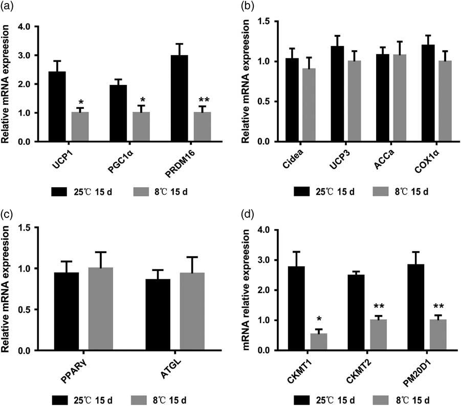
To understand the reasons behind the morphological results presented above, qRT-PCR was performed to measure mRNA abundance of browning marker genes, including UCP1, PGC1α and PRDM16 in porcine sWAT. The mRNA levels of these genes showed significant decline following chronic cold exposure (Figure 4a). However, no significant change was evident in mitochondrial thermogenic genes like Cidea, UCP3, ACCα and Cox1α (Figure 4b), nor in lipogenic or lipolytic genes, such as PPARγ and ATGL, respectively (Figure 4c). Interestingly, expression of genes associated with UCP1-independent mitochondrial thermogenesis, such as CKMT1, CKMT2 and PM20D1 also showed significant decline following chronic cold exposure (Figure 4d).
Chronic cold exposure of porcine subcutaneous white adipose tissue (sWAT) impairs the expression of browning genes. (a) Relative mRNA expression of browning marker genes (UCP1, PGC1α and PRDM16); (b) mitochondrial thermogenic genes (Cidea, UCP3, ACCα and COX1α); and (c) lipogenic (PPARγ) or lipolytic (ATGL) genes; and (d) UCP1-independent thermogenesis-related genes (CKMT1, CKMT2 and PM20D1) following chronic cold exposure (n=5, values shown are mean+SEM, *P<0.05, **P<0.01).

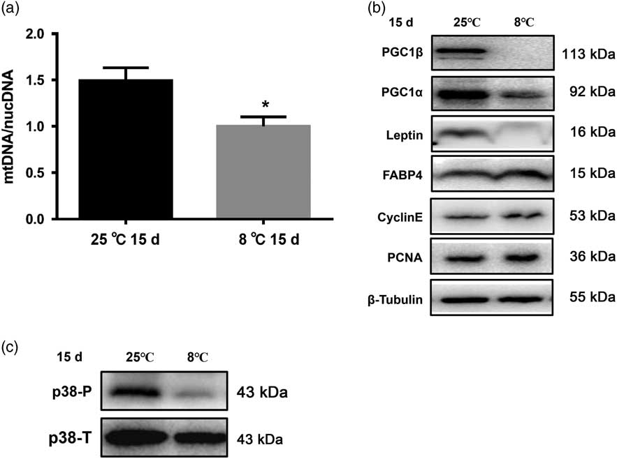
Because WAT browning was associated with an increase in the number of mitochondria, the mtDNA copy numbers in sWAT were tested. Despite the lack of significant change in the expression of the mitochondrial thermogenesis genes, the number of mitochondria was reduced by about 30% following chronic cold exposure (Figure 5a), which was consistent with our findings based on the mitochondrial biogenesis gene PGC1a (Figures 4a, 5b). These results collectively show that chronic cold stress could not upregulate mitochondrial gene expression, but reduce mtDNA copy numbers, suggesting a decline in mitochondrial biogenesis.
Chronic cold exposure of porcine subcutaneous white adipose tissue (sWAT) impairs the expression of browning proteins and inhibits mitochondrial biogenesis. (a) Mitochondrial number in sWAT. (b) Expression of PGC1β, PGC1α, Leptin, FABP4, CyclinE and PCNA proteins. Β-Tubulin was used as a reference gene. (c) Phosphorylated changes of p38 mitogen-activated protein kinase (MAPK) following chronic cold exposure (n=5; values shown are mean+SEM, *P<0.05).

Consistent with the decline in mRNA levels, chronic cold stress also markedly reduced levels of PGC1β and PGC1α proteins, but effected no changes in the adipogenesis marker FABP4, and proliferation markers PCNA and CyclinE (Figure 5b and Supplementary Material Figure S5). Leptin is a hormone produced by adipocytes that plays a key role in the regulation of food intake and energy expenditure of animals. Our results show that the levels of leptin in sWAT declined following chronic cold exposure, indicating a reduction in energy expenditure (Figure 5b and Supplementary Material Figure S5). As shown in Figure 5c and Supplementary Material Figure S6, levels of phospho-p38 also declined following chronic cold exposure. Altogether, our data provides evidence that chronic cold exposure impairs the browning process and inhibits mitochondrial biogenesis and the expression of browning marker genes.
Discussion
The purpose of this study was to investigate whether browning of porcine sWAT could be induced under cold conditions. Here, acute cold exposure could induce browning of porcine sWAT, whereas chronic cold exposure could not, suggesting that browning was dependent on the duration of cold exposure. Humans have BAT depots between the shoulder blades that are analogous to beige adipocytes in mice (Jespersen et al., Reference Jespersen, Larsen, Peijs, Daugaard, Homoe, Loft, de Jong, Mathur, Cannon, Nedergaard, Pedersen, Moller and Scheele2013). Furthermore, activation of these cells declines with age (Yoneshiro et al., Reference Yoneshiro, Aita, Matsushita, Okamatsu-Ogura, Kameya, Kawai, Miyagawa, Tsujisaki and Saito2011). Thus, piglets were selected to study the browning of WAT in the present study. Moreover, sWAT is more highly susceptible to browning than visceral WAT in mice (Seki et al., Reference Seki, Hosaka, Lim, Fischer, Honek, Yang, Andersson, Nakamura, Naslund, Yla-Herttuala, Sun, Iwamoto, Li, Liu, Samani and Cao2016; Wang and Seale, Reference Wang and Seale2016). Accordingly, browning of sWAT of piglets subjected to cold exposure was studied.
Our results showed that the sWAT from the group subjected to acute cold exposure (4°C for 10 h) appeared more reddish in colour than that from the control group, suggesting more intensive vascularisation and possibly increased cellular contents within adipocytes (Xue et al., Reference Xue, Petrovic, Cao, Larsson, Lim, Chen, Feldmann, Liang, Zhu, Nedergaard, Cannon and Cao2009 and Reference Xue, Xu, Zhang, Farokhzad and Langer2016; Seki et al., Reference Seki, Hosaka, Lim, Fischer, Honek, Yang, Andersson, Nakamura, Naslund, Yla-Herttuala, Sun, Iwamoto, Li, Liu, Samani and Cao2016). Histological staining indicated shrinkage transition of adipocytes. Moreover, the group subjected to acute cold exposure also showed elevated expression of browning, thermogenic genes and p38 MAPK phosphorylation compared with the control. Collectively, acute cold exposure induced browning of porcine sWAT that was analogous to that which occurred in humans and mice.
In contrast, following chronic cold exposure, porcine sWAT showed no evidence of browning, which is inconsistent with observations in mice (Rosenwald et al., Reference Rosenwald, Perdikari, Rulicke and Wolfrum2013). The expression of browning marker genes declined following chronic cold exposure, and no morphological differences were noted between the control and treatment groups. We found that the mtDNA copy number was reduced by about 30% and the expression of PGC1a, which was related to mitochondrial biogenesis, also showed a decline following chronic cold exposure (Wu et al., Reference Wu, Puigserver, Andersson, Zhang, Adelmant, Mootha, Troy, Cinti, Lowell, Scarpulla and Spiegelman1999; Puigserver and Spiegelman, Reference Puigserver and Spiegelman2003). The decline in expression of the browning genes could be attributed to the inhibition of mitochondrial biosynthesis during chronic cold exposure. Furthermore, because of the constant expression of lipogenic genes, such as PPARγ and FABP4, the protein expression of proliferative genes PCNA and CyclinE were evaluated further; however, no significant changes were observed. Altogether, chronic cold exposure inhibited mitochondrial biogenesis of porcine sWAT, and had no effect on lipolysis and cell proliferation.
The UCP1 plays a thermogenic role in the mitochondria of browning adipose tissue. The mRNA levels of UCP1 and its independent pathway genes showed significant increase following acute cold exposure, and decline following chronic cold exposure. In addition, UCP1-independent pathways of mitochondrial thermogenesis have been evaluated in previous studies (Kajimura et al., Reference Kajimura, Spiegelman and Seale2015; Kazak et al., Reference Kazak, Chouchani, Jedrychowski, Erickson, Shinoda, Cohen, Vetrivelan, Lu, Laznik-Bogoslavski, Hasenfuss, Kajimura, Gygi and Spiegelman2015; Long et al., Reference Long, Svensson, Bateman, Lin, Kamenecka, Lokurkar, Lou, Rao, Chang, Jedrychowski, Paulo, Gygi, Griffin, Nomura and Spiegelman2016). The rising expression of CKMT1, CKMT2 and PM20D1 following acute cold exposure indicated that both creatine and N-acyl amino acids played a role in mitochondrial thermogenesis. Therefore, the pig could be used as a potential model to study UCP1-independent thermogenesis.
In summary, acute cold exposure could induce browning of porcine sWAT, resulting in smaller adipocytes and upregulated expression of browning genes that are similar to those of humans and mice. Chronic cold exposure impaired the browning process with a decline in both mitochondrial biogenesis and the expression of browning marker genes that were different from those found in mice. Thus, porcine browning could be induced by acute cold exposure, but impaired by chronic cold exposure. Overall, our findings present a novel model to study WAT browning that could potentially aid in combatting human obesity. Here, the existence of porcine WAT browning was found. Furthermore, in swine industry, activating WAT browning by some browning activators may contribute to maintaining a favourable balance in the body temperature of piglets.
Acknowledgements
This work was supported by the National Key Basic Research and Development Programme (2015CB943102), National Natural Science Foundation of China (31572366), Major Projects for Genetically Modified Organisms Breeding (2014ZX08009-047B) and the National Science and Technology Major Project of China (2016ZX08006003).
Supplementary material
To view supplementary material for this article, please visit https://doi.org/10.1017/S1751731117002981