Introduction
The hemipteran superfamily Hylicelloidea Evans, Reference Evans1956, as the basal lineage of the clade Clypeata Qadri, Reference Qadri1967, is the ancestral group of all the three modern cicadomorphan lineages (i.e., froghoppers [Cercopoidea Leach, Reference Leach and Brewster1815], hair and singing cicadas [Cicadoidea Latreille, Reference Latreille1802], and treehoppers and allies [Membracoidea Rafinesque, Reference Rafinesque1815, s.l.]). To date, this superfamily is divided into four families: Hylicellidae Evans, Reference Evans1956, from the upper Paleozoic to the Lower Cretaceous of Australia and Eurasia (Lambkin, Reference Lambkin2020); Chiliocyclidae Evans, Reference Evans1956, from the Triassic of Australia, and some fossils from Argentina with disputed systematic position (Lara and Bashkuev, Reference Lara and Bashkuev2020); Mesojabloniidae Storozhenko, Reference Storozhenko1992, from the Triassic of Kyrgyzstan, Argentina, and Australia (Lambkin, Reference Lambkin2019; Lara et al., Reference Lara, Bustos-Escalona, Mancuso and Arcucci2021); and Minlagerrontidae Chen, Szwedo, and Wang in Chen et al. (Reference Chen, Szwedo, Wang, Zheng, Jiang, Jiang, Wang and Zhang2019a), exclusively recorded in the mid-Cretaceous Kachin amber of northern Myanmar (Chen et al., Reference Chen, Wang, Zhang, Jiang, Jiang, Zheng and Wang2020). Among the four hylicelloid families, Hylicellidae, with ~15 genera ascribed to three subfamilies (Hylicellinae Evans, Reference Evans1956, Vietocyclinae Shcherbakov, Reference Shcherbakov1988b, and Conjucellinae Shcherbakov, Reference Shcherbakov2012), was dominant and widespread from the Triassic to Early Cretaceous (Lambkin, Reference Lambkin2019; Chen et al., Reference Chen, Wang, Zhang, Jiang, Jiang, Zheng and Wang2020).
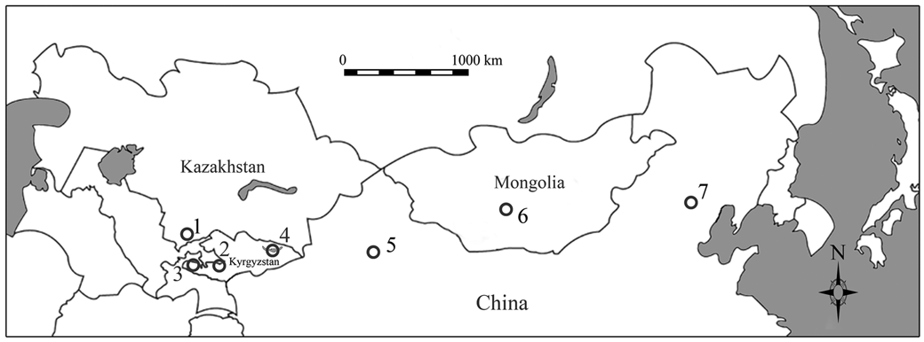
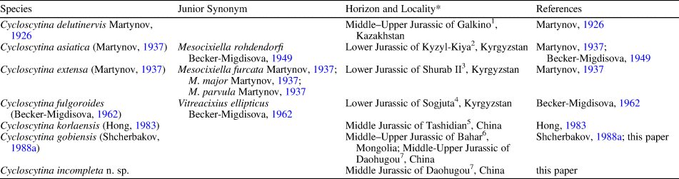
Abundant true hoppers (Fulgoromorpha + Cicadomorpha) with high biodiversity have been recorded in the Mesozoic of China; documentation of hylicelloid fossils in China, however, is relatively scarce. Up to now, only four isolated tegmina have been reported, and attributed to the subfamily Vietocyclinae of Hylicellidae: Cycloscytina korlaensis (Hong, Reference Hong1983) from the Middle Jurassic of Tashidian, Xinjiang; Jiphara wangi Ren, Reference Ren, Ren, Lu, Guo and Ji1995; and J. reticulata Ren, Reference Ren, Ren, Lu, Guo and Ji1995; and Vietocycla katyae Fu and Huang, Reference Fu and Huang2019b, from the Lower Cretaceous of Fangshan, Beijing. We herein report some new whole-bodied fossils from the well-known Jurassic Daohugou beds of Inner Mongolia, China. These specimens assuredly belong to the genus Cycloscytina Martynov, Reference Martynov1926 (see Systematic paleontology), but their systematic position at a species level is not easily determined due to the high variability and complexity of tegminal venation for Cycloscytina, as well as the high species-level diversity of this genus (Table 1; Fig. 1). To objectively elucidate the systematic position of these new hylicellids, we executed the geometric morphometric analysis (GMA) based on tegminal characters in this study.
Locations of Mesozoic hylicellid fossils ascribed to the genus Cycloscytina Martynov, Reference Martynov1926. See Table 1 for detailed information on geographical and stratigraphical distribution of each fossil locality.

Mesozoic hylicellid species currently ascribed to the genus Cycloscytina Martynov, Reference Martynov1926.

*Superscript numbers indicate localities labeled in Figure 1.
Most hylicelloids have been described solely based on the information of isolated forewings or hind wings. To date, just a few body structures of the extinct Hylicelloidea have been revealed. Hylicellid fossils reported herein, with most of them preserved as whole-bodied fossils with forewing and hind wing attached, provide some novel insights on the evolution of basal Clypeata.
Geological setting
All hylicellid specimens described herein were collected from the Daohugou deposits, Haifanggou Formation, Chifeng, Inner Mongolia, China at new Daohugou Village (41°18′N, 119°13′E). Our hylicellid fossils, preserved in tuffaceous mudstones with abundant conchostracans (Triglypta haifanggouensis [Chen, in Zhang, Chen, and Shen, Reference Zhang, Chen and Shen1976]), are from the middle and upper section of the Daohugou Bed. The detailed stratigraphic column for the Haifanggou Formation near Daohugou Village was presented in Fu and Huang (Reference Fu and Huang2019a, fig. 1). The Daohugou beds, yielding abundant and diverse fossil plants and animals, are now considered among the most important insect Lagerstätten (Rasnitsyn et al., Reference Rasnitsyn, Zhang and Wang2006). The age of the Daohugou beds is somewhat controversial (e.g., Zhang, Reference Zhang2002; Shen et al., Reference Shen, Chen and Huang2003; Rasnitsyn and Zhang, Reference Rasnitsyn and Zhang2004; Wang et al., Reference Wang, Li, Zhang, Fang, Wang and Zhang2015; Chen and Wang, Reference Chen and Wang2016; Xu et al., Reference Xu, Zhou, Sullivan, Wang and Ren2016). Recent radiometric dating results (Liu et al., Reference Liu, Liu and Zhang2006, Reference Liu, Kuang, Jiang, Peng, Xu and Sun2012; Wang et al., Reference Wang, Hu, Zhang, Zhang, He, Deng, Wang, Zhou and Zhu2013) and paleontological studies (Wang et al., Reference Wang, Fang, Wang and Zhang2018, and references therein), however, suggest that the Daohugou beds are of latest Middle Jurassic–earliest Late Jurassic age (Callovian–Oxfordian). The insect fossils from the Daohugou beds are not only taxonomically diverse but also contain some representatives with significant information on the evolutionary history and ecological habits of Mesozoic insects (e.g., Huang et al., Reference Huang, Engel, Cai, Wu and Nel2012; Chen et al., Reference Chen, Wang, Engel, Wappler, Jarzembowski, Zhang, Wang, Zheng and Rust2014).
Material and methods
Materials
Eleven new Jurassic fossil specimens were collected from the uppermost Middle–lowermost Upper Jurassic of Daohugou, Chifeng, Inner Mongolia, China, and now are housed in the Nanjing Institute of Geology and Palaeontology, Chinese academy of Sciences in Nanjing, China, and the Shandong Tianyu Museum of Nature in Pingyi, China.
Methods
Fossil specimens were photographed using a Nikon D800 digital camera or Nikon SMZ1000, and examined and/or micro-photographed dry or under ethanol using Zeiss SteREO Discovery V8 stereomicroscopes. Illustrations and line drawings were prepared with CorelDraw 12.0 and Adobe Photoshop CS3. All measurements were made in ImageJ 1.42q (National Institutes of Health, Bethesda, MD, USA).
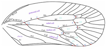
The terminology of wing venations and cells used in the present study follows Chen et al. (Reference Chen, Jiang, Shang, Zhang, Zheng, Zhang and Zhang2021), which is mainly modified from Nel et al. (Reference Nel, Prokop, Nel, Grandcolas, Huang, Roques, Guilbert, Dostal and Szwedo2012) and Bourgoin et al. (Reference Bourgoin, Wang, Asche, Hoch, Soulier-Perkins, Stroinski, Yap and Szwedo2015) (Fig. 2). The independent median vein is treated as vein M, following Kukalova-Peck (Reference Kukalova-Peck and Naumann1991) and Nel et al. (Reference Nel, Prokop, Nel, Grandcolas, Huang, Roques, Guilbert, Dostal and Szwedo2012), rather than median posterior vein (MP) in Bourgoin et al. (Reference Bourgoin, Wang, Asche, Hoch, Soulier-Perkins, Stroinski, Yap and Szwedo2015). Schubnel et al. (Reference Schubnel, Desutter-Grandcolas, Legendre, Prokop, Mazurier, Garrouste, Grandcolas and Nel2020) demonstrated the presence of an independent main postcubital vein in all Neoptera between the cubital and anal veins, so we herein treat the first longitudinal vein on clavus as the postcubital vein (Pcu) rather than the first anal vein (A1). The nomenclature of body structures mainly follows Evans (Reference Evans1966) and Moulds (Reference Moulds2005).
Line drawing of right tegmen of hylicellid specimen STMN48-1827 reported in this study, with landmarks (1–14) used in geometric morphometric analyses, and tegminal vein and cell terminology labeled. Pc = precosta; CP = costa posterior; bScP = basal part of subcosta posterior; R = radius; RA = radius anterior; RP = radius posterior; M = media; M1+2 = media anterior; M3+4 = media posterior; CuA = cubitus anterior; CuA1 = first branch of cubitus anterior; CuA2 = second branch of cubitus anterior; CuP = cubitus posterior; Pcu = postcubitus; A1 = first anal vein; cua-cup = crossvein between CuA and CuP; ir = crossvein between RA and RP; r-m = crossvein between RP and M; im = crossvein between M1+2 and M3+4; m-cua = crossvein between M3+4 and CuA; C1 = cell between forking of RA and RP; C2 = cell between R and M1+2; C3 = cell between forking of stem M; C4 = cell between M3+4; C5 = cell between forking of CuA.

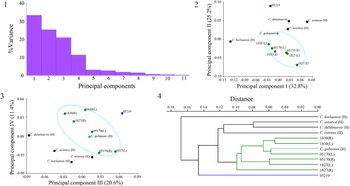
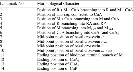
The geometric morphometric analysis was applied to the study of phenetic relationships of wing venations among known Cycloscytina and our new specimens. All Cycloscytina species were included in GMA herein, except C. fulgoroides (Becker-Migdisova, Reference Becker-Migdisova1962) whose known specimens are poorly preserved and lack key landmarks chosen in the present study. The holotypes of C. asiatica (Martynov, Reference Martynov1937), C. extensa (Martynov, Reference Martynov1937), C. korlaensis (Hong, Reference Hong1983), and C. gobiensis (Shcherbakov, Reference Shcherbakov1988b) were redrawn following line drawings in the original papers (Martynov, Reference Martynov1937; Hong, Reference Hong1983; Shcherbakov, Reference Shcherbakov1988b). The holotype of C. delutinervis Martynov, Reference Martynov1926, was redrawn following line drawings in Shcherbakov (Reference Shcherbakov and Rozanov1988a). The well-preserved right and left tegmina of STMN48-1827, STMN48-1830, NIGP05170, and the isolated tegmen of NIGP05219 were chosen in GMA (Supplemental Fig. 1). Based on the criterion for GMA that all specimens must have the same number of landmark points, we selected 14 landmarks (Table 2; Supplemental Fig. 1), which represent key points on the wing veins and can be clearly observed in all the above-mentioned Cycloscytina specimens. CorelDraw 12.0 was used to re-draw the wing venation of Cycloscytina specimens used in GMA. JPEG images of the line drawings were exported to the software tps-UTIL (Rohlf, Reference Rohlf2006b) to build an empty TPS file for these images. The software tps-DIG (Rohlf, Reference Rohlf2006a) was used for digitization of the 14 landmarks. The xy coordinates of landmarks were standardized to the Procrustes distances. Cluster analysis was performed in PAST 3.06 (Hammer et al., Reference Hammer, Harper and Ryan2001) and principal component analysis (PCA) was executed in MorphoJ 1.06d (Klingenberg, Reference Klingenberg2011). Results of morphometric statistics are shown in Figures 3 and 4.
Results of geometric morphometric analysis. (1) Percentage of variance related to the principal components; (2) graph showing principal components 1 and 2; (3) graph showing principal components 3 and 4; (4) tree showing phenetic relationships among studied specimens, obtained using UPGMA. L = left tegmen; R = right tegmen; H = holotype; 05170 = NIGP05170; 05219 =|NIGP05219; 1827 =|STMN48-1827; 1830 = STMN48-1830.

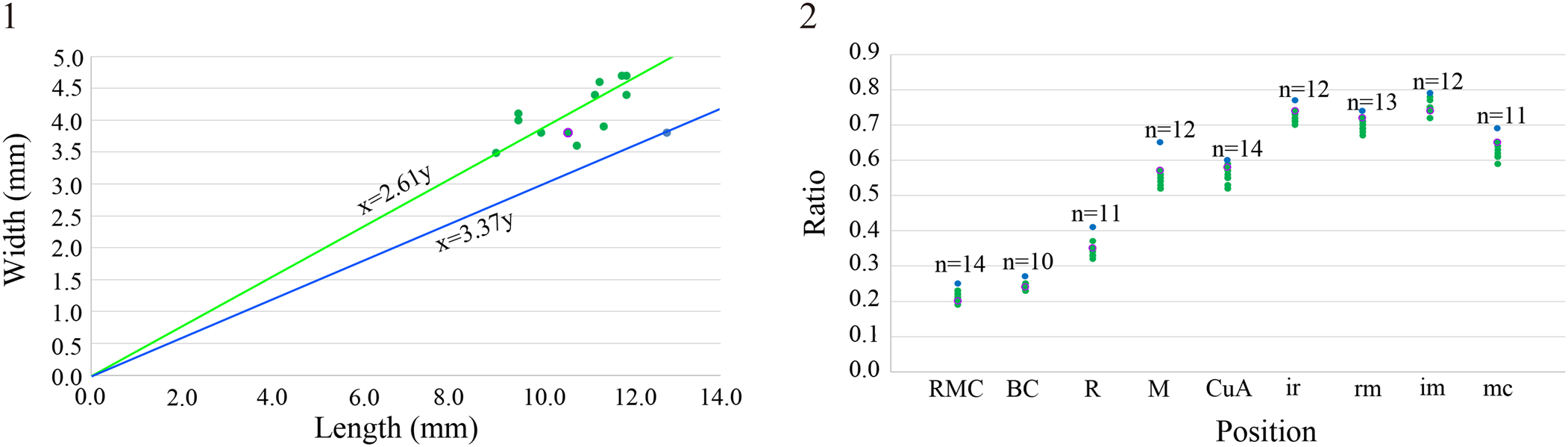
Scatter plots showing morphometric statistics of tegmina of holotype of Cycloscytina gobiensis and 11 new hylicellid fossils reported herein. (1) Bivariate plots of wing length and width; (2) ratios of branching position of longitudinal veins and mid-point position of crossveins to tegminal length. Green-filled circles represent specimens of Cycloscytina gobiensis, purple-outlined circles represent the holotype of Cycloscytina gobiensis, and blue-filled circles represent the holotype of Cycloscytina incompleta n. sp. Green line represents average tegminal length/width ratio of Cycloscytina gobiensis and blue line represents tegminal length/width ratio of Cycloscytina incompleta n. sp. RMC, BC, R, M, CuA: branching position of vein R + M + CuA, M + CuA (basal cell length), R, M, CuA/tegminal length; ir, rm, im, mc: mid-point position of basal crossvein ir, r-m, im, m-cua/tegminal length.

Landmarks and their corresponding morphological characters used in geometric morphometric analysis.

Repositories and institutional abbreviations
Type and other specimens examined in this study are deposited in the following institutions: the Nanjing Institute of Geology and Palaeontology, Chinese academy of Sciences in Nanjing, China (NIGP), the Shandong Tianyu Museum of Nature in Pingyi, China (STMN), and the Paleontological Institute of the Russian Academy of Sciences in Moscow, Russia (PIN).
Systematic paleontology
Order Hemiptera Linnaeus, Reference Linnaeus1758
Suborder Cicadomorpha Evans, Reference Evans1946
Superfamily Hylicelloidea Evans, Reference Evans1956
Family Hylicellidae Evans, Reference Evans1956
Subfamily Vietocyclinae Shcherbakov, Reference Shcherbakov1988
Genus Cycloscytina Martynov, Reference Martynov1926
Type species
Cycloscytina delutinervis Martynov, Reference Martynov1926, from the Middle–Upper Jurassic of Galkino, Kazakhstan, by original designation.
Species included
Seven species from the Jurassic of Asia: the type species; Cycloscytina asiatica (Martynov, Reference Martynov1937) (= Mesocixiella rohdendorfi Becker-Migdisova, Reference Becker-Migdisova1949) from the Lower Jurassic of Kyzyl-Kiya, Kyrgyzstan; C. extensa (Martynov, Reference Martynov1937) (= M. furcata Martynov, Reference Martynov1937, M. major Martynov, Reference Martynov1937, and M. parvula Martynov, Reference Martynov1937) from the Lower Jurassic of Shurab II, Kyrgyzstan; C. fulgoroides (Becker-Migdisova, Reference Becker-Migdisova1962) (= Vitreacixius ellipticus Becker-Migdisova, Reference Becker-Migdisova1962) from the Lower Jurassic of Sogjuta, Kyrgyzstan; C. korlaensis (Hong, Reference Hong1983) from the Middle Jurassic of Tashidian, Korla, China; C. gobiensis (Shcherbakov, Reference Shcherbakov and Rozanov1988a) from the Middle–Upper Jurassic of Bayan-Khongor, Mongolia, and the uppermost Middle–lowermost Upper Jurassic of Daohugou, Inner Mongolia, China; and the new species C. incompleta n. sp., described below, from the uppermost Middle–lowermost Upper Jurassic of Daohugou, Inner Mongolia, China.
Cycloscytina gobiensis (Shcherbakov, Reference Shcherbakov and Rozanov1988)
Figures 5–7
Holotype
PIN 3791/2100, from Middle–Upper Jurassic of Bahar, Mongolia.
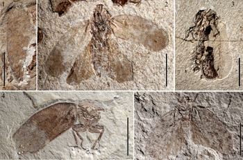
General habitus of new specimens of Cycloscytina gobiensis reported herein. (1) STMN48-1826, an isolated tegmen with clavus missing; (2) STMN48-1827, a whole-bodied male adult in dorsoventral aspect with tegmina and hindwings outspread; (3) STMN48-1828, a whole-bodied male adult in lateral aspect covered with left tegmen; (4) STMN48-1829a, an adult in frontal aspect, with right tegmen partly preserved; (5) STMN48-1830a, a whole-bodied adult in dorsoventral aspect with tegmina and hindwings outspread. Scale bars = 5.0 mm.

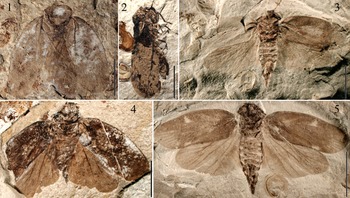
General habitus of new specimens of Cycloscytina gobiensis reported herein. (1) STMN48-1831a, a whole-bodied adult in dorsoventral aspect with tegmina and hindwings outspread; (2) STMN48-1832a, a female adult in lateral aspect covered with right tegmen; (3) NIGP05111, a whole-bodied female adult in dorsoventral aspect with tegmina and hindwings outspread; (4) NIGP05169, a whole-bodied adult in dorsoventral aspect with tegmina and hindwings outspread; (5) NIGP05170, a whole-bodied female adult in dorsoventral aspect with tegmina and hindwings outspread. Scale bars = 5.0 mm.

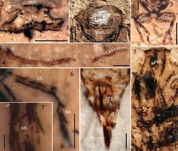
Morphological details of representatives of new specimens of Cycloscytina gobiensis reported herein. (1) Crown of STMN48-1830a, showing three ocelli; (2) pronotum and mesonotum of STMN48-1831a; (3) pro- and mesothoracic legs of STMN48-1829b; (4) right antenna of NIGP05169, with white triangles indicating flagellomeres; (5) rostrum of STMN48-1827; (6) right metathoracic leg of STMN48-1832a; (7) enlarged lateral spine on right metatibia of STMN48-1832a; (8) pygofer of NIGP05170. All photographs were taken with specimens wetted with ethanol, except STMN48-1831a. Symbols: cla. = claw; cox. = coxa; cro. = crown; fem. = femur; msn. = mesonotum; oce. = ocellus; ovi. = ovipositor; prn. = pronotum; psc. = postclypeus; pyg. = pygofer; rst. = rostrum; tar. = tarsus; tib. = tibia; tro. = trochanter. (1, 2, 6, 8) Scale bar = 1.0 mm; (3–5) scale bar = 2.0 mm; (7) scale bar = 0.1 mm.

Revised diagnosis
Body length ~12 mm. Flagellum with basal five or six segments long, and other segments extremely short. Pro-coxae not modified. Ovipositor long, slightly extending beyond tip of anal tube. Tegmen ~10.5 mm long, 4.0 mm wide, with length/width ratio ~2.6; basal cell ~0.24 of tegminal length; C3 distinctly longer than C2; R <5 times as long as M + CuA; M + CuA present, branching into M and CuA at connection with crossvein cua-cup; M forking slightly apicad of middle of wing, slightly basad or at the same level of branching position of CuA and much basad of claval apex; m-cua basad of claval apex. Hind wing with M1+2 and M3+4 with two or three terminal branches.
Description
Body cylindric, dark in color, ~12 mm long (Figs. 5, 6).
Head somewhat semicircular in dorsal view (Fig. 7.1), with length in midline ~0.4 times width, much narrower than pronotum. Crown flat, with anterior margin almost straight, tylus not visible in dorsal view. Compound eyes large, fusiform. Ocelli three, relatively large and very distinct, close to each other, and near anterior margin of crown. Antenna with ~15 segments; flagellum with basal five or six segments long, and remaining ones extremely short (Fig. 7.4). postclypeus inflated, ornamented with a distinct mid-ridge and thick transverse grooves, with length/width ratio ~1.8; anteclypeus much smaller than postclypeus; rostrum very long, extending to metathoracic coxae (Fig. 7.5).
Pronotum somewhat hexagonal, distinctly expanded, much larger than head, and covering most mesonotum, with length in midline ~0.4 times width; disc with posterior two-thirds area transversely rugose; anterior margin straight, in close contact with coronal posterior margin; anterior angles rounded; anterolateral margins smoothly curved, nearly as long as anterior margin; lateral angles sharp; posterolateral margins nearly straight, almost as long as anterior and anterolateral margins; posterior angles widely angulate; posterior margin almost straight, but sightly concave near midline of pronotum (Fig. 7.2). Mesonotum somewhat isosceles triangular, with length/width ratio ~0.4; lateral margins smoothly concave; posterior angle very sharp; disc with posterior one-third area weakly rugose (Fig. 7.2).
Prothoracic legs with coxae thick, somewhat coniform, and close to each other; trochanter much smaller than coxa; femur not very thick, about four-fifths length of tibia; tibia slender, covered with distinct setae; tarsus with apical tarsomere enlarged and much longer than basi- and midtarsomere; tarsal claws well developed (Fig. 7.3). Mesothoracic legs with trochanter slightly longer than pro-trochanter; femur almost as long as, but slightly slenderer than, pro-femur; tibia ~1.7 times long as femur, covered with tiny setae, slightly enlarged apically, with one row of tiny apical teeth; tarsus with apical tarsomere enlarged and much longer than basi- and midtarsomere; tarsal claws well developed (Fig. 7.3). Metathoracic legs largely destroyed for all specimens reported herein, tibia partly preserved in STMN48-1832, slender, with a lateral spine preserved, apical teeth likely absent or at least not well developed; tarsus with basi- and midtarsomere much longer than those of pro- and meso-tarsus, apical tarsomere longer than basi- and midtarsomere (Fig. 7.6).
Abdomen with each segment almost of similar width in dorsoventral view, but sharply shrinking for pygofer. Female with pygofer much longer than that of male. Pygofers and genitals of male specimens reported herein not preserved well, with morphological details not visible. Female pygofer with ovipositor very large, ensiform in lateral view and curved upwards, slightly extending beyond tip of anal tube; anal tube stout, with a dark-stained apical process (Figs. 6, 7).
Tegmen ~10.5 mm (9.0–11.9 mm) long, 4.0 mm (3.5–4.7 mm) wide, with average length/width ratio 2.61 (2.32–3.00). Dark-stained surface, with some irregular white speckles; upper surface densely covered with very tiny granules for basal half. Costal margin strongly arched; anterior margin smoothly arched or slightly concave; apical margin rounded; posterior margin slightly concave near claval apex; costal and claval margins slightly thickened. Basal cell relatively short, ~0.24 of tegminal length; costal area distinct, not extending beyond apex of postcostal cell; postcostal cell broad, with apex slightly basad of or almost the same level as claval apex; radial, median, and cubital cells prolonged; C1 long and narrow; C2 closed by two crossveins r-m, broad; C3 distinct, but almost as wide as C2; C4 and C5 subequal in length. Pc + CP long, ending at costal margin. Independent bScP very thick basally, then disappearing, not fused with R + M + CuA. Stem R + M + CuA branching into R and M + CuA at about basal 0.21 of tegminal length. Stem R <5 × stem M + CuA length, forking at about basal 0.34 of tegminal length; stem RA with 6–10 terminal branches; stem RP sinuous at connection with crossveins ir and r-m, with 2–4 terminal branches. Stem M + CuA present, branching into M and CuA at connection with crossvein cua-cup, near about basal 0.24 of tegminal length. Stem M nearly straight, forking slightly apicad of middle of wing, slightly basad or at the same level of branching position of CuA and much basad of claval apex, with 6–8 terminal branches. Stem CuA obviously curved at base, then nearly straight, and forking at about basal 0.55 of tegminal length. Stem CuP nearly straight. Pcu long and sinuous. A1 short and smoothly curved. Crossvein ir single or two in number; r-m commonly two; im commonly single, apicad of basal r-m, and basad of apical r-m; m-cua single, basad of claval apex; cua-cup singly, long, almost transverse.
Hind wing slightly shorter than tegmen, ~9.0 mm (8.5–9.7 mm) long, with distinct narrow appendix near wing apex. Outer margin with a very distinct incision at ending position of CuA2. RA single, very long, subparallel to RP. RP fused with branch M1+2 for a short distance. Stem M long and nearly straight, branching into M1+2 and M3+4 near middle of wing; M1+2 forking at about basal two-thirds wing length, with two or three terminal branches; M3+4 forking position variable, basad, apicad, or at same level of bifurcation of M1+2, with two or three terminal branches. Stem CuA long, slightly curved at base, then nearly straight, forking into CuA1 and CuA2 slightly apicad of bifurcation of M; CuA1 slightly curved at connection with crossvein m-cua, then almost straight; CuA2 apparently longer than CuA1, strongly curved at about its one-third length, then straight and gradually tending to CuA1, with ending point at incision of wing outer margin and extremely close to that of CuA1. CuP smoothly curved. Pcu very long and sinuous, closest to CuP near same level of bifurcation of M. A1 slightly curved apically. Crossvein m-cua long, slightly apicad of middle of wing.
New material
STMN48-1826, an isolated tegmen with clavus missing; STMN48-1827 and NIGP05169, whole-bodied male adults in dorsoventral aspect with tegmina and hindwings outspread; STMN48-1828, a whole-bodied male adult in lateral aspect covered with left tegmen; STMN48-1829a, b (part and counterpart), an adult in frontal aspect, with right tegmen partly preserved; STMN48-1830a, b (part and counterpart), NIGP05111, and NIGP05170, whole-bodied adults in dorsoventral aspect with tegmina and hindwings outspread; STMN48-1831a, b (part and counterpart), a whole-bodied adult in dorsoventral aspect with tegmina and hindwings outspread; STMN48-1832a, b (part and counterpart), a female adult in lateral aspect covered with right tegmen.
Remarks
The holotype of this species was from Middle–Upper Jurassic of Bahar, Mongolia. We herein report ten new specimens from Middle–Upper Jurassic of Daohugou, Inner Mongolia, China. Those specimens display a series of intraspecific variation (Figs. 5–7).
Cycloscytina incompleta new species
Figure 8
Holotype
NIGP05219, an isolated tegmen with clavus missing.
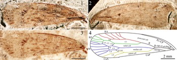
Holotype of Cycloscytina incompleta n. sp. (NIGP05219a, b). (1) Photograph of NIGP05219a; (2) photograph of NIGP05219b; (3) photograph of NIGP05219a, wetted with ethanol; (4) line drawings based on NIGP05219a and mirrored NIGP05219b, with tegminal vein and cell terminology labeled. See Figure 2 for abbreviations. Scale bars = 2.0 mm.

Diagnosis
Tegmen ~13 mm long and 4 mm wide, with length/width ratio ~3.4; basal cell ~0.27 of tegminal length; cell C3 almost as long as C2; R ~7 × stem M + CuA length; M + CuA present, branching into M and CuA at connection with crossvein cua-cup; M forking at about two-thirds of tegminal length, much apicad of branching position of CuA, and almost at the same level as claval apex; m-cua slightly apicad of claval apex.
Occurrence
Daohugou Village, Ningcheng County, Inner Mongolia, China; latest Middle–earliest Upper Jurassic.
Description
Tegmen 12.8 mm long, 3.8 mm wide, with length/width ratio 3.37. Dark-stained surface; upper surface densely covered with very tiny granules for basal half. Costal margin slightly thickened and smoothly arched; anterior margin slightly concave; apical margin rounded. Basal cell relatively long, ~0.27 of tegminal length; costal area distinct, not extending beyond apex of postcostal cell; postcostal cell broad and very long, with apex basad of claval apex; radial, median, and cubital cells prolonged; C1 long and narrow; C2 closed by two crossveins r-m, broad; C3 almost as long as, but slightly narrower than, C2; C4 and C5 subequal in length. Pc + CP long, ending at costal margin. Independent bScP very thick basally, then disappearing, not fused with R + M + CuA. Stem R + M + CuA branching into R and M + CuA at basal 0.25 of tegminal length. Stem R ~7 × stem M + CuA length, forking at basal 0.41 of tegminal length; stem RA with ~5 terminal branches; stem RP sinuous at connection with crossveins ir and r-m, with 3 terminal branches. Stem M + CuA present, branching into M and CuA at connection with crossvein cua-cup, at basal 0.27 of tegminal length. Stem M nearly straight, forking at about basal two-thirds of tegminal length, much apicad of branching position of CuA and almost at the same level as claval apex, with ~6 terminal branches. CuA obviously curved at base, then nearly straight, and forking at basal 0.60 of tegminal length. CuP nearly straight. Crossvein ir single; r-m two in number; im single, apicad of basal r-m, and basad of apical r-m; m-cua single, slightly apicad of claval apex; cua-cup singly, long, slightly inclined.
Etymology
The specific name is from the Latin ‘incompletus’ (incomplete), indicating its holotype with clavus unpreserved.
Remarks
The new hylicellid species can be attributed to the genus Cycloscytina of the subfamily Vietocyclinae based on the following combined tegminal characteristics: RA and M multi-branched, but just with five and six terminal branches, respectively; RP with three terminal branches; crossvein r-m two in number; and crossveins ir, im, and m-cua single. The new taxon is different from other known congenerics by possessing the tegmen with C3 almost as long as and slightly narrower than C2, and the forking position of M distinctly migrating towards wing apex and much apicad of CuA bifurcating. In addition, Cycloscytina incompleta n. sp. differs from C. asiatica, C. extensa, C. fulgoroides, and C. korlaensis by bearing the tegmen with stem M + CuA present and/or connecting to crossvein cua-cup at its bifurcation; from C. delutinervis and C. gobiensis by bearing a slenderer tegmen with relatively longer basal cell and m-cua slightly apicad of claval apex.
Results
The GMA dataset of Cycloscytina used in this study contains 12 tegminal samples and 14 landmarks. For GMA, matrices with missing data (landmarks) cannot be used, therefore specimens of C. fulgoroides and seven of the new specimens (STMN48-1826, STMN48-1828, STMN48-1829, STMN48-1831, STMN48-1832, NIGP05111, and NIGP05169) are not included in the analysis because they are too poorly preserved to display some key morphological characters. The 14 landmarks represent critical morphological traits for hylicellids (Table 2). The branching position of RA is a valuable feature among most cicadomorphans. For most Cycloscytina fossils, however, basal RA terminal branches are extremely thin, and hard to observe accurately, therefore this characteristic was not included.
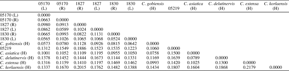
Relative Procrustes distances among 12 tegminal samples are shown in Table 3. The Procrustes distances among new specimens STMN48-1827, STMN48-1830, NIGP05170, and the holotype of C. gobiensis are commonly lower than 0.1 or just slightly higher than 0.1. The remaining Procrustes distances, however, are >0.1, with just four exceptions (Table 3). PCA analysis recognizes 11 principal components, with four most informative principal ones representing ~90% of variation (Fig. 3.1). Both graphs of principal components, I vs. II and III vs. IV, show that specimens STMN48-1827, STMN48-1830, NIGP05170, and the holotype of C. gobiensis are close to each other, but the new specimen, NIGP05219, is far away from them (Fig. 3.2, 3.3). The result of cluster analysis using arithmetic averages (UPGMA) and ‘Euclidean’ similarity index is shown in Figure 3.4. Within the UPGMA tree, specimens STMN48-1827, STMN48-1830, NIGP05170, and the holotype of C. gobiensis form an independent clade, C. asiatica, C. delutinervis, and C. extensa constitute another clade, and NIGP05219 and C. korlaensis occupy the basal position.
Relative Procrustes distance of geometric morphometric analysis of specimens of Cycloscytina Martynov, Reference Martynov1926. L = left tegmen; R = right tegmen; H = holotype; 05170 = NIGP05170; 05219 =|NIGP05219; 1827 =|STMN48-1827; 1830 = STMN48-1830.

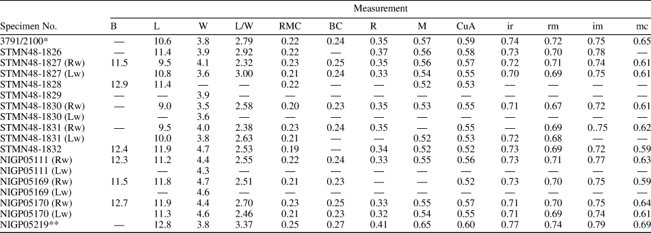
Our GMA results indicate that the new specimens STMN48-1827, STMN48-1830, and NIGP05170 can be compared to the holotype of C. gobiensis, but specimen NIGP05219 is likely new to science. To further explore the similarity/difference among our new specimens and C. gobiensis, morphometric statistics of some critical tegminal characters of all new specimens reported herein, as well as the holotype of C. gobiensis, was executed, (results shown in Table 4; Fig. 4). The results suggest that 10 new specimens (STMN48-1826, STMN48-1827, STMN48-1828, STMN48-1829, STMN48-1830, STMN48-1831, STMN48-1832, NIGP05111, NIGP05169, and NIGP05170) can be assigned to C. gobiensis, and NIGP05219 represents a new hylicellid species. A series of discriminatory morphological characters further confirm the result of morphometric statistics.
Morphometric data of tegmina of new hylicellid specimens in this study and holotype of Cycloscytina gobiensis. Abbreviations: Rw = right tegmen; Lw = left tegmen; B = body length (mm); L = tegminal length (mm); W = tegminal width (mm); L/W = tegminal length/width ratio; RMC, BC, R, M, CuA = branching position of vein R + M+ CuA, M + CuA (basal cell length), R, M, CuA/tegminal length; ir, rm, im, mc = middle point position of basal crossvein ir, r-m, im, m-cua/tegminal length.

*holotype of Cycloscytina gobiensis; **holotype of Cycloscytina incompleta n. sp.
Discussion
The highly diversified family Hylicellidae, containing the oldest representatives of the cicadomorphan clade Clypeata, is likely the basal group of Clypeata and ancestral to all modern cicadomorphan lineages (Wang et al., Reference Wang, Szwedo and Zhang2012; Lambkin, Reference Lambkin2019). Up to now, three subfamilies (Hylicellinae, Vietocyclinae, and Conjucellinae) have been erected within Hylicellidae (Shcherbakov, Reference Shcherbakov1988b, Reference Shcherbakov2012). The subfamily Vietocyclinae, with the somewhat reticular tegmen closed by multiple crossveins and much proliferated longitudinal venations, can be easily discriminated from its confamilial relatives (Lambkin, Reference Lambkin2020). Conjucellinae is characterized by a specialized tegmen with the terminal branches of longitudinal veins distinctly reduced in number (Shcherbakov, Reference Shcherbakov2012). The known representatives of Hylicellinae possess some common tegminal features (e.g., RP single, M four-branched, and crossveins [ir, r-m, im, m-cua] single) that are shared with other primitive cicadomorphan lineages, as well as with basal groups of living cicadomorphans. Therefore, this subfamily might be transitional between ancient Paleozoic Cicadomorpha and modern descendants.
The taxonomic history of Hylicellidae is confusing, with a mass of synonyms and incorrect systematic positions. During the last 30 years, extensive taxonomic reviews have been executed in a series of studies (Shcherbakov, Reference Shcherbakov1988b, Reference Shcherbakov2012; Lambkin, Reference Lambkin2019, Reference Lambkin2020; Lara and Bashkuev, Reference Lara and Bashkuev2020; Lara et al., Reference Lara, Bustos-Escalona, Mancuso and Arcucci2021), aiding us to better understand the components of Hylicellidae. Two Cycloscytina species, C. plachutai Becker-Migdisova, Reference Becker-Migdisova1949, and C. reducta Becker-Migdisova, Reference Becker-Migdisova1949, from the Lower Jurassic of Kyrgyzstan were transferred to Procercopidae Handlirsch, Reference Handlirsch1906, by Shcherbakov (Reference Shcherbakov1988b), with the former species treated as incertae sedis and the latter species transferred to Anthoscytina Hong, Reference Hong1983. Cycloscytina plachutai bears a tegmen with crossveins not numerous and M three-branched, which are shared by the type species of Procercopina Martynov, Reference Martynov1937. Thus, this species is herein transferred to the genus Procercopina, resulting in P. plachutai new combination.
The hylicellid classification and taxonomy are based almost exclusively on tegminal characters because most hylicellid specimens are just represented by isolated tegmina. Hylicellids, especially Hylicellinae and Vietocyclinae, possess a tegmen with considerable intraspecific or even intra-individual variation in venation, due to its proliferated longitudinal veins and crossveins. The variation might be considered to be diagnostic at a species-level, leading to many synonyms in previous publications (see Shcherbakov, Reference Shcherbakov1988b, Reference Shcherbakov2012; Lambkin, Reference Lambkin2019, Reference Lambkin2020). Geometric morphometric analysis, with great statistical power on shape continuum variations, has shown its effectiveness in objectively resolving taxonomic uncertainties and disentangling morphologically confusing groups, and so has widely been used in the study of phenetic relationships for extinct organisms (Evin et al., Reference Evin, Baylac, Ruedi, Mucedda and Pons2008; Lin et al., Reference Lin, Shih, Labandeira and Ren2016; Li and Zhang, Reference Li and Zhang2017). The GMA results of Cycloscytina presented herein suggest that most of our new specimens can be compared to the holotype of C. gobiensis, and the specimen NIGP05219 likely belongs to a new species (Fig. 3). Our subsequent morphometric statistics with more specimens and tegminal characters confirm the GMA conclusion (Fig. 4). On the basis of a series of discriminatory morphological characters, the diagnosis of C. gobiensis is revised and the new hylicellid, C. incompleta n. sp., is erected.
Although GMA has been used widely in systematic studies of extinct organisms, its application on fossil insects is relatively scarce (e.g., Lin et al., Reference Lin, Shih, Labandeira and Ren2016; Shih et al., Reference Shih, Li and Ren2017; Jouault and Nel, Reference Jouault and Nel2021). We herein provide a new case study on GMA for fossil insects, especially on the data with only wing venations included. Our GMA results suggest that this method is sensitive to explore new species: the holotype of C. incompleta n. sp. (NIGP05219) is located far away from the remaining known Cycloscytina spp. in PCA analysis (Fig. 3.2, 3.3) and forms an independent clade in cluster analysis (Fig. 3.4).
Our results also indicate that it might be unpractical to solely use GMA to define species boundaries. Four specimens of C. gobiensis were included in our MGA, along with three of our new specimens with both right- and left-wing information (Table 3). Although these seven wings are located in a single zone in our PCA analysis, they are very dispersive (Fig. 3.2, 3.3); the cluster analysis also shows that the distances among individuals of C. gobiensis are not distinctly shorter than those between different species (Fig. 3.4). To date, GMA studies of wing-bearing fossil insects are all with limited individuals and limited landmarks, and the effectiveness of GMA application for fossil insect species delimitation should be further examined in future studies using large data matrices.
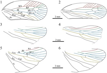
Our GMA and morphometric statistics analyses indicate that some wing characters, which are commonly chosen as critical traits for the diagnosis of extinct cicadomorphans and other fossil insects, are not very reliable. The tegminal shape (length/width ratio) is treated as an important discriminating character for fossil cicadomorphans, but specimens of C. gobiensis display an extremely high intraspecific or even intra-individual variation (Figs. 4.1, 5.2, 9.1, 9.2). The variation is likely strongly influenced by taphonomic factors, therefore deformation should be considered in taxonomic studies (Chen et al., Reference Chen, Wang, Zheng, Jarzembowski, Jiang, Wang, Zheng and Zhang2019b). The venation on hind wings has simplified topology and reduced terminal branches, and is commonly very conservative at species, genus, or even family levels (Chen et al., Reference Chen, Zheng, Wei and Wang2017). The relative forking position of M1+2 and M3+4 of the hind wing is considered one of diagnostic characters of the hylicellid Vietocycla in Shcherbakov (Reference Shcherbakov1988b). The new specimens of C. gobiensis reported herein, however, show that the character is even unstable for the left and right hind wings of the same individual (Figs. 5.2, 6.3, 9.3–9.6). In addition, the number of the terminal branches of hind wing M1+2 and M3+4 display a great intra-individual variation (Figs. 5.2, 6.3, 9.3–9.6). Our findings suggest that Hylicellidae is likely not as stable as other cicadomorphan groups in hind wing venation.
Reconstructed line drawings of tegmen and hind wing of Cycloscytina gobiensis reported herein, showing high morphological variation. (1, 2) Right tegmen and left tegmen of STMN48-1827; (3, 4) left hind wing and right hind wing of STMN48-1827; (5, 6) right hind wing and left hind wing of NIGP05111. See Figure 2 for abbreviations.

Most known hylicellid species have been erected solely on the basis of isolated tegmina or hind wings, and their body structures are little known. The nine whole-bodied specimens of C. gobiensis reported herein display some well-preserved morphological traits (Figs. 5–7). Our new fossil data show that C. gobiensis bears a median ocellus, which is a plesiomorphic character for Cicadomorpha and is retained in modern Cicadoidea, Tettigarctidae and Cicadidae, as well as in the ancient groups of two other modern cicadomorphan lineages (Cercopoidea and Membracoidea s.l.). Our new specimen NIGP05169 reveals that C. gobiensis possesses the specialized ~15-segmented antennae with basal five or six flagellomeres long, and remaining segments extremely short (Fig. 7.4). The nine-segmented antenna is a primitive condition for Clypeata, which is also shared by modern Cicadoidea and Myerslopiidae of Membracoidea s.l. (Anufriev and Emeljanov, Reference Anufriev, Emeljanov and Ler1988; Hamilton, Reference Hamilton1999; Chen et al., Reference Chen, Szwedo, Wang, Zheng, Jiang, Jiang, Wang and Zhang2019a). The distinctly expanded pronotum with posterior area transversely rugose in C. gobiensis (Fig. 7.2) is widely shared by the basal groups of Cicadoidea and Cercopoidea (Tettigarctidae and Procercopidae, respectively) (Wang et al., Reference Wang, Szwedo and Zhang2012; Chen et al., Reference Chen, Wang, Zhang, Wang and Zheng2015; Chen and Wang, Reference Chen and Wang2016).
Subterranean and accordingly root-sucking nymphs have been postulated for hylicelloids, with some adults displaying distinctly modified prothoracic legs (Shcherbakov, Reference Shcherbakov1988b; Shcherbakov and Popov, Reference Shcherbakov, Popov, Rasnitsyn and Quicke2002; Chen et al., Reference Chen, Szwedo, Wang, Zheng, Jiang, Jiang, Wang and Zhang2019a). Our new specimens of C. gobiensis with normal prothoracic legs (Fig. 7.3) suggest that nymphs of some hylicelloid groups might be not subterranean, so avoided predators in other ways. In addition, the new specimen STMN48-1832 indicates that the metatibiae of C. gobiensis likely possess lateral spine(s) (Fig. 7.7), as do Cicadoidea and Cercopoidea. This character, however, should be further confirmed since the metathoracic legs of our specimens are all largely destroyed and very obscure.
The type locality of C. gobiensis is the Bahar outcrop of the Togo-Khuduk Formation in Mongolia. The outcrop has yielded ~20 fossil genera and species of insects, with most of them exclusively recorded from this stratum. The snipe fly genus Palaeobolbomyia Kovalev, Reference Kovalev1982, with seven known species, is documented at Bahar and Daohugou, and the Upper Jurassic Karabastau Formation of Kazakhstan, as well as the Upper Jurassic Khaya and Uda formations of Russia. The hump-winged cricket genus Bacharaboilus Gorochov, Reference Gorochov1988, and lacewing genus Chorilingia Shi et al., Reference Shi, Wang, Yang and Ren2012, with three and five known species, respectively, are only recorded in the Bahar outcrop and the Daohugou Biota. In addition, our new fossils reveal that the hylicellid species C. gobiensis was distributed in both the Bahar and Daohugou outcrops. The available paleontological data suggest that the two assemblages are undoubtedly related, and the Bahar Lagerstätte is likely late Middle to early Late Jurassic in age.
Acknowledgments
Our sincere gratitude is offered to S. Zamora and C. Cai for editing and two anonymous reviewers for the very useful comments on the earlier version of the manuscript. This research was supported by the National Natural Science Foundation of China (42172033; 42125201; 41502007), Strategic Priority Research Program of the Chinese Academy of Sciences (XDB26000000), and Natural Scientific Foundation of Shandong Province (ZR2020YQ27).
Data availability statement
Data available from Zenodo: https://zenodo.org/record/6460025.