





Prologue
Since the publication of Myers’ foundational work in Reference Myers2012, which positioned biodesign as a novel design paradigm integrating living organisms to generate novel functions, forms and sustainable modes of production, the field has expanded rapidly. This expansion has been driven by technological advances and the acceleration of interdisciplinary collaboration (Atzmon Reference Atzmon2023; Karana Reference Karana2020; Marseglia et al. Reference Marseglia, Cantini, Celli, Brunelli and Lotti2025). Today, biodesigners increasingly realise, upscale and disseminate their ideas through education and public engagement, broadening the field’s scope (Grushkin et al. Reference Grushkin, Vijayakumar and Eastman2022; Toivonen and Van Ditmar Reference Toivonen and Van Ditmar2024; Walker et al. Reference Walker, Barrera, Perez-Piza, Acquah, Grushkin and Vijayakumar2023). Alongside these developments, ethical awareness and critical discourse surrounding biodesign have evolved in parallel, including calls for more holistic and regenerative approaches (Armstrong Reference Armstrong, Lockton, Lenzi, Hekkert, Oak, Sádaba and Lloyd2022; Collet Reference Collet2020; Karana et al. Reference Karana, McQuillan, Rognoli and Giaccardi2023). These perspectives advocate fostering creative assemblages; environments where humans and non-humans actively participate and coevolve within a dynamic ecology of interconnected living and non-living entities (Karana et al. Reference Karana, McQuillan, Rognoli and Giaccardi2023).
As biodesign is applied more substantively in real-world contexts, these considerations have shifted from foresight concerns to practical and urgent questions for the near-term development of the field. Biodesigners are increasingly called upon to engage with the full lifecycle of living artefacts, while attuning to more-than-human (MTH) temporalities and scales (Ikeya et al. Reference Ikeya, Barati and Wensveen2025; Ofer and Alistar Reference Ofer and Alistar2023; Zhou et al. Reference Zhou, Doubrovski, Giaccardi and Karana2024), and multispecies interactions that unfold across these stages (Groutars et al. Reference Groutars, Kim and Karana2024). Yet most biodesign practices, particularly at the microbial scale and within applied research contexts, remain dominated by heavily controlled scientific workflows (Ihls and Pollini Reference Ihls and Pollini2025). While these approaches ensure scientific rigour, technical feasibility and quality control, they often entail implicit instrumentalisation (Zhou Reference Zhou2025). Even with notable advancements in MTH design methods (Giaccardi et al. Reference Giaccardi, Redström and Nicenboim2024; Nicenboim et al. Reference Nicenboim, Lindley, Zaga, Berger, Forlano, Giaccardi, Gray, Ciliotta Chehade, Hekkert, Forlano, Ciuccarelli and Lloyd2024), their implementation in biodesign laboratory settings remains limited. To address this gap, we engage directly with biodesign practice to explore whether and how MTH sensibilities can be cultivated and enacted alongside conventional laboratory conditions.
The first author (biodesigner) approached this inquiry as a designer with an artistic background, newly engaging with laboratory-based biological research. Prior to the three-year engagement described here, their experience with biology and microbial systems was limited, having developed an interest in ecological and microalgal sustainability through design but only beginning to learn biological fundamentals through a graduate biodesign course at Delft University of Technology. Immediately following this foundational exposure, the first author entered the biodesign lab to explore scientific protocols and cultivate hands-on familiarity with living systems. The lab trajectory commenced at this point, focusing on in-depth exploration of a specific cyanobacterial strain – Synechocystis sp. PCC 6803 (Stanier et al. Reference Stanier, Kunisawa, Mandel and Cohen-Bazire1971) – which formed the basis for subsequent design inquiry. Over a period of three years, the account traces a process shaped by both in-situ, contemporaneous recording and note-taking and retrospective synthesis. Experimental milestones, laboratory notebooks, logs, photographs and ongoing personal and dialogic reflections collectively scaffolded the inquiry, weaving together immediate impressions and reflective insights. Its biological traits, particularly phototactic behaviour, prompted experiments focused on motility and scale. These investigations developed along two parallel trajectories: one, the macroscopic manifestations of motility, guided by a biological protocol, and two, the speculation on light sensing in relation to movement through an experiential mode of becoming-with cyanobacteria (Kim et al. Reference Kim, Kim, Martins, Karana, Gray, Ciliotta Chehade, Hekkert, Forlano, Ciuccarelli and Lloyd2024). The speculative trajectory emerged after a period of laboratory immersion, as the first author began to work more specifically with cyanobacteria and grappled with the question of how these organisms might sense the environment around them. While Kim et al. (Reference Kim, Kim, Martins, Karana, Gray, Ciliotta Chehade, Hekkert, Forlano, Ciuccarelli and Lloyd2024) laid the conceptual groundwork for cultivating MTH sensibilities towards microbes in biodesign, the present study introduces and demonstrates specific operational scenarios for enacting and examining these sensibilities in the laboratory context.
The first author’s experiences are articulated through two autoethnographic accounts that foreground moments of self-reflective (un)learning within laboratory and studio–lab hybrid settings. These accounts make visible the methodological boundaries of contemporary biodesign practice and explore how biodesigners can embody MTH sensibilities from within existing regulated conditions. Many MTH works use decentring methods that immerse practitioners in shared environments and enable them to access knowledge often inaccessible to humans alone (Giaccardi et al. Reference Giaccardi, Cila, Speed and Caldwell2016; Özgen et al. Reference Özgen, Li, Ploehn, Chang, Chen and Hsu2025; Rossellini Reference Rossellini2010; Thwaites Reference Thwaites2016), and the first author similarly positioned themselves alongside microbes to gain a deeper, more acclimatised understanding within their experimental context. The first trajectory investigates cyanobacterial motility through laboratory studies supported by a bio-digital setup (phototaxis toolbox) for tuning light conditions, along with an optical see-through head-mounted display (HMD) that was primarily used to access scientific literature during the lab experiments. The second trajectory involved a video see-through HMD to visually speculate on cyanobacterial light response.
Through this journey, we critically examine how engaging with cyanobacteria involves both learning established scientific protocols and unlearning anthropocentric assumptions embedded within them. Early in the process, moments arose that unsettled the first author’s prior approaches: for instance, repeated failures to induce cyanobacterial motility in the laboratory prompted a reevaluation of seemingly minor environmental factors, and illuminated the need to let go of human-centred expectations of control. Similarly, the attempt to simulate cyanobacterial sensing through immersive digital tools highlighted the limits of translating microbial sensory experience into human terms, underscoring the need to unlearn ingrained visual biases. These early experiences prompted a reconsideration of ingrained habits and exposed a broader methodological limitation: conventional laboratory documentation rarely makes space for the complex relational or contextual factors in biodesign. Together, they point to something more fundamental – practising with MTH sensibilities demands not just new methods, but a shift in perspective that requires letting go of control, unlearning established conventions and cultivating new modes of attention, articulation and recognition.
Negotiating between instrumental and MTH persepectives
What follows are two parallel trajectories of a biodesign research journey, presented side by side across these pages. On the left, the laboratory trajectory unfolds through biological experiments following established research protocols. On the right, the design studio trajectory moves outside the lab, where speculative explorations take centre stage. While each begins from different starting points, both ultimately encounter moments of (un)learning as their trajectories unfold. Both are narrated from the autoethnographic perspective of the first author. Autoethnography is an ethnography focused on the self where the author is both informant and investigator (Cunningham and Jones Reference Cunningham and Jones2005). Also known as first-person research, it enables designers to create for themselves and refine their work through personal use (Höök et al. Reference Höök, Caramiaux, Erkut, Forlizzi, Hajinejad, Haller, Hummels, Isbister, Jonsson, Khut, Loke, Lottridge, Marti, Melcer, Müller, Petersen, Schiphorst, Márquez Segura, Ståhl, Svanæs, Tholander and Tobiasson2018; Lucero et al. Reference Lucero, Desjardins, Neustaedter, Höök, Hassenzahl and Cecchinato2019; Neustaedter and Sengers Reference Neustaedter and Sengers2012). Given that it has proven to be useful for capturing everyday nuances and is well-suited to design’s hands-on, embodied nature (Adams et al. Reference Adams, Jones and Ellis2013), while encouraging critical reflection (Desjardins and Wakkary Reference Desjardins and Wakkary2016; Tomico et al. Reference Tomico, Wakkary and Andersen2023), it is an excellent methodology for this inquiry. In biodesign, autoethnographic approaches have been used to foreground first-hand laboratory experiences, revealing the interdisciplinary dynamics and the drifting boundaries of knowledge in this field (Kuznetsov et al. Reference Kuznetsov, Barrett, Fernando and Fowler2018; Sawa Reference Sawa2016; Stefanova Reference Stefanova2021). Past efforts have shown the value of integrating laboratory observations and lived experience, merging personal standpoints with empirical insights (Pollini Reference Pollini2024). Building on this foundation, we use an autoethnographic lens to probe actionable MTH modes of practice within the biodesign lab operating under regulated scientific backdrops (see Figure 1). At its core, our autoethnography is a self-reflective journey marked by uncertainty, vulnerability and permeability. Our accounts intentionally foreground these qualities, adopting a direct and subjective voice to reflect the lived experience of inquiry. The tone and subheadings are intentionally unpolished, capturing the first author’s ongoing tensions and negotiations between instrumental aims and MTH perspectives throughout the process. Visual cues throughout the narrative are used to highlight and trace the first author’s nuanced moments of ⚠ tension, ⚖ negotiation,  unlearning,
 perspective shifts and
 human-centred assumptions.
Annotations on images of the Biosafety Level 1 Biodesign Lab at TU Delft, the starting point of the autoethnographic journey.

So, what is Synechocystis sp. PCC 6803?
As someone trained in design and learning about biology, my encounter with Synechocystis sp. PCC 6803 (Figure 2) was not intentional. PCC 6803 is a unicellular freshwater cyanobacterium capable of phototrophic growth via oxygenic photosynthesis in light and heterotrophic growth in darkness (Heidorn et al. Reference Heidorn, Camsund, Huang, Lindberg, Oliveira, Stensjö and Lindblad2011). Recommended by the biodesign lab technician (Joren Wierenga) and the fourth author, a cyanobacteria specialist, my initial reaction was negative: memories of toxic algal blooms, the acrid smell of out-of-control eutrophic water and green slime coating surfaces in my fish tank. Yet, seeing these organisms under controlled laboratory conditions changed my perspective. Instead of foul odours, I found vivid green liquid, quietly supporting the biodesign work at hand. Cyanobacteria are ancient world-makers, having existed for approximately 2.7 billion years and contributing to the Great Oxygenation Event (Demoulin et al. Reference Demoulin, Lara, Lambion and Javaux2024). Beginning this research became my first positive interaction with cyanobacteria. In my lab explorations, I learned to culture PCC 6803 in BG11 medium under cool-white light at ≈20 μmol photons m−2 s−1 and at room temperature (20–22°C) (Rippka Reference Rippka1988; Rippka et al. Reference Rippka, Deruelles, Waterbury, Herdman and Stanier1979; Williams Reference Williams1988), observing the gradual intensification of the green in the culture flasks as their photosynthetic pigments accumulated. All these qualities made it an intriguing, if humbling, entry point for my laboratory journey.
Singlet oxygen imaging in Synechocystis cells. Cells were exposed to red light for 30 min at 37 °C with 50 μM Singlet Oxygen Sensor Green (SOSG). Images show combined Nomarski differential interference contrast (DIC) and SOSG-endoperoxide (EP) fluorescence (λem = 505–525 nm). Scale: 5 μm. (Adapted from Prasad et al. Reference Prasad, Sedlářová and Pospíšil2018).

Trajectory A
What, they can move?!
I had no idea they could move, as shown in Figure 3. The discovery arrived unexpectedly while exploring basic biological practices with PCC 6803; I encountered a phototaxis assay conducted at both macroscopic and microscopic scales (Jakob et al. Reference Jakob, Schuergers and Wilde2017). The images showed green streaks spreading across agar surfaces, microscopic living beings moving in ways visible even to the human naked eye. The moment felt improbable, marked by a temporal dissonance between microbial movement and human perception, motion unfolding at a scale I was not expected to notice. The technical explanations came later, grounding my wonder in this movement. They use retractile type IV pili (T4P) to traverse moist surfaces in a jerky motion called twitching motility (Bhaya et al. Reference Bhaya, Bianco, Bryant and Grossman2000), and individual cells can directly detect unidirectional illumination by focusing light in a sharp focal point on their distal side (Schuergers et al. Reference Schuergers, Lenn, Kampmann, Meissner, Esteves, Temerinac-Ott, Korvink, Lowe, Mullineaux and Wilde2016). But what captivated me was simpler: the possibility of observing their movement towards light at a macroscopic scale, like plants bending towards windows or insects drawn to bulbs. This visibility – from cellular machinery to behaviour potentially observable without complex instrumentation – collapsed the distance I had assumed between myself and microbial life. This encounter with phototaxis became the foundation for what would follow in my laboratory practice.
Synechocystis cells accumulating in finger-like projections while moving towards a directional light source at 49 h. White rectangle (left) indicates the region highlighted on the right. Scale bars: 10 µm. (Adapted from Ursell et al. Reference Ursell, Chau, Wisen, Bhaya and Huang2013).

Okay, let’s make them move
I was certain it would be simple to make PCC 6803 move. Full of excitement and with minimal protocol details, I began growing the cells in the lab to comprehend and test their phototaxis behaviour firsthand. I watched the culture flasks deepen from pale to vivid green, showing this organism’s growth and readiness for experimentation. I prepared Petri dishes with their nutritional requirements (BG11 agar medium), folded black paper into makeshift light barriers and inoculated the cultures I judged “green enough” onto the prepared surfaces. My understanding was reductive: healthy cyano, BG11 agar medium, directional light. I centrifuged cells to concentrate them into a deep, dark inoculum, confident that having more cells would help them to thrive and subsequently amplify their movement (see Figure 4).
Culturing Synechocystis sp. PCC 6803 in liquid media flasks (left) and inoculated as a concentrated 3 × 3 grid on an agar Petri dish for a phototaxis experiment (right).

⚠ They never moved. Sometimes they bleached out, pale apparitions of their former green. Sometimes contamination bloomed, unknown fungi, heterotrophic bacteria claiming the plates I thought I’d prepared carefully. Sometimes they simply stayed green at the initial inoculation site, looking perfectly healthy under the microscope yet utterly immobile, as if refusing to meet my expectations (Figure 5). I had anticipated one month for this experiment. It took far longer than I anticipated. The failure was relentless, repetitive, humbling. Each unsuccessful plate became a lesson in what I had overlooked, dismissed or misunderstood about the conditions these organisms required to move.
Microscope setup displaying a real-time microscopic image of inoculated Synechocystis sp. PCC 6803 cells on an agar Petri dish via a connected monitor.

So, I built an augmented setup
The failures forced me into conversation. I met repeatedly with the biodesign lab technician, who guided me through the lab experiments and with the fourth author, a microbiologist, cataloguing what I had overlooked: temperature, humidity, light intensity, sterile conditions, precise medium type, agar concentration and even strain accuracy. Each variable I had dismissed as minor could derail everything. Meticulousness, I realised, was not pedantry but attentiveness, a form of respect for microbial requirements I had failed to extend. From that moment, I sought to understand the shared environment at their microbial scale, attuning my experimental setting to their level of sensibilities. I began augmenting conventional lab tools with digital instruments: a customised phototaxis setup we had built from 3D-printed containers, LED strips and a Raspberry Pi (Figures 6 and 7); a photometer and an infrared thermometer; and a HoloLens 2 to visualise data spatially during practice.
Custom-built phototaxis assay toolbox featuring a 3D-printed Petri dish container lined with an RGBW LED strip, connected to a housed Raspberry Pi control unit.

Close-up view of the custom phototaxis assay toolbox in operation, showing an LED strip surrounding a Petri dish inoculated with Synechocystis sp. PCC 6803 colonies.

I also learned that PCC 6803 has several known wild-type variants that can differ greatly in characteristics such as motility. Sadly, many studies used distinct substrains, often without specifying them (Schmelling and Bross Reference Schmelling and Bross2024), leaving me confused. As the protocol I was referring to also used a particular substrain, I requested the exact motile “Moscow” substrain, “PCC-M,” from the protocol’s author. The organisms I sought carried their own lineage and history of care.
Let’s make their motility controllable!
I felt ready. The failures had transformed me into someone who engaged with protocol as microbial requirements, not rules to follow, but sensitivities to respect. I had adapted the protocol to reflect both their needs and my available equipment. As shown in Figures 8 and 9, the augmented setup allowed me to attend to multiple scales simultaneously: automated light control, real-time environmental monitoring and hands-free check of scientific details on existing literature while maintaining sterility.
The first author using a HoloLens 2 mixed reality headset to examine a Synechocystis sp. PCC 6803 liquid culture flask during laboratory practice.

The first author’s point-of-view perspective via the HoloLens 2 headset, showing multiple digital windows displaying reference protocols and microscopic imagery floating above the physical laboratory bench during a phototaxis experiment.

Everything felt aligned. I was genuinely excited, anticipatory in a way that made the waiting feel charged with possibility. Would I finally see them move? My team shared this excitement. We gathered around the subtle promise of movement – not merely to witness motility under the lens, but to dwell on what such a quality might come to mean for biodesign. The conversations had already leapt ahead: if we could make them move in response to light cues, could we scale this up? Programme patterns? Create living displays? The instrumental possibilities felt tantalisingly close. I stood at the threshold between failure and hoped-for success, between observation and application. I carried all my preparation towards organisms I believed would finally perform for me. I was unaware that I was crossing from learning to see their autonomous behaviour towards seeking to make them move, to control their movement, to instrumentalise their capacities for human-determined ends.
Wait, does this happen in nature?
A question interrupted my instrumental momentum: Does this motility happen in nature? I began searching beyond the protocol. What I found exceeded my laboratory imagination. Synechocystis phototaxis in nature is questionable, since their lower light sensitivity means they respond only to bright sources like sunlight in shallow water, and microphysical factors further obscure their movement (Nilsson and Colley Reference Nilsson and Colley2016). Yet, other phototactic cyanobacteria offered insights at scales I could witness. Some Oscillatoria species form massive conical mat structures through gas-filled buoyancy, and their collective phototactic movement can create visible architecture (Biddanda et al. Reference Biddanda, McMillan, Long, Snider and Weinke2015). In nature, cyanobacteria can grow to form visible green mats at the water’s surface, as illustrated in Figures 10 and 11. This phenomenon, known as cyanobacterial blooms, is influenced by several factors, including light, which directs cells towards optimal photosynthetic conditions. As a result, biomass becomes concentrated, nutrient cycles shift and water chemistry and colour are transformed (Dervaux et al. Reference Dervaux, Mejean and Brunet2015; Wu et al. Reference Wu, Wu, Rovelli and Lorke2024; Xue et al. Reference Xue, Ma, Shen, Wu, Hu, Guo, Cao and Xiong2023).
Synechocystis bloom in the pristine Nadgee Lake, November 2008. (Adapted from Scanes et al. Reference Scanes, Ferguson and Potts2020).

Cyanobacterial mat covering the lake floor at Middle Island Sinkhole, Lake Huron, displaying a panoramic overview (top; photo: Tane Casserly, NOAA) and a rising gas-buoyant conical finger (bottom; photo: Joe Hoyt, NOAA) (left; adapted from Biddanda et al. Reference Biddanda, McMillan, Long, Snider and Weinke2015). Eutrophication during summer in the canals of the author’s neighbourhood (right; photo: Fanzhe Zeng).

What I had pathologised as environmental contamination revealed itself as ecological agency. Stepping outside the laboratory, I began to recognise their dynamic presence everywhere: seasonal cyanobacterial blooms coloured canals and lakes, even pushing people away from the water during summer. They were not performing for my apparatus; they were actively reshaping ecosystems, mediating planetary chemistry, operating as world-making agents whose movements affect and shift ecological processes across scales I was only beginning to comprehend.
I still couldn’t get them to move, but…
Returning to the laboratory, I confronted an irony: after nearly three years, I still could not make them move towards the light. Technical mastery remained elusive. Yet something more consequential had shifted. The repeated failures, the augmented instruments and the oscillation between controlled laboratory conditions and in-situ ecological observation fundamentally reconfigured my relationship to these organisms. Navigating the gap between strain and substrain taught me that even subtle genetic differences can yield distinct forms of livingness, highlighting the complexity and variability inherent to microbial life. Observing local wild cyanobacteria further emphasised that strain-level classification is a fragile, human-maintained construct, nearly impossible to pinpoint in nature. This tension unsettled the boundaries I had assumed between lab organism and ecological being, between experimental subject and autonomous world-maker. The friction between protocol-driven practice and field-based encounters surfaced ethical questions that biodesign often brackets: What does “contamination” mean from multispecies perspectives? How do practices of “waste” and “sterilisation,” essential for biosafety and technical rigour, constrain the epistemic bearing and operational legitimacy of MTH sensibilities that regenerative futures demand (Figures 12 and 13)?
Two culture flasks containing Synechocystis sp. PCC 6803 prepared for autoclaving as biological waste (left). A liquid culture of the motile Synechocystis sp. PCC 6803 substrain ‘PCC-M’ displaying distinctive colony traits with biofilm formation (right).

Microscopic view of an unknown fungal mycelium growing on a Synechocystis sp. PCC 6803 agar Petri dish.

⚖ I remain committed to technical mastery, but I now understand failure, augmentation, and scale-shifting not as obstacles to biodesign but as methodological conditions, ways of holding biodesign practices accountable to the microbial worlds they seek to engage.
Trajectory B
What, they can see?!
While studying their motility, I became captivated by the mechanism enabling it: micro-optics. Each Synechocystis cell functions as a microscopic eyeball. Directional illumination produces a sharply focused spot of light – resembling a photonic nanojet – at the cell periphery opposite the light source (Schuergers et al. Reference Schuergers, Lenn, Kampmann, Meissner, Esteves, Temerinac-Ott, Korvink, Lowe, Mullineaux and Wilde2016). This lensing effect creates an intensity gradient that is much stronger than simple shading, allowing the cell to perceive light direction and control its movement accurately. As shown in Figure 14, at 3 µm in diameter – half a billion times smaller than the human eye – these cells perceive inverted, low-resolution images, discerning only blurred outlines of objects (Schuergers et al. Reference Schuergers, Lenn, Kampmann, Meissner, Esteves, Temerinac-Ott, Korvink, Lowe, Mullineaux and Wilde2016). The comparison, especially with a commentary illustration,Footnote 1 inspired my design imagination and prompted me to verify its scientific validity with the study’s authors and publisher. They “see”, not metaphorically, but optically. Their entire “cell” operates as a sensory organ, collapsing the distinction between perceiving and being. This recognition shifted my experimental curiosity: if they possess “vision” – however alien to my own – could I technically inhabit their visual perspective? Could I design a way to see as similar as they seem to “see”, to experience light not as ambient illumination but as directional information guiding movement? The question moved me beyond observing their behaviour towards attempting something more intimate: becoming-with cyanobacteria through shared sensorial experience.
Diagram comparing optical imaging resolution between a camera lens, a human eye, and a single Synechocystis cell, where the bacterium acts as a microscopic lens producing a highly blurred, inverted image of its surroundings. (Adapted from Schuergers et al. Reference Schuergers, Lenn, Kampmann, Meissner, Esteves, Temerinac-Ott, Korvink, Lowe, Mullineaux and Wilde2016).

Okay, let’s see like them
This question pulled me out of the laboratory and into design speculation. I began by depicting comparative visions: if they see through their entire cell as a lens, what would colour, resolution, focal length and field of view mean from their perspective? The literature offered sparse visual information, mostly microscope-enhanced imagery and an illustrative figure (Schuergers et al. Reference Schuergers, Lenn, Kampmann, Meissner, Esteves, Temerinac-Ott, Korvink, Lowe, Mullineaux and Wilde2016), which I relied on heavily. As can be seen in Figure 15, I sketched possibilities: an apparatus that would let me perceive surroundings through cyanobacterial light sensitivity, experiencing illumination not as an ambient backdrop but as a directional imperative. Could I design a wearable system that translates their phototactic response into a human sensory experience? More provocatively: if they move collectively in response to light gradients, could I create a human assemblage – a nexus of people wearing synchronised devices – that behaves like a colony? The speculation felt simultaneously absurd and necessary.
The first author’s early design ideation sketch from a laboratory notebook, exploring immersive digital tools to simulate cyanobacterial collective behaviour and light-sensing mechanisms.

I was attempting to imagine MTH modes of knowing the organisms I worked with, not through observation but through embodied simulation. Immersive digital tools – specifically, video see-through HMDs – offered technical means to construct perceptual experiences beyond human sensory norms. The question was no longer whether I could observe their vision, but whether I could inhabit it, however partially and imperfectly (see Figure 16).
Conceptual side-by-side rendering comparing standard human vision of laboratory glassware (left) with a simulated, low-resolution cyanobacterial perspective of the same environment (right).

So, I built an immersive setup
I lacked the technical skills to implement this vision, so I sought collaboration with the third author, a spatial computing researcher curious about HMD applications in biodesign. I brought them into the laboratory – showed them the incubators, sterile workflows and cultures I tended – and explained what I was attempting. Figure 17 shows how we discussed translating speculative vision into technical possibility and chose accessible tools: Meta Quest 3 for hardware and Unity for development. Although the Quest 3 primarily supports fully immersive virtual environments, its passthrough capability offered sufficient see-through quality to function as a controllable perceptual filter rather than complete immersion. This mattered. I wanted to remain situated in the biodesign workplace while perceiving them through cyanobacterial sensory parameters, not escape into virtual abstraction.
The first author discussing the laboratory integration of a Meta Quest 3 head-mounted display (HMD) with the third author, utilising a blackboard timeline to map research stages.

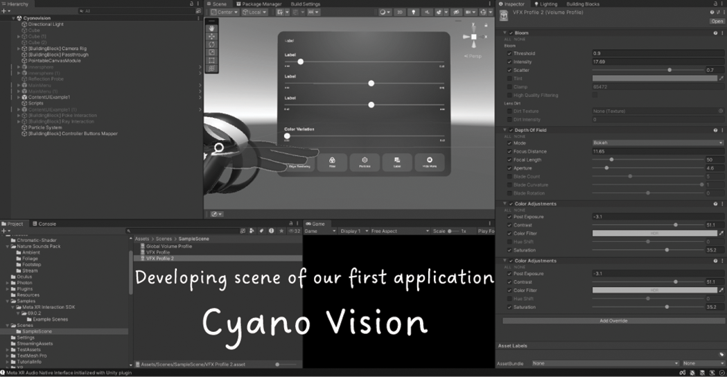
We aimed to create a cyano-vision simulation I could wear in the lab and beyond. The timeline forced compromises; we had to prioritise which visual parameters to simulate and which to defer. Yet through iterative co-ideation and their technical support, the concept gradually materialised into something experienceable: a wearable apparatus that would mediate my perception, allowing me to move through familiar spaces while sensing light as directional information rather than ambient illumination, which we called Cyano Vision (Figure 18).
Software user interface screenshot showing the development scene of the custom mixed reality application, Cyano Vision, built within the Unity game engine by the third author.
Let’s make their vision simulatable!
When I first wore the prototype of Cyano Vision (Figure 19), I felt astonished. After two years of speculation, the collaboration had produced something experienceable. The mixed reality simulator immersed me in a liquid-like environment: blurry, monochromatic blue-green vision, floating particles suspended in aquatic space (Figure 20). All parameters required manual adjustment; I could attune the filter towards microbial sensibilities (Kim et al. Reference Kim, Kim, Martins, Karana, Gray, Ciliotta Chehade, Hekkert, Forlano, Ciuccarelli and Lloyd2024), making it more alien or clarify it for instrumental utility so I could continue laboratory work while wearing it. The simulation relied entirely on the Quest 3’s passthrough cameras, making calibration subjective, guided by imagination rather than measurement. I was excited by what this gradual realisation enabled.
The first author testing the custom Cyano Vision mixed reality application using a Meta Quest 3 headset while handling a cell culture flask inside the biodesign laboratory.

In-application, first-person view of Cyano Vision during testing by the first author, showing the digital interface for calibrating simulation parameters within the laboratory environment.

Moving through the laboratory wearing the apparatus made the ontological distance between my sensory world and theirs palpable. Each parameter adjustment forced me to articulate what I imagined their perception might be. I began proposing technical upgrades to make the simulation more scientifically accurate and immersive: sensors to measure light intensity, directional algorithms to respond to photon gradients and features grounding the experience in empirical data. I imagined amplifying the somatic becoming experience, making the filter responsive to actual light conditions. The possibilities multiplied. If I could calibrate this apparatus accurately, it could become a tool for embodied understanding across different microbial scales and sensory modes.
Wait, do they really “see” the world?
My collaborator (third author) asked a question that stopped me: “Why are you focusing on visualising their perception, when their understanding of seeing fundamentally differs from human vision? It’s actually about sensing light and direction, not vision in the human sense.” The question exposed my assumption. I returned to the literature (e.g., Figures 21 and 22). The cell from Synechocystis does act as a lens and the surrounding membrane as retina, but this does not constitute a “real eye” in biological terms, because these structures have other essential functions that are not primarily devoted to vision (Nilsson and Colley Reference Nilsson and Colley2016). In fact, the simplest organisms with real eyes are warnowiid dinoflagellates, unicellular eukaryotic algae measuring 50–100 μm, far larger than the 3 μm Synechocystis (Nilsson and Colley Reference Nilsson and Colley2016). I had been translating their sensory world into human visual terms, privileging sight as the primary mode of knowing. But cyanobacteria don’t “see” in the ocular sense I had been simulating. They sense light. Their entire cell structure responds to directional photon flux as information guiding movement. The recognition troubled my entire approach. I had requested designing visual spectacle – immersive imagery, monochromatic filters, particle effects – when what I needed was sensory reorientation towards directional light response. The apparatus I had shaped mediated the wrong modality. My human-centredness shaped not only the simulation’s parameters but also its fundamental premise. In that moment, I realised that attuning to micro-scale conditions requires more than applying novel techniques; it demands unlearning deeply ingrained human ways of sensing.
Micro-optic lensing effects in a Synechocystis cell with a 3 µm scale bar (left; adapted from Schuergers et al. Reference Schuergers, Lenn, Kampmann, Meissner, Esteves, Temerinac-Ott, Korvink, Lowe, Mullineaux and Wilde2016), compared with the eye-like ocelloid structure within a warnowiid dinoflagellate, Proterythropsis sp., with a 10 µm scale bar (right; adapted from Hoppenrath et al. Reference Hoppenrath, Bachvaroff, Handy, Delwiche and Leander2009).

Fundus photograph of a human retina, highlighting the bright circular optic disc where the optic nerve exits the eye. Scale bars: 1,500 µm and 10,000 µm. (Adapted from Churkin Reference Churkin2008).

I still couldn’t see like them, but
Returning to the design studio, I confronted what the prototype had revealed: I had designed for visual immersion when I needed sensory translation. With my collaborator (third author), I began experimenting with light sensors and haptic feedback, assembling accessible components to translate light-related data into tactile cues rather than relying on visual-only sensing (Figure 23). We tinkered with different sensors and motors to improve our response when light conditions matched the biologically relevant range of Synechocystis, which is about 10 µmol photons m–2 s–1 of directional illumination (Schuergers et al. Reference Schuergers, Lenn, Kampmann, Meissner, Esteves, Temerinac-Ott, Korvink, Lowe, Mullineaux and Wilde2016). The setup was almost becoming like a health monitor (e.g., Figure 24), but for cyanobacterial well-being rather than human metrics. Yet the immersive simulation had generated its own insights. The difficulty of calibrating Cyano Vision to actual cyanobacterial vision became methodologically valuable in itself; each failed parameter adjustment exposed how deeply my sensory assumptions were embedded in human visual norms.
The first author prototyping hardware modifications for Cyano Vision, testing the integration of infrared sensors and haptic vibration motors on a Seeed Studio development board.

Conceptual rendering of a future iteration of Cyano Vision, featuring a wearable, spherical helmet designed to provide immersive environmental light-sensing feedback.

The apparatus functioned as an unlearning tool, revealing how persistently human-centred my imagination remained. Moving forward, I recognised that Cyano Vision, rather than serving solely as an exploratory artefact, operated as a speculative probe. Its value lay in its exposure of anthropocentric assumptions and in unsettling habitual modes of perceiving microbial worlds. By foregrounding the limitations and frictions in simulating cyanobacterial sensing, Cyano Vision functioned as a device for unlearning; it prompted reconsideration of what it meant to encounter difference and invited ongoing reflection on human interpretative frames. The technical challenge was also an ethical one: how to design for becoming-with rather than becoming-as.
Between these two trajectories…
These parallel trajectories – one pursuing controlled motility, the other attempting perceptual translation – converged on three interrelated insights about practising biodesign with MTH sensibilities.
Different perspectives on the laboratory
Wearing the prototype of Cyano Vision forced me to confront what I had assumed as an individual territory was a singular shared space. The laboratory exists at radically different scales; what appears to me as benchtop and incubator constitutes an entirely different spatial reality for microbes inhabiting gradients I cannot perceive with my naked eye. This scalar difference, present in everyday laboratory encounters, shaped how I navigated my relationship with the environment and its inhabitants. Each parameter adjustment made the familiar environment increasingly alien, revealing how profoundly my understanding privileges human-scale perception. Designing with microbial awareness requires learning to hold these incommensurable perspectives together, not collapsing their alterity into human frameworks, but developing approaches that acknowledge differences of scale as constitutive of shared environments themselves.
Single cell, 70 trillion cells and others
The augmented setup made visible the entangled agencies constituting even the simplest protocol. I had imagined myself as the sole actor orchestrating movement, but repeated failures revealed a dense multispecies ecology: my body maintaining temperature and humidity through breath and presence; the organisms, each an autonomous agent; contaminating fungi and bacteria claiming inadequately sterilised plates; and non-living instruments mediating our encounter. The protocol was never a human directing microbes, but a contingent assemblage of bodies, technologies and environmental conditions. The relational texture of these encounters became apparent through situated practice: every attempt to shape experimental conditions demanded attention to the web of influences at play, as mapped in Figure 25. Recognising this entanglement transformed my practice from instrumental control towards response-ability: learning to attune to the micro-scale requirements that make cyanobacterial agency possible, understanding that biodesign always involves negotiating among multiple, often conflicting, forms of livingness.
Actor-network mapping tracing the multispecies relationships, technical apparatuses and environmental conditions spanning the human and non-human actors involved across the first author’s autoethnographic research journey.

Laboratory as a unique ecosystem
Both trajectories exposed the laboratory as operating across temporalities radically divergent from human and microbial lifeworlds. My struggle to observe motility unfolded against their circadian rhythms, evolutionary lineage and the minutes-to-hours scale of their phototactic response. Each failed experiment represented weeks on my timeline but encompassed entire lifecycles of contaminating organisms and subtle physiological shifts in the cyanobacteria I sought to understand. The lab ecology operates simultaneously across multiple temporal registers, from photosynthetic processes occurring in microseconds to culture growth measured across days, strain maintenance spanning decades and evolutionary processes extending across deep time. These overlapping temporalities revealed the laboratory as a space where rhythms and durations intermingle, demanding attentiveness to how biodesign practice is shaped by encounters with time at many scales. Observing beyond the laboratory revealed them as agents reshaping planetary chemistry across geological scales. This temporal diversity demands practices that acknowledge temporal differences as fundamental to MTH engagement.
So, how can biodesigners integrate MTH sensibilities into their practice?
What did I learn along the way?
The two autoethnographic accounts conducted in a biodesign laboratory demonstrate that cultivating MTH sensibilities within environments typically governed by sterility, regulation and meticulous microbial handling is not only possible but can unfold along multiple trajectories. MTH learning can emerge not only through protocol-driven laboratory practice but also through experimental and speculative inquiry in hybrid studio–laboratory settings.
Before the divergence into two trajectories, my approach was grounded in a fundamental understanding of organisms that resonated with traditional biological training. For designers lacking a background in biology, particularly those accustomed to working with inert materials such as plastic, metal or wood, the bottom-up biodesign approach that starts from a scientific understanding of organisms can initially feel unfamiliar, even coercive. Living materials, however, resist predictability; they demand technical precision, ethical attentiveness and continuous responsiveness. This scientific rigour, while preserving biological integrity, also creates the conditions for a distinctive form of MTH learning that is unique to biodesign.
Scientific knowledge plays a crucial role in supporting MTH sensibilities in biodesign. Biodesign necessarily involves working with living systems across theoretical and applied regenerative contexts, so biological and ecological knowledge helps designers move beyond anthropocentric assumptions. It provides both a technical foundation and a cultivated respect for livingness at the microbial scale.
In biological experiments, neglecting the precise environmental requirements for cyanobacterial phototactic motility – such as temperature, humidity and agar concentration – revealed how easily anthropocentric assumptions fill epistemic gaps. In studio-lab explorations, defining the optical qualities of cyanobacteria primarily through human visual perception led prematurely into speculative design. In both cases, scientific knowledge provided a corrective scaffold for MTH learning, helping prevent misdirection and enabling more responsible design possibilities.
However, blind assimilation into scientific workflows carries its own risks. As demonstrated in the laboratory journey, the bio-digital setup initially developed to understand phototaxis gradually became instrumentalised for control and application. In laboratories prioritising sterility and scientific equivalence to conventional biology labs, their borrowed instrumental practices can dominate the environment, subtly compelling biodesigners to conform to utilitarian and technocentric logics.
While scientific tools illuminate mechanisms, they rarely address the broader entangled relationships in which organisms are embedded. For instance, although biodesigners may claim to “collaborate” with microbes, any “uninvited” microorganism is quickly labelled as “contamination” and excluded from the process. When unknown fungal or bacterial colonies appear on plates, they are typically discarded immediately, without consideration of their possible relevance or relationship. Echoing Anna L. Tsing’s observation that “As contamination changes world-making projects, mutual worlds – and new directions – may emerge” (Tsing Reference Tsing2015, 27), this prompts a crucial question: Do such selective monoculture biodesign practices truly support regenerative ecologies and nurture MTH sensibilities? While cultivating Staphylococcus aureus, Alexander Fleming (Reference Fleming1929) observed a contaminating fungus inhibiting bacterial growth. Recognising its potential significance, he preserved and studied it, ultimately leading to the discovery of penicillin. The pressure to translate living qualities into socially applicable artefacts further reinforces anthropocentric design paradigms and restricts opportunities to enact MTH-oriented values, for example, by encouraging attunement to microbial conditions rather than technical optimisation.
To unlearn this intrinsic instrumentalisation, the laboratory journey suggested the value of shaping more exploratory and self-reflective design tools for biodesigners. Within the strict confines of laboratory environments, overhauling established scientific infrastructures may be unrealistic. However, by starting with familiar laboratory artefacts and questioning their taken-for-granted assumptions, I found that even subtle reconfigurations could be significant, for instance, by adapting existing protocols to facilitate closer observation of microbial behaviour or by creating opportunities for ethical reflection within everyday experimental routines. Instead of focusing on spectacular MTH applications, such as high-tech installations or colourful displays, these efforts open opportunities for situated reflection and the expression of MTH thinking within real constraints.
In contrast, when design inquiry begins with exploratory imagination outside the conventional laboratory workflow, MTH approaches can be more actively foregrounded. Hybrid environments that blend laboratory work with external studio spaces enable engagement with speculative and HCI-informed methods. Immersive digital tools, such as Meta Quest 3, supported the development of Cyano Vision, facilitating embodied and sensory experiences that narrow the perceptual distance from microbial entities. For organisms whose cognitive or sensory attunement is otherwise inaccessible, extended or virtual sensory mediation can serve as a bridge, much like scientific knowledge itself.
Yet another tension emerges here. As shown, speculative freedom may inadvertently marginalise scientific rigour, which can be detrimental when working with living systems. Drawing from the challenges I encountered in prototyping, biodesigners’ need for mechanisms to continually assess whether their design processes accurately reflect the biological and ecological qualities of the organisms involved became noticeable. These mechanisms do not suppress ethical awareness; rather, they counteract implicit anthropocentrism and encourage critical unlearning.
Along the way, the process of unlearning began to manifest in subtle but tangible shifts within my design practice. Rather than approaching laboratory work as a matter of imposing control, I found myself increasingly attuned to unfolding conditions and emergent dynamics as they arose. Encounters with contamination, once met with the instinct to eradicate, became occasions to reconsider boundaries and interactions; still attentive to safety, but open to the possibility of learning through these interruptions. In developing sensory simulations, my focus gradually shifted from purely replicating microbial phenomena to translating across sensory modalities, allowing new forms of understanding to emerge. These changes, while not always dramatic, signal how unlearning can quietly recalibrate the designer’s everyday actions and sensibilities.
Looking back: What became visible through my self-reflective (un)learning?
Throughout the preceding accounts, the narration has moved between immediacy and reflection, not simply to chronicle events but to foreground how personal encounters with uncertainty, adaptation and failure become sources of insight. Rather than offering a purely descriptive record, the autoethnographic voice is used to draw out the underlying patterns and shifts in orientation that emerged from these experiences; patterns that speak not just to my own process, but to broader dynamics at play in biodesign practice. This mode of narration thus serves to distil, from the lived detail, those recurring tensions and inflexion points that shape the trajectory of MTH engagement.
Adopting an explicitly subjective voice enabled the surfacing of relational dynamics that conventional laboratory documentation tends to conceal. Previously, my descriptions of cyanobacteria were confined to strain codes, italicised taxonomies and binary states: alive, dead, dormant, viable. Such documentation, shaped by a controlled laboratory culture, left little room to articulate MTH reflections.
Through autoethnographic narration, I was able to articulate the network of relations among microbes, tools, protocols and myself. Cyanobacteria emerged not merely as research subjects but as living organisms encountered in situated interaction. This reframing unsettled habitual modernist separations between object and subject and opened space for unlearning entrenched laboratory hierarchies.
Importantly, the value of this approach lies not in presenting a universal MTH tool but in offering a nuanced account of tensions, frictions and negotiations. Because biodesign contexts vary widely – in organisms, purposes, infrastructures and institutional cultures – such positioned narratives provide relatable entry points for other biodesigners seeking to apply MTH thinking within their own constraints.
Did my journey happen in solitude?
While this inquiry is narrated from my perspective and adopts an autoethnographic voice, its realisation was fundamentally collective. Over the past three years, my team – spanning microbiology, HCI, MTHD and (bio)design – has consistently engaged in reflective sessions on my journey that we referred to as “retreats.” These gatherings became forums for sharing diverse disciplinary perspectives, critically examining methodologies and collaboratively navigating the evolving process. They not only surfaced and negotiated interdisciplinary tensions within the field but also provided the scaffolding needed to move beyond methodological boundaries. Without such sustained, dialogic reflection, I might have remained confined within my own assumptions, unable to fully engage with or appreciate the scientific and MTH dimensions of biodesign.
Epilogue and Impact statement for biodesign
This study demonstrates that MTH sensibilities in biodesign are not confined to speculative or activist settings but can also emerge within scientifically rigorous laboratory environments. Instead of positioning scientific precision and relational awareness as oppositional, this autoethnographic account illustrates how their dynamic interplay fosters productive reflection and innovation. By tracing moments of instrumentalisation, misalignment and recalibration, the study illuminates the often-unspoken negotiations and tensions that shape everyday biodesign practice, offering a richer understanding of how MTH perspectives can be integrated into the discipline.
Methodologically, this work extends biodesign research by employing autoethnography as a critical tool for self-reflection and analysis. Articulating the first author’s relationship with cyanobacteria beyond conventional scientific labelling reveals the underlying power dynamics and epistemic hierarchies present in laboratory culture. This reflective approach does not dismiss scientific rigour; rather, it positions science and technology as active collaborators in cultivating MTH sensibilities. By broadening the methodological vocabulary of biodesign, this perspective suggests that meaningful transformation can arise not only from new materials or artefacts but also from reconfiguring the ways practitioners narrate, interpret and relate to their engagements with living systems.
Practically, this study provides insight into how existing laboratory tools and workflows can be subtly reoriented to support working with cyanobacteria without compromising safety or scientific precision. It encourages interdisciplinary collaboration among technicians, microbiologists and laboratory managers to collectively reshape the conditions under which biodesign is practised. By fostering this collaborative reorientation, the study expands the methodological boundaries of the field and strengthens biodesign’s capacity to engage regenerative futures with greater vigilance, responsibility and adaptability.
Data availability statement
The primary experimental materials generated and used in this study comprise (1) an adapted macroscopic phototaxis assay of Synechocystis sp. PCC 6803 and (2) a Unity application package for Cyano Vision.
(1) Data associated with the adapted phototaxis assay is available at https://doi.org/10.4121/d3466d24-9012-4b35-8818-e4535cda60bd.
(2) The Unity application package for Cyano Vision is available at https://github.com/OEOxr/CyanoVision-MR.
Please note: Both items were developed for exploratory research and have not undergone formal validation. For further information or access to experimental materials, contact the corresponding author.
Acknowledgements
We extend our sincere gratitude to Joren Wierenga, our research technician at the Biodesign Lab, Faculty of Industrial Design Engineering, Delft University of Technology, for his invaluable technical assistance throughout this research. We are also grateful to Nils Schürgers, research fellow in the Group of Molecular Genetics of Prokaryotes at the Institute of Biology III, University of Freiburg, for generously sharing his expertise on the Synechocystis sp. PCC 6803 phototaxis experiment and for providing the motile strain.
Author contributions
Conceptualisation: Jiho Kim, Elvin Karana. Data curation: Jiho Kim. Formal analysis: Jiho Kim. Investigation: Jiho Kim. Methodology: Jiho Kim, Elvin Karana. Project administration: Elvin Karana, Joana Martins. Resources: Elvin Karana. Software: Okan Efe Öğretmen. Supervision: Elvin Karana, Joana Martins, Iohanna Nicenboim. Visualisation: Jiho Kim. Writing original draft: Jiho Kim. Writing review & editing: All authors.
Financial support
This research received no specific grant from any funding agency, commercial or not-for-profit sectors.
Open access funding provided by Delft University of Technology.
Competing interests
The authors have no conflicts of interest to declare for this publication.
Ethical standards
Ethical approval and consent are not relevant to this article type.


 Open access


