

Introduction
Indoor mold counts typically increase after flooding and water damage,Reference Johanning, Auger, Morey, Yang and Olmsted 1 and cases of respiratory diseases usually increase after earthquakes.Reference Pegues, Daar and Murthy 2 The most common cause of allergic bronchopulmonary mycosis (ABPM) is Aspergillus fumigatus. Aspergilli other than A. fumigatus that have been reported to cause ABPM include A. niger, A. sydowii, and A. terreus.Reference Matsuse, Tsuchida and Fukahori 3 Eurotium herbariorum is the name usually given to the sexual stage of A. glaucus. Exposure of children living on farms to Eurotium species decreases their risk of developing atopy and asthma.Reference Ege, Mayer and Normand 4 In contrast, increases in A. versicolor and Eurotium counts in school buildings with moisture damage have been associated with increased prevalence of asthma.Reference Haverinen, Husman and Toivola 5 Eurotium herbariorum has not previously been reported to cause ABPM.
Case Presentation
A 66-year-old, Japanese male, ex-smoker (packs per year: 75.25) was initially diagnosed with bronchial asthma at the age of five years, and he achieved remission at the age of 13 years. He was displaced from his home during the Great East Japan Earthquake on March 11, 2011 and moved to temporary housing in Miyagi Prefecture in June 2011 at the age of 62 years. He experienced the first episode of chest tightness, coughing, and wheezing in February 2012, and he was diagnosed as having bronchial asthma in March 2012. He continued to experience asthma exacerbations, even after the initiation of fluticasone (100 μg) plus salmeterol (50 μg) twice daily; notably, he experienced no symptoms during a 10-day absence from the temporary housing.
From June 2013, time-course surveillance was performed of fungal mycoflora in the temporary housing. Fungal cells per 10 L of air were sampled on a single agar plate by using an Air IDEAL 3P air-sampling system (bioMérieux; Marcy l’Etoile, France) placed approximately one meter above floor level. Air samples were collected at each sampling point of the bedroom, living room, kitchen, and outdoors for a total of 10 culture plates, including five plates of dichloran – rose bengal – chloramphenicol agar (DRBC; Thermo Fisher Scientific; Braintree, Massachusetts USA) and five plates of dichloran –glycerol agar (DG18; Thermo Fisher Scientific). Total fungal counts were obtained; in addition, colonies belonging to the genera Aspergillus, Cladosporium, Penicillium, or Eurotium were counted separately by each genus according to the macroscopic and microscopic features of the colonies. Total and genus-specific fungal counts were expressed as the number of colony-forming units per cubic meter (CFU/m3), as estimated by using the most-probable-number method.Reference Fannin, Spendlove, Cochran and Gannon 6
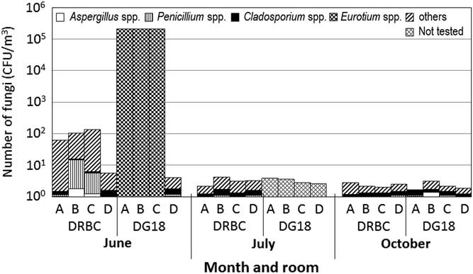
The initial round of mycofloral surveillance detected high counts of Eurotium in the air of all three indoor areas sampled (the bedroom, kitchen, and living room), with a maximal fungal count of 163,200 CFU/m3 compared with fungal counts of no more than 1000 CFU/m3 in normal housing (Figure 1). This result was noteworthy because Cladosporium and Penicillium fungi typically predominate in the indoor air of normal dwellings, including the homes of allergic patients.Reference Garrett, Rayment, Hooper, Abramson and Hooper 7 In view of these results, the patient was instructed on the use of allergen avoidance techniques. Molecular methods were applied to confirm the species-level identification of isolates of E. herbariorum. Partial nucleotide sequences of the β-tubulin gene were determined as previously described.Reference Glass and Donaldson 8 The nucleotide sequences that were obtained here were used as query sequences in a BLAST search of sequences previously registered in GenBank (National Center for Biotechnology Information, National Institutes of Health; Bethesda, Maryland USA). The results of the BLAST were consistent with those from morphologic identification and confirmed that the organism was E. herbariorum.
Time Course of Surveillance of Fungal Mycoflora in the Patient’s Temporary Housing Before and After the Incorporation of Allergen Avoidance Techniques; A: Living Room; B: Bedroom; C: Kitchen; D: Outdoors. Note: In June, Eurotium predominated and completely covered the plates, and other genera could not be counted. In July and October, Eurotium colonies were included in the counts for Aspergillus. Abbreviations: CFU, colony-forming units per cubic meter; DG18, dichloran – glycerol agar; DRBC, dichloran – rose bengal – chloramphenicol agar.

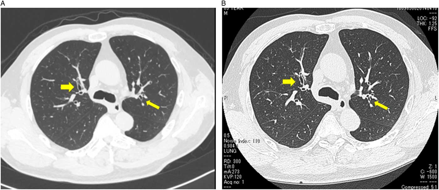
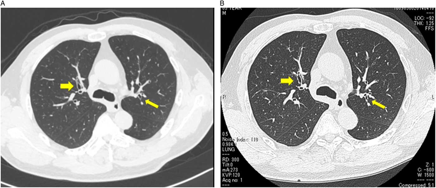
Written informed consent was obtained from the patient to perform skin prick tests against A. fumigatus and E. herbariorum. The wheal and flare sizes were evaluated at 15 minutes after A. fumigatus or E. herbariorum pricked compared with that of wheal size after histamine done. He confirmed positive reactions to both A. fumigatus (20 x 20 mm; Torii Pharmaceutical; Tokyo, Japan) and E. herbariorum (18 x 16 mm; equivalent to A. glaucus; the antigen used was a pure culture of an isolate from his temporary housing) compared with 12 x 10 mm after pricked histamine. In addition, the total serum IgE level and presence of antigen-specific IgE antibodies were evaluated by ELISA; the total IgE level was 112.0 IU/mL, and antigen-specific IgE antibodies against A. fumigatus but not A. glaucus were detected. Ouchterlony double-immunodiffusion testingReference Ouchterlony 9 of the patient’s serum confirmed the presence of antigen-specific precipitating antibodies to A. glaucus (Greer; Lenoir, North Carolina USA) and E. herbariorum, but not to A. fumigatus (Torii Pharmaceutical). Double diffusion between A. glaucus and E. herbariorum was confirmed by the Ouchterlony assay, which suggested the presence of common antigenicity between them. Central bronchiectasis in right B3 and in left B2 (arrows) was present more clearly in April 2014 than in August 2013 (Figure 2), and greater bronchial wall thickening was seen in April 2014 than in August 2013. He did not have any other known causes of bronchiectasis
Computed Tomography Scan of the Upper Lungs of Patient, Performed in A: August 2013 and B: April 2014. Note: Central bronchiectasis in right B3 and in left B2 (arrows) was more prominent in April 2014 than in August 2013, and bronchial wall thickness was greater in April 2014 than in August 2013.
After obtaining written informed consent from the patient, he was performed bronchial provocation tests with A. fumigatus and E. herbariorum. Ten minutes after antigen-specific provocation with A. fumigatus or E. herbariorum at 10 mg/mL, the patient developed wheezing (E. herbariorum) and chest tightness (A. fumigatus), and his forced expiratory volume in one second (FEV1) decreased by 18.7 % (FEV1: 1.71 L/s to 1.39 L/s; A. fumigatus) or 14.4 % (FEV1: 1.87 L/s to 1.60 L/s; E. herbariorum) of that before provocation. In addition, he experienced late reactions, with symptoms such as wheezing or chest tightness and the decrease of peak expiratory flow at seven hours (E. herbariorum) to 20 hours (A. fumigatus) after the last antigen provocation.
The diagnosis of this patient was consistent with ABPM given his positive skin tests to both E. herbariorum and A. fumigatus, the presence of antigen-specific precipitating antibodies to A. glaucus and E. herbariorum, and central bronchiectasis with bronchial wall thickening. He did not have any other known causes of bronchiectasis. This patient experienced the first episode of chest tightness, coughing, and wheezing in February 2012. The case was unusual because the duration for sensitization to onset was so short (ie, only eight months). It is possible that this patient had a low total IgE level owing to the short duration of exposure to E. herbariorum before his ABPM illness. From the data obtained, he was diagnosed as having ABPM caused by E. herbariorum during temporary housing after the Great East Japan Earthquake.
The patient’s twice-daily fluticasone dose was increased from 100 to 250 μg; he continued to receive salmeterol (50 μg) twice daily; and 10 mg of a leukotriene antagonist was added to his treatment regimen. The successful avoidance of exposure for E. herbariorum might be able to be treated without receiving systemic corticosteroids. In addition, all tatami was removed from his living quarters. The tatami mat is made from rushes and is to be laid on the floor in the room.
Total and E. herbariorum-specific fungal counts obtained one and four months after these allergen avoidance measures had been initiated were markedly lower than the initial counts (Figure 1). He did not have further exacerbations in temporary housing over the four months after the avoidance of allergens.
Conclusion
This report presents the rare case of a patient who developed ABPM due to exposure to E. herbariorum during temporary housing after the Great East Japan Earthquake.