Introduction
The classical dynamins belong to a superfamily of GTPases that include dynamin-like proteins (Ferguson & De Camilli, Reference Ferguson and De Camilli2012). Dynamins participate in diverse membrane dynamics and vesicle trafficking functions, contributing mechanical force to excise newly formed vesicles from membranes of cellular components (McNiven et al., Reference McNiven, Cao, Pitts and Yoon2000). Three genes in mammals encode the dynamin proteins: DNM1, DNM2, and DNM3 (Cao et al., Reference Cao, Garcia and McNiven1998). Of these, dynamin-2 is the only member that is ubiquitously expressed. Dynamin-1 is neuron-specific, whereas dynamin-3 expression is tissue-specific, reportedly found in the brain, testis, lung, and heart (Cao et al., Reference Cao, Garcia and McNiven1998). All three dynamin isoforms are present in neuronal tissue. Determining the physiological role of the dynamins is a significant challenge, as there are overlapping functions of the isoforms (Ferguson et al., Reference Ferguson, Brasnjo, Hayashi, Wolfel, Collesi, Giovedi, Raimondi, Gong, Ariel, Paradise, O’Toole, Flavell, Cremona, Miesenbock, Ryan and De Camilli2007; Raimondi et al., Reference Raimondi, Ferguson, Lou, Armbruster, Paradise, Giovedi, Messa, Kono, Takasaki, Cappello, O’Toole, Ryan and De Camilli2011) yet splice variants of each isoform may perform separate cellular tasks (Cao et al., Reference Cao, Garcia and McNiven1998; McNiven et al., Reference McNiven, Cao, Pitts and Yoon2000).
Dynamin-1, dynamin-2, and dynamin-3 proteins have been identified in mouse photoreceptor cells of the retina (Liu et al., Reference Liu, Tan, Levenkova, Li, Pugh, Rux, Speicher and Pierce2007); however, the extent of their expression in the retina has not been fully investigated. Only two diseases are associated with mutations in dynamin genes and both have a visual defect. Mutations in DNM2 underlie an autosomal dominant form of intermediate Charcot–Marie–Tooth disease, a peripheral neuropathy that can result in vision loss (Zuchner et al., Reference Zuchner, Noureddine, Kennerson, Verhoeven, Claeys, De Jonghe, Merory, Oliveira, Speer, Stenger, Walizada, Zhu, Pericak-Vance, Nicholson, Timmerman and Vance2005) and autosomal dominant centronuclear myopathy, which can induce a paralysis of the extraocular muscles (Jeub et al., Reference Jeub, Bitoun, Guicheney, Kappes-Horn, Strach, Druschky, Weis and Fischer2008). Given the importance of dynamins in the central nervous system, it would not be surprising if additional defects in dynamin gene products affect retinal physiology and induce ocular phenotypes. The photoreceptor cells have an extremely high-energy requirement due in part to tonic synaptic signaling and extensive vesicular protein transport (Linton et al., Reference Linton, Holzhausen, Babai, Song, Miyagishima, Stearns, Lindsay, Wei, Chertov, Peters, Caffe, Pluk, Seeliger, Tanimoto, Fong, Bolton, Kuok, Sweet, Bartoletti, Radu, Travis, Zagotta, Townes-Anderson, Parker, Van der Zee, Sampath, Sokolov, Thoreson and Hurley2010). To meet this high demand, photoreceptors have developed specialized mechanisms to handle the continuous cycling of vesicles. How the dynamins contribute to these specialized features remains to be determined.
We previously identified a splice variant of dynamin-1, termed dynamin-1 (a,c), which colocalizes and interacts with the photoreceptor-specific Tubby-like protein 1 (Tulp1) (Xi et al., Reference Xi, Pauer, Ball, Rayborn, Hollyfield, Peachey, Crabb and Hagstrom2007). Mutations in TULP1 cause a form of autosomal recessive retinitis pigmentosa, a type of inherited retinal degeneration, which leads to blindness (Hagstrom et al., Reference Hagstrom, North, Nishina, Berson and Dryja1998). As an approach to better understanding dynamin function in photoreceptor cells, we report here the characterization of dynamin isoform expression and localization in the mouse retina and the identification of possible dynamin-1 interacting proteins.
Methods
Mice
Generation and maintenance of tulp1−/− mice on a C57BL/6 background has been described previously (Hagstrom et al., Reference Hagstrom, Duyao, North and Li1999). Control C57BL/6 mice were obtained from The Jackson Laboratory. Mice were euthanized by CO2 inhalation followed by cervical dislocation. All animal experiments were approved by the Institutional Animal Care and Use Committee of the Cleveland Clinic and were performed in compliance with the National Institutes of Health guidelines.
Immunohistochemistry
Eyes from P16 mice were prepared as previously described (Xi et al., Reference Xi, Pauer, Ball, Rayborn, Hollyfield, Peachey, Crabb and Hagstrom2007; Grossman et al., Reference Grossman, Watson, Pauer, Bollinger and Hagstrom2011). Briefly, after removal of the cornea and lens, the posterior poles were fixed in 4% paraformaldehyde in phosphate buffered saline (PBS) for 3 h. The eyecups were then immersed through a graded series of sucrose solutions: 10% for 1 h, 20% for 1 h, and 30% overnight. The posterior pole was embedded in Optimal Cutting Temperature Compound (OCT) freezing medium, flash frozen on powderized dry ice, and immediately transferred to −80°C. The tissue was sectioned at 10-μm thickness using a cryostat (Leica, Wetzlar, Germany) at −30°C. For each genotype (wt and tulp1−/−), a minimum number of four sections from five different mice were examined for each antibody. Retinal sections were blocked in 5% bovine serum albumin (BSA) and 1% normal goat serum with 0.1% Triton X-100 for 1 h before incubation with primary antibodies overnight at 4°C. A panel of well-characterized antibodies was used for immunostaining. Table 1 contains a complete list of antibodies, immunogens, sources, host species, and dilutions used. After washing three times in PBS, sections were incubated in fluorescent secondary antibodies at room temperature for 1 h. Secondary antibodies were: Alexa Fluor® 488 goat anti-rabbit IgG and goat anti-mouse IgG, Alexa Fluor® 594 goat anti-rabbit IgG and goat anti-mouse IgG (Invitrogen, Carlsbad, CA). The sections were then rinsed three times with PBS followed by a dH2O rinse and coverslipped with Vectashield® mounting media with 4',6-diamidino-2-phenylindole (DAPI) (Vector Laboratories, Burlingame, CA). The sections were imaged using an Olympus BX-61 fluorescent microscope (Olympus, Tokyo, Japan), equipped with a charge-coupled device (CCD) monochrome camera (Hamamatsu Photonics, Bridgewater, NJ).
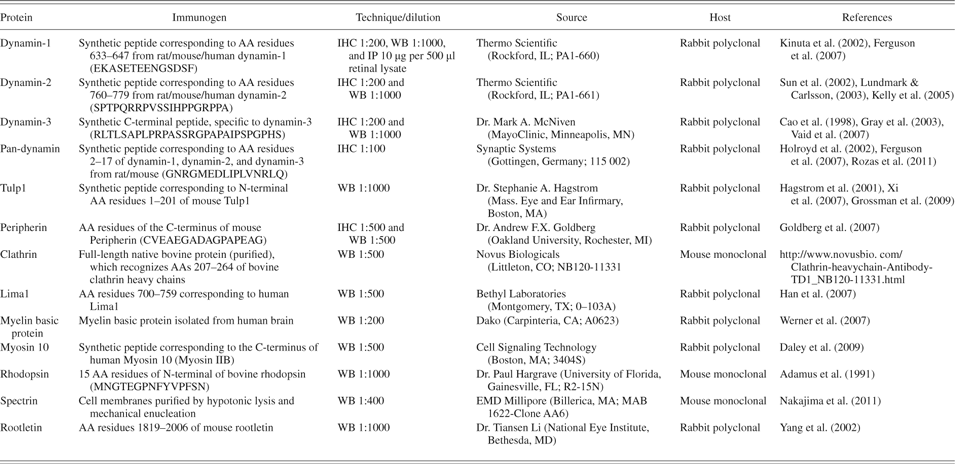
Primary antibody characterization

Western blot
Western blot analysis was performed as previously described with P16 mouse tissues from wt and tulp1−/− mice (n = 2 for each genotype) (Hagstrom et al., Reference Hagstrom, Adamian, Scimeca, Pawlyk, Yue and Li2001; Xi et al., Reference Xi, Pauer, Marmorstein, Crabb and Hagstrom2005, Reference Xi, Pauer, Ball, Rayborn, Hollyfield, Peachey, Crabb and Hagstrom2007). Briefly, proteins were separated on SDS-PAGE gels and electroblotted to polyvinylidene fluoride (PVDF) membranes. Membranes were incubated with primary antibodies (Table 1), followed by peroxidase-conjugated secondary antibodies and were detected by chemiluminescence.
Immunoprecipitation and protein identification
Immunoprecipitation (IP) with Dynabeads® was performed according to the manufacturer (Dynabeads® Protein A 100.06D, Invitrogen, Carlsbad, CA). IPs followed by protein identification by liquid chromatography tandem mass spectrometry (LC MS/MS) were performed using mouse retina and brain tissue. In separate experiments, IPs followed by western blot analysis were performed using bovine retinal tissue or mouse brain and retinal tissue. Briefly, adult bovine retina (600 mg: ∼1 retina), P16–P60 mouse retinas (300 mg: 30 retinas), or P16–P60 mouse brains (300 mg: ∼1 brain) were homogenized in 3 ml of lysis buffer (50 mM Tris pH 8.0, 150 mM NaCl, 10% glycerol, 0.5% Triton X-100, 0.1% NP40) supplemented with protease inhibitors (Roche Applied Science, Indianapolis, IN). Homogenization was accomplished by freezing the tissue and manually grinding with a disposable pestle within a microcentrifuge tube. This was repeated for two additional cycles. The suspension was centrifuged at 9000 rpm for 15 min, the supernatant containing the retinal lysates was transferred to a separate tube, and the protein concentration (bovine: 4 μg/μl; mouse: 1.2 μg/μl) was determined by the bicinchoninic acid assay (Thermo Scientific, Rockford, IL). For IP, 10 μg of polyclonal anti-dynamin-1 antibody (Thermo Scientific, Rockford, IL) or nonspecific rabbit IgG (SouthernBiotech, Birmingham, AB) was used for each 500 μl of retinal lysate. Immediately prior to IP, the antibody was crosslinked to the beads using BS³ (Bis[sulfosuccinimidyl] suberate) (Thermo Scientific, Rockford, IL) for 30 min at room temperature. The IP binding reactions proceeded overnight at 4°C, followed by three washes with buffer. Proteins were then eluted with 20 μl of SDS sample buffer. Immunoprecipitated antigens were fractionated by SDS-PAGE on a 4–20% Tris–glycine 5-well gel (Invitrogen) with 60 µl of each IP product loaded per gel lane. The gel was then stained with colloidal Coomassie blue (Gel Code Blue, Pierce, Rockford, IL) and a disposable gridcutter (Gel Company Inc., San Francisco, CA) was used to excise 2-mm serial sections from each lane containing the IP products. Each gel sample was digested in situ with trypsin and proteins identified by LC MS/MS using a quadrupole time-of-flight instrument and a CapLC system (CapLC system; Waters Corporation, Milford, MA) as described previously (Crabb et al., Reference Crabb, Miyagi, Gu, Shadrach, West, Sakaguchi, Kamei, Hasan, Yan, Rayborn, Salomon and Hollyfield2002; Xi et al., Reference Xi, Pauer, West, Crabb and Hagstrom2003, Reference Xi, Pauer, Marmorstein, Crabb and Hagstrom2005, Reference Xi, Pauer, Ball, Rayborn, Hollyfield, Peachey, Crabb and Hagstrom2007). Protein identifications utilized ProteinLynx Global Server software (Waters Corporation), Mascot 2.2 (Matrix Science, Boston, MA) and the Swiss-Protein murine sequence database (May 2012 release, 16,529 total mouse sequences). Bioinformatic functional analyses were performed with Ingenuity Pathway Analysis (Summer Release, June 2012, Ingenuity Systems, Redwood City, CA, www.ingenuity.com) and the UniProtKB/Swiss-Prot protein database (web.expasy.org/docs/).
Results
All dynamin isoforms are expressed in the mouse retina
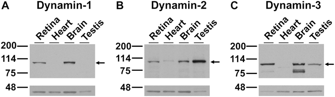
Western blot analysis shows that all three dynamin isoforms are expressed in the mouse retina (Fig. 1). All studies were conducted at P16, an age at which all the cell types of the retina are present in wt mice but which precedes photoreceptor cell death in tulp1−/− mice (Hagstrom et al., Reference Hagstrom, Duyao, North and Li1999; Grossman et al., Reference Grossman, Pauer, Narendra, Peachey and Hagstrom2009, Reference Grossman, Watson, Pauer, Bollinger and Hagstrom2011). Dynamin-1, the neuronal-specific isoform, was detected in the retina and brain at its known molecular weight of 100 kDa but not in testis (Fig. 1A). This is in agreement with previous findings (Cao et al., Reference Cao, Garcia and McNiven1998; Ferguson et al., Reference Ferguson, Brasnjo, Hayashi, Wolfel, Collesi, Giovedi, Raimondi, Gong, Ariel, Paradise, O’Toole, Flavell, Cremona, Miesenbock, Ryan and De Camilli2007). In western blots probed with dynamin-2 antibodies, immunoreactive bands were present at ∼100 kDa in all tissues examined (Fig. 1B), in agreement with studies supporting the ubiquitous expression of this isoform (Cao et al., Reference Cao, Garcia and McNiven1998; Ferguson et al., Reference Ferguson, Brasnjo, Hayashi, Wolfel, Collesi, Giovedi, Raimondi, Gong, Ariel, Paradise, O’Toole, Flavell, Cremona, Miesenbock, Ryan and De Camilli2007). We detected dynamin-3 as a component of the retina as well as the brain and testis (Fig. 1C).
Multi-tissue analysis indicating that all three dynamin isoforms are present in the P16 mouse retina. (A) Western blot probed with dynamin-1 antibodies. (B) Western blot probed with dynamin-2 antibodies. (C) Western blot probed with dynamin-3 antibodies. Actin is shown in lower blots (∼45 kDa) as a control for loading equivalency.
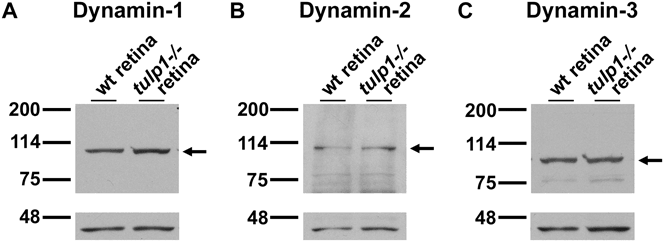
Because of the association between Tulp1 and dynamin-1 (a,c) in the retina (Xi et al., Reference Xi, Pauer, Ball, Rayborn, Hollyfield, Peachey, Crabb and Hagstrom2007), we examined whether dynamin-1 expression is altered in the absence of Tulp1. Western blot analysis of wt and tulp1−/− retinal lysates probed with a dynamin-1-specific antibody identified one band at ∼100 kDa in both lysates (Fig. 2A). Probing with antibodies against actin supported equal protein loading across lanes. Our results suggest that dynamin-1 protein expression levels are not severely altered in the absence of Tulp1. Western blots of wt and tulp1−/− retinal lysates probed with dynamin-2 (Fig. 2B) and dynamin-3 (Fig. 2C) antibodies also showed that their levels were not grossly affected by the absence of Tulp1.
All three dynamin isoforms are expressed in the P16 wt and tulp1−/− mouse retina at similar levels, respectively. (A) Western blot of wt and tulp1−/− retinal homogenates probed with dynamin-1 antibodies. In both samples, a single band is present at ∼100 kDa at similar levels. (B) Western blot of wt and tulp1−/− retinal homogenates probed with dynamin-2 antibodies. In both samples, a major band is present at ∼100 kDa at similar levels. (C) Western blot of wt and tulp1−/− retinal homogenates probed with dynamin-3 antibodies. In both samples, a single band is present at ∼100 kDa at similar levels. Actin is shown in lower blots (∼45 kDa) as a control for loading equivalency.
Differential localization of dynamin isoforms in the mouse retina
The distribution of the dynamin isoforms in P16 mouse retinas was examined by immunohistochemistry (IHC) (Fig. 3). A minimum of four sections from five different mouse retinas were examined for each experiment. Figure 3A shows that dynamin-1 is localized to the inner segment (IS), cell bodies and axons of the outer nuclear layer (ONL), and synaptic terminals of the photoreceptor cells in wt mouse retina. Immunoreactivity was also detected in other portions of the retina, including the outer plexiform layer (OPL), around cell bodies in the inner nuclear layer (INL), the inner plexiform layer (IPL), and ganglion cell layer (GCL) (Fig. 3A). High magnification shows that staining was present throughout the OPL, indicating that dynamin-1 is localized to the photoreceptor terminals as well as the postsynaptic dendrites of the second-order neurons.
Immunofluorescent localization of dynamin isoforms in P16 mouse retinal sections. Images of dynamin proteins (first column and pseudocolored red in the merged images) and nuclei labeled with DAPI (second column and pseudocolored blue in the merged images) compare the localization of dynamin isoforms. (A) In the wt retina, dynamin-1 staining is present in the IS, ONL, OPL, INL, IPL, and GCL. In the fourth column, showing a magnified view, staining is present throughout the OPL, indicating that dynamin-1 is localized to the photoreceptor terminals as well as in the postsynaptic dendrites of the second-order neurons. (B) In the tulp1−/− retina, dynamin-1 staining exhibits the same pattern as in the wt, signifying that dynamin-1 localization is unaffected in the absence of tulp1. (C) Dynamin-2 immunoreactivity is detected in the IS, OPL, IPL, and GCL; however, the magnified view shows that the signal is most concentrated above the myoid region of the IS. (D) Dynamin-3 expression is predominantly localized to the IPL. Less signal is detected in the IS, OPL, INL, and GCL. (E) A pan-dynamin antibody that was raised against an epitope shared by all three isoforms shows a staining pattern similar to that of dynamin-1, with immunoreactivity in the IS, ONL, OPL, INL, IPL, and GCL. Scale bars = 50 µm.
Since an association exists between Tulp1 and dynamin-1 (Xi et al., Reference Xi, Pauer, Ball, Rayborn, Hollyfield, Peachey, Crabb and Hagstrom2007), we examined dynamin-1’s localization in the tulp1−/− retina (Fig. 3B). Figure 3B indicates that the localization of dynamin-1 in the tulp1−/− retina appears similar to that in the wt retina with immunoreactivity seen in the IS, ONL, OPL, INL, IPL, and GCL. These results imply that the absence of Tulp1 does not grossly affect the retinal distribution of dynamin-1.
In contrast to the broad staining pattern of dynamin-1 in the wt retina, dynamin-2 immunoreactivity was found predominantly above the myoid region of the IS (Fig. 3C). The myoid region is the proximal portion of the IS, which houses the machinery for protein translation, including the Golgi apparatus and endoplasmic reticulum. High magnification indicates that dynamin-2 localizes near the region of the connecting cilium (CC), the intracellular link between the functionally different compartments of the IS and OS (Fig. 3C). This is in agreement with proteomic data from CC preparations (Liu et al., Reference Liu, Tan, Levenkova, Li, Pugh, Rux, Speicher and Pierce2007). There was also dynamin-2 staining in the OPL, IPL, and GCL but much less pronounced as compared with dynamin-1. Dynamin-3 localization in the wt retina was most apparent in the IPL (Fig. 3D). Immunoreactivity was also detected in the IS, OPL, INL, and GCL. Finally, a pan-dynamin antibody appears to label much of the neural retina similar to dynamin-1 (Fig. 3E). The localization patterns of dynamin isoforms in wt and tulp1−/− mice were similar at earlier (P13) and later ages (P21) (data not shown).
Dynamin-1 interacting proteins in the mouse retina
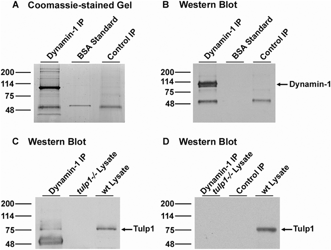
To identify dynamin-1 interacting proteins, we performed IPs from wt mouse retinal homogenates using a dynamin-1 specific antibody. Two independent IP experiments were performed, and Fig. 4 shows a representative SDS-PAGE result. The IP and control lanes were excised into 31 fractions and proteins in each were identified by LC MS/MS. The major Coomassie blue band at ∼100 kDa (Fig. 4A) was identified in both independent experiments as dynamin-1 (based on 83% sequence coverage). In addition, western blot analysis with anti-dynamin-1 also identified the 100 kDa band as dynamin-1 (Fig. 4B). Subsequent western blot analysis with anti-Tulp1 supported Tulp1 as a constituent of a dynamin-1 protein complex in the wt mouse retina but not in the tulp1−/− retinal lysate (Fig. 4C). In a separate experiment, Tulp1 was also detected by western blot analysis of dynamin-1 IP products from bovine retinal homogenates (data not shown). Tulp1 was not identified in an IP from the wt retina using nonspecific IgG antibodies or in an IP from tulp1−/− retina using dynamin-1 antibodies (Fig. 4D).
IP of a dynamin-1 complex from the mouse retina. (A) Gel Code Blue stain of an SDS-PAGE gel separating retinal lysates incubated with dynamin-1 antibodies. Lane 1: proteins immunoprecipitated with dynamin-1 antibodies. Lane 2: BSA standard. Lane 3: proteins immunoprecipitated with rabbit IgG antibodies, serving as a negative control. Lanes 1 and 3 were excised into 2-mm segments, digested with trypsin, and the peptides identified by LC MS/MS. (B) Western blot of IP experiment shown in A probed with dynamin-1 antibodies. An arrow points to a major band at ∼100 kDa, corresponding to the correct size of the dynamin-1 protein. This band was identified as dynamin-1 by LC MS/MS analysis. The band in Lanes 1 and 3 at ∼50 kDa was identified as IgG heavy chain. (C) Western blot of a dynamin-1 IP experiment probed with Tulp1 antibodies. Lane 1: proteins immunoprecipitated with dynamin-1 antibodies. Lane 2: tulp1−/− retinal lysate. Lane 3: wt retinal lysate. An arrow points to a major band at ∼78 KDa, corresponding to the correct size of the Tulp1 protein, in both the IP product and the wt retinal lysate. This band is not seen in the tulp1−/− retinal lysate. (D) Western blot of control IP experiments probed with Tulp1 antibodies. Lane 1: proteins immunoprecipitated with rabbit IgG antibodies from wt retina. Lane 2: proteins immunoprecipitated with dynamin-1 antibodies from tulp1−/− retina. Lane 3: wt retinal lysate.
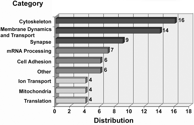
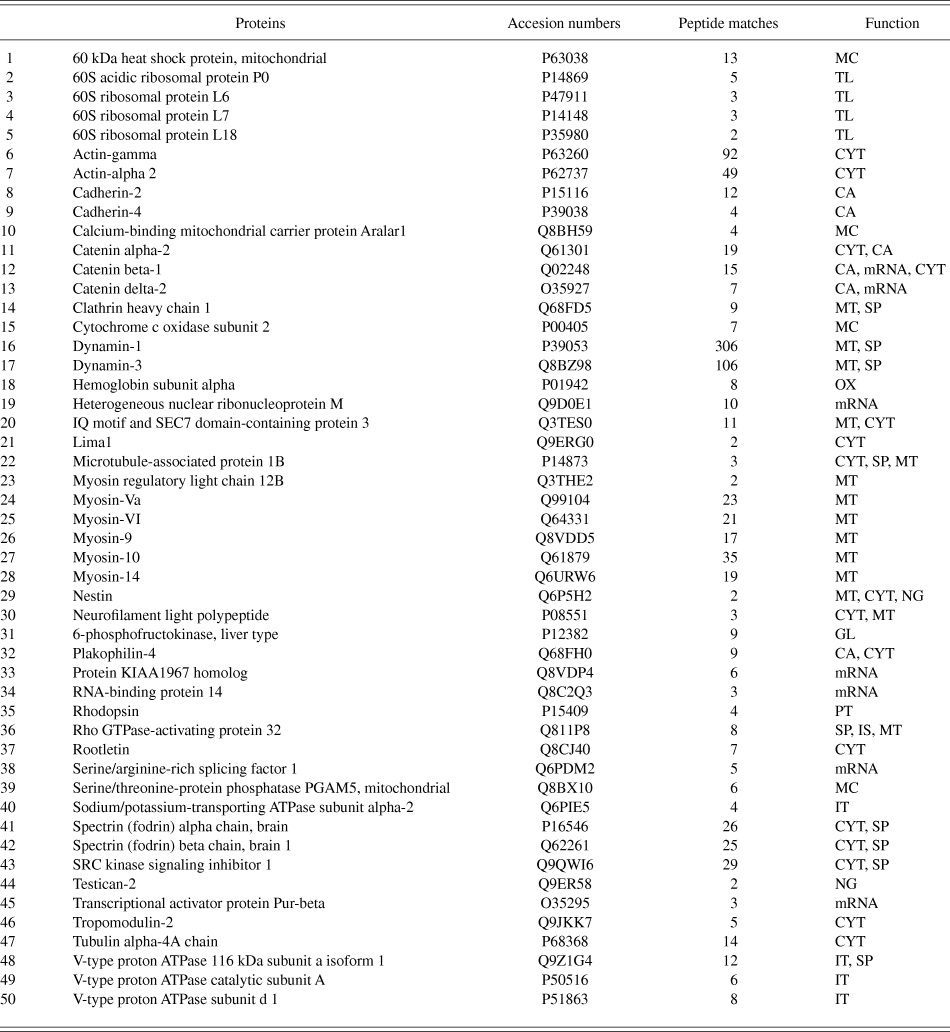
Proteomic analyses of the anti-dynamin-1 IP products from the mouse retina identified in 128 proteins (Supplementary Table 1). Table 2 presents a summary of the 50 proteins identified in two IP experiments that were not identified in control IP experiments using nonspecific IgG. These 50 proteins were classified into nine functional groups using bioinformatic methods, with several proteins categorized into more than one group (Fig. 5). The three most prevalent functional categories were cytoskeletal-associated membrane dynamics and transport and synaptic processes (Fig. 5). Sixteen proteins were classified as members of the cytoskeleton, fourteen were involved in membrane dynamics and vesicular protein transport, and nine were synaptic proteins. Other categories containing fewer proteins included mRNA processing, cell adhesion, ion transport, translation and mitochondrial-associated.
Bar graph representing the potential dynamin-1 interacting proteins organized by cellular function. Classifications were determined using IPA software and review of protein databases. Each category originates from Table 2 and represents the proteins that were identified in both IP experiments but not in the control experiment. Some proteins are encompassed in more than one functional class. The three most prevalent categories were cytoskeletal, membrane dynamics and transport, and synaptic-associated.
Proteins identified in two anti-dynamin-1 IPs from mouse retina
Peptide hits that did not reach statistical significance as determined by Matrix Science Mascot were omitted from the results. All Keratins and Histone H2B types (10 types) were omitted from the results. Highest matches within experiments reported: AM, anti-microbial; AP, apoptosis; CA, cell adhesion; CM, cellular metabolism; CJ, cell junction; CP, complement; CYT, cytoskelton; GL, glycolysis; IS, intracellular signaling; IT, ion transport; MC,Mitochondria; mRNA, mRNA processing; MT, membrane dynamics and transport; NG, neurogenesis; OX, oxygen binding; PT, phototransduction, SP: synpase, SR: stress response, TL: translation.
To determine whether these potential dynamin-1 interactions are retina-specific or ubiquitous, we conducted parallel IPs in wt mouse brain tissue. Two independent brain IP experiments were performed and nonspecific IgG was used as a control. As performed in retinal experiments, the IP and control lanes were excised into 31 fractions and proteins in each were identified by LC MS/MS. Proteomic analyses of the anti-dynamin-1 IP products from the mouse brain identified in 132 proteins (Supplementary Table 2). Nineteen proteins were identified in two IP experiments that were not identified in control IP experiments using nonspecific IgG (Supplementary Table 3). Of these, seven proteins were identical between the retina and the brain. These include actin-alpha 2, cadherin 2, catenin alpha-2, catenin beta-1, dynamin-1, dynamin-3, and sarcoma (SRC) kinase signaling inhibitor 1. All seven proteins can be classified into the functional categories of cytoskeletal-associated or membrane dynamics and transport.
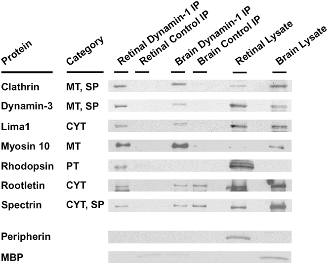
We further validated seven identified protein partners of dynamin-1 in the retina: clathrin, dynamin-3, lima1, myosin 10, rhodopsin, and spectrin by western blot analysis of IP complexes (Fig. 6). These are representative proteins of the three major functional classes of dynamin-1 interacting partners—cytoskeletal, membrane dynamics and transport, and synaptic. All seven proteins were clearly detected in the retinal IP samples but not in the control IP samples, indicating that these proteins interact with dynamin-1. The abundant brain protein, myelin basic protein (MBP), was used as a brain-specific control; peripherin was used as retina-specific control. In agreement with our proteomic data, peripherin and MBP were not detected in the IP product lanes of their respective tissues; however, they were present in the appropriate lysate preparations (Fig. 6).
Western blot analysis validating seven dynamin-1 protein-binding partners identified in proteomic analysis. The selected proteins from the three most prevalent functional categories are highlighted. The brain and retinal IP complexes were separated by SDS-PAGE and then immunoblotted with the different antibodies as indicated. The category for each protein is indicated. MBP was used as a brain-specific control and peripherin was used as a retina-specific control. CYT: cytoskeleton, MT: membrane dynamics and transport, PT: phototransduction, and SP: synapse.
Discussion
Dynamins play important roles in neurons of the central nervous system, where they generate vesicles from membranes (Ferguson & De Camilli, Reference Ferguson and De Camilli2012). Herein, we confirm that dynamin-1, dynamin-2, and dynamin-3 are expressed in the retina and are present in multiple classes of retinal neurons. Furthermore, our results indicate that each isoform has a distinct pattern of distribution throughout the retina.
The broad cellular localization of dynamin-1 is similar to that seen using a pan-dynamin antibody (Fig. 3A and 3E), indicating that dynamin-1 is the dominant isoform in the mouse retina as has been postulated for the mouse brain (Ferguson et al., Reference Ferguson, Brasnjo, Hayashi, Wolfel, Collesi, Giovedi, Raimondi, Gong, Ariel, Paradise, O’Toole, Flavell, Cremona, Miesenbock, Ryan and De Camilli2007). In contrast to dynamin-1, dynamin-2 localization is highly concentrated above the myoid region of the IS near the CC (Fig. 3C). The CC represents a transition zone between the IS and OS and plays a key role in the transport of proteins as they are synthesized in the IS and trafficked to the OS (Insinna & Besharse, Reference Insinna and Besharse2008). This suggests that dynamin-2 plays a role in intracellular transport in the photoreceptor cell as has been proposed for other cell types (Cao et al., Reference Cao, Garcia and McNiven1998). Dynamin-3 expression is most pronounced in the IPL, where synaptic terminals of second-order bipolar neurons synapse with amacrine and ganglion cells (Fig. 3D). Dynamin-3 is also present throughout the OPL and appears to be expressed in photoreceptor terminals and the dendrites of second-order neurons. These results suggest that dynamin-3 plays a critical role in retinal synaptic processing.
Dynamin-1 and Tulp1 interact in photoreceptor cells and colocalize in the IS, ONL, and OPL (Xi et al., Reference Xi, Pauer, Ball, Rayborn, Hollyfield, Peachey, Crabb and Hagstrom2007). Although Tulp1 was below detection limits by mass spectroscopy in the mouse retina, we confirm the interaction of these two proteins by western blot analysis in both mouse (Fig. 4) and bovine retinas. Tulp1 is a photoreceptor-specific protein, which associates with the cytoskeleton and plays a role in intracellular protein transport (Hagstrom et al., Reference Hagstrom, Duyao, North and Li1999; Xi et al., Reference Xi, Pauer, Marmorstein, Crabb and Hagstrom2005, Reference Xi, Pauer, Ball, Rayborn, Hollyfield, Peachey, Crabb and Hagstrom2007; Grossman et al., Reference Grossman, Pauer, Narendra, Peachey and Hagstrom2009). Tulp1−/− mice develop early onset photoreceptor degeneration with defects in protein vesicular transport (Hagstrom et al., Reference Hagstrom, Duyao, North and Li1999, Reference Hagstrom, Adamian, Scimeca, Pawlyk, Yue and Li2001; Grossman et al., Reference Grossman, Pauer, Narendra, Peachey and Hagstrom2009, Reference Grossman, Watson, Pauer, Bollinger and Hagstrom2011). Several proteins are not transported correctly to the OS in tulp1−/− retinas (Grossman et al., Reference Grossman, Watson, Pauer, Bollinger and Hagstrom2011); therefore, we investigated whether dynamin-1 localization was influenced in the absence of Tulp1. Surprisingly, dynamin-1 expression and localization did not appear to be affected in tulp1−/− retinas (Figs. 2 and 3). In fact, all three dynamin isoform protein levels were comparable in wt and tulp1−/− retinas, thus excluding dynamin-1 mislocalization as part of the retinal degenerative process observed in tulp1−/− mice.
Fifty proteins were identified in two retinal anti-dynamin-1 IPs, suggesting that these proteins may interact with dynamin-1 in the mouse retina in vivo (Table 2). Of these, 30 were either constituents of the cytoskeleton or involved in membrane dynamics and transport (Fig. 5). This is not surprising given that there is a bidirectional interaction between cytoskeleton proteins and cellular membranes (Doherty & McMahon, Reference Doherty and McMahon2008). Furthermore, the dynamin proteins have been shown to function in vesicle movement in association with cytoskeleton elements in neurons of the central nervous system (Ferguson et al., Reference Ferguson, Brasnjo, Hayashi, Wolfel, Collesi, Giovedi, Raimondi, Gong, Ariel, Paradise, O’Toole, Flavell, Cremona, Miesenbock, Ryan and De Camilli2007; Jaiswal et al., Reference Jaiswal, Rivera and Simon2009). Our protein interaction results suggest that a similar model applies to retinal neurons. Although there are differences between the dynamin-1 protein constituents between retina and brain, the majority of the proteins identified in both retina and brain are involved in either membrane dynamics and transport or associated with the cytoskeleton (Supplementary Tables 2 and 3). The cytoskeleton is the cellular scaffolding composed of microfilaments, intermediate filaments, and microtubules that is critical in membrane dynamics, such as endocytosis at the synapse (Mooren et al., Reference Mooren, Galletta and Cooper2012), vesicle generation at the trans-Golgi face (Stow & Heimann, Reference Stow and Heimann1998), and motor-driven intracellular vesicular protein transport (Anitei & Hoflack, Reference Anitei and Hoflack2012). We identified several potential retinal dynamin-1 interacting proteins that comprise the filamentous scaffolding of the cytoskeleton including actin, tubulin, neurofilament polypeptide, rootletin, and lima1, and proteins that modulate or stabilize these networks, such as nestin, spectrin, microtubule-associated protein 1B (MAP1B), and catenin. Several of these proteins were validated by western blot analysis indicating that these proteins likely interact with dynamin-1 in vivo (Fig. 6).
A graphic depiction of the highly polarized and compartmentalized photoreceptor cell is shown in Fig. 7. The three regions where dynamin-1 and Tulp1 colocalize (Fig. 7, shaded in orange) exhibit high levels of cytoskeleton-dependent vesicular transport and extensive membrane dynamics. The compartment-specific biological processes include OS protein transport (Fig. 7A), axonal transport (Fig. 7B), and synaptic vesicle transport, and membrane retrieval (Fig. 7C).
A graphic depiction of the photoreceptor cell, highlighting the three areas where there are high levels of cytoskeletal-dependent vesicular transport and extensive membrane remodeling. These compartment-specific functions represent the largest categories of dynamin-1 interacting partners and are active in the regions where dynamin-1 and Tulp1 colocalize (shaded in orange). (A) IS is the compartment in which OS proteins are processed and packaged into post-Golgi vesicles for transport through the IS and CC to the OS disks. (B) Anterograde and retrograde axonal transport allows nascent protein complexes to be trafficked to the photoreceptor synapse and cellular components to be transported back toward the cell body for degradation. (C) At the terminal, endocytosis replenishes synaptic vesicles and compensates for increases in membrane area due to exocytosis.
Photoreceptor OS-specific proteins are packaged into vesicles generated at the trans-Golgi face and transported through the CC (Fig. 7A). All three dynamin isoforms have been shown to function in vesicle generation at the trans-Golgi face (Cao et al., Reference Cao, Garcia and McNiven1998). In support of this role, we identified members of the spectrin protein family as dynamin-1 binding partners. Spectrins associate with the cytoskeleton and play a role in protein transport from the trans-Golgi network (Fath et al., Reference Fath, Trimbur and Burgess1997). In mammalian photoreceptors, there appears to be several distinct post-Golgi vesicle transport pathways (Grossman et al., Reference Grossman, Watson, Pauer, Bollinger and Hagstrom2011), some of which are linked to myosin-based movement (Liu et al., Reference Liu, Udovichenko, Brown, Steel and Williams1999). The myosin family is composed of actin-based motor proteins that are responsible for many diverse functions including intracellular transport (Syamaladevi et al., Reference Syamaladevi, Spudich and Sowdhamini2012). We identified several members of the myosin family as potential dynamin-1 retinal binding partners, including myosin VI. Myosin VI is localized to the IS region and has been implicated in cargo sorting and vesicle formation at the trans-Golgi face (Warner et al., Reference Warner, Stewart, Luzio, Steel, Libby, Kendrick-Jones and Buss2003; Kitamoto et al., Reference Kitamoto, Libby, Gibbs, Steel and Williams2005). Important to both vesicle generation and transport in the IS, the non-muscle cell α- and γ-actin isoforms were identified in our IP analyses. Actin microfilaments perform many cellular functions not the least of which is to provide tracks for the myosins to transport cargo. In addition to the actin-based transport pathways, there are distinct microtubule networks in the IS and CC of the photoreceptor, which are critical in the transport of OS proteins (Whitehead et al., Reference Whitehead, Wang, Bost-Usinger, Hoang, Frazer and Burnside1999). Previous studies suggest that the dynamins stabilize microtubule networks (Tanabe & Takei, Reference Tanabe and Takei2009). We identified α-tubulin, a component of microtubules, as a possible dynamin-1 binding partner.
A second region, where there is vesicular transport in the photoreceptor cell, is throughout the axon (Fig. 7B). Protein transport in the axon is bidirectional and relies on microtubule-based motor proteins (Schwartz, Reference Schwartz1979). Here too, the dynamins may regulate the stability of axonal microtubule networks (Tanabe & Takei, Reference Tanabe and Takei2009). Pertinent to this role, we identified MAP1B as a potential dynamin-1 interacting protein. MAP1B is expressed primarily in neurons where it functions in stabilizing microtubules (Halpain & Dehmelt, Reference Halpain and Dehmelt2006). We have localized MAP1B to the ONL (Grossman et al., Reference Grossman, Beight, Ebke and Hagstrom2012a), where it may interact with Tulp1 (Xi et al., Reference Xi, Pauer, West, Crabb and Hagstrom2003). Interestingly, Tulp1 exhibits a genetic interaction with a gene for the related protein, MAP1A, whereby an allele of the gene significantly attenuates photoreceptor degeneration in Tulp1 mutant mice (Maddox et al., Reference Maddox, Ikeda, Ikeda, Zhang, Krebs, Nishina and Naggert2012). Both MAP1A and Tulp1 have been shown to bind tubulin and actin, linking the microtubule networks to the microfilament cytoskeleton (Noiges et al., Reference Noiges, Eichinger, Kutschera, Fischer, Nemeth, Wiche and Propst2002; Xi et al., Reference Xi, Pauer, West, Crabb and Hagstrom2003, Reference Xi, Pauer, Marmorstein, Crabb and Hagstrom2005). These results indicate that interactions between dynamin-1, MAPs, and Tulp1 may contribute to the stability of these networks and modulate protein movement along these networks.
The synaptic terminal is also reliant upon continuous membrane remodeling and vesicle cycling (Fig. 7C). Synaptic membrane events are highly coordinated by the cytoskeleton to replenish synaptic vesicles and compensate for increases in membrane area due to exocytosis. Synaptic proteins were the third largest category of potential dynamin-1 interacting proteins in our study and included proteins exclusive to synaptic vesicle transport and membrane retrieval, such as SRC kinase signaling inhibitor 1 and V-type proton ATPase 116 kDa subunit a isoform 1.
The dynamins have long been associated with clathrin-mediated endocytosis (CME) and clathrin-independent endocytosis (van der Bliek & Meyerowitz, Reference van der Bliek and Meyerowitz1991). The dynamin-1 (a,c) retinal splice variant has a frameshift mutation, which terminates the protein at amino acid 814 (Xi et al., Reference Xi, Pauer, Ball, Rayborn, Hollyfield, Peachey, Crabb and Hagstrom2007), eliminating the last 53 amino acids in the proline-rich domain (PRD). Many proteins of the CME pathway bind to the PRD of dynamin, including the amphiphysins, endophilins, and syndapins (Anggono & Robinson, Reference Anggono and Robinson2007). In agreement with this model, none of these proteins were detected in this study. However, rho GTPase-activating protein 32 was identified, which has been shown to regulate clathrin-independent endocytic pathways (Qualmann & Mellor, Reference Qualmann and Mellor2003; Lundmark et al., Reference Lundmark, Doherty, Howes, Cortese, Vallis, Parton and McMahon2008).
Although our study focused primarily on dynamin-1, dynamin-3 may also perform important functions in the retina. Dynamin-3 staining was strongest in the IPL, but immunoreactivity was also seen in the IS and at the photoreceptor terminal (Fig. 3D). Self-assembly of dynamin polymers has been shown to involve the GTPase, middle, and GTPase effector domain (GED) domains (Smirnova et al., Reference Smirnova, Shurland, Newman-Smith, Pishvaee and van der Bliek1999), all of which are present in the retina-specific dynamin-1 splice variant (Xi et al., Reference Xi, Pauer, Ball, Rayborn, Hollyfield, Peachey, Crabb and Hagstrom2007). Therefore, we hypothesize that this dynamin-1 isoform maintains this essential ability. In our study, dynamin-3 was identified and confirmed as a dynamin-1 binding partner, suggesting that different isoforms may have in vivo interactions in the retina. Indeed, there is recent evidence that dynamin-1 and dynamin-3 isoforms co-operate in promoting activity-dependent synaptic vesicle endocytosis in neurons of the brain (Raimondi et al., Reference Raimondi, Ferguson, Lou, Armbruster, Paradise, Giovedi, Messa, Kono, Takasaki, Cappello, O’Toole, Ryan and De Camilli2011).
Twenty other apparent dynamin-1 interacting proteins fell into additional categories. Several are important to mitochondrial function. Although not previously attributed to dynamin-1, members of the dynamin superfamily are known to be involved in mitochondrial fission and fusion (Ferguson & De Camilli, Reference Ferguson and De Camilli2012). Proteins involved in cell adhesion were also identified. This is not surprising, given that there is an association between the cytoskeleton and the cell adhesion matrices (Kaibuchi et al., Reference Kaibuchi, Kuroda and Amano1999). Finally, although the dynamins are not known to function in translation or transcription, we identified several proteins in these groups, perhaps indicating expanded retinal roles for this isoform.
In conclusion, all three dynamin isoforms are expressed in the mouse retina with distinct localization patterns. Dynamin-1 exhibits the broadest localization and appears to be the primary isoform in the retina. Dynamin-1 localizes to specialized compartments of the photoreceptor and many of the potential interacting proteins can be grouped into cytoskeleton and membrane-modeling categories, participating in intracellular transport processes relevant to endocytic and post-Golgi pathways. In addition, dynamin-1 may be important for photoreceptor cellular stability. Nevertheless, several of these proteins are specific to distinct pathways, suggesting that dynamin-1 may have compartment-specific interactomes. This has recently been proposed for Tulp1 (Grossman et al., Reference Grossman, Pauer, Hoppe and Hagstrom2012b). Additional analyses are necessary to support this idea and to expand upon the role of dynamins in the retina, including differentiating cell-type-specific functions and splice variant contributions.
Acknowledgements
We thank Dr. Andrew F.X. Goldberg for his gift of peripherin/rds antibodies, Dr. Tiansen Li for his gift of rootletin antibodies, Dr. Mark A. McNiven for his gift of Dynamin-3 antibodies, Dr. Paul Hargrave for his gift of rhodopsin antibodies, and Dr. Neal S. Peachey for his insightful comments on this manuscript. This work is supported by NIH EY15638 (SAH), Foundation Fighting Blindness, and a Research to Prevent Blindness Center Grant. None of the authors have any conflicts of interest to declare.