The incidence of venous thromboembolism in children has steadily increased in the past decade, by nearly 10% per year, reaching currently 5 out of 100,000 hospitalised children.Reference Raffini, Huang, Witmer and Feudtner1,Reference Vidal, Sharathkumar, Glover and Faustino2 Improved survival of critically ill children associated with prolonged hospital stay is the main reason. Central venous lines represent the main risk factor for deep venous thrombosis in children, especially in the neonatal population (<1 month old). However, this risk depends on the type of catheter, with a high incidence of central catheter- related thrombosis in tunneled catheters and a rather low incidence of thromboembolic events in peripherally inserted central catheters.Reference Vidal, Sharathkumar, Glover and Faustino2 A significant number of catheter-related thromboembolic events may remain undiagnosed in children during the acute phase. According to Menéndez et al, nearly 20% of thrombosis related to peripherally inserted central catheters are asymptomatic in the paediatric population.Reference Menéndez, Verdú and Calderón3 However, complications related to thromboembolic events in peripherally inserted central catheters have been rarely reported in pediatrics.
We report a 6-month-old child with undiagnosed neonatal catheter-related venous thrombosis, resulting in a severe cyanosis.
Case report
A 6-month-old female patient known to have bronchopulmonary dysplasia was referred by the pulmonologist to the paediatric cardiology clinic because of a refractory and unusual cyanosis.
This patient was born at 25 weeks and 6 days of gestation; birth weight was 875 grams. The mother had received steroid treatment more than 48 hours before delivery. The newborn exhibited respiratory distress, requiring surfactant instillation and prolonged ventilation. Parenteral nutrition was administered during the 2 first weeks of life, using a peripheral inserted central catheter in the right brachial vein. The catheter was removed at day 18 without any complications. Non-invasive ventilation was continued until day 23 and the baby was discharged from hospital at day 86 with, according to the current definition, severe bronchopulmonary dysplasia treated with oxygen therapy and spironolactone.
During the following 6 months, cyanosis worsened but remained unresponsive to an increase of at-home oxygen treatment with a minimum oxygen saturation of 78%. Clinically, the child had no tachypnea, no retraction signs, and the capillary blood gas test remained normal (pH = 7.39; PCO2 = 40 mmHg). Chest computed tomography (CT) scan showed no pulmonary lesions. An echocardiography confirmed the absence of cyanotic CHD but showed unusual venous structures at the posterior side of the left atrium. Contrast echocardiography was not conclusive because of the presence of a patent foramen ovale. Blood clotting was normal. CT angiography with injection through a venous access on the left foot (Revolution GSI General Electric; intravenous injection of 20 ml Xenetic 350 ml/ml, no sedation required) revealed an obstruction of the superior vena cava and the left brachiocephalic (e.g. innominate) venous trunk with multiple collateral vessels between the venomous trunks and the right and left inferior pulmonary veins (Supplementary material: 3D reconstruction).
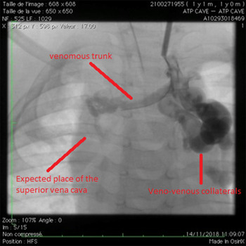
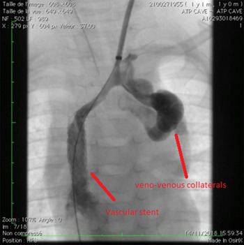
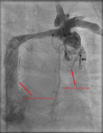
The patient underwent a first cardiac catheterization with double venous access (left jugular and right femoral). The existence of a narrow lumen within the thrombus allowed progressive balloon dilation and final deployment of two balloon-expandable stents (Valeo®, Bard Inc., New Providence, NJ, USA) dilated up to 8 mm (Figs 1 and 2). Oxygen saturation normalised immediately after the intervention. Oral anticoagulation (warfarin) was prescribed for a period of 6 months and anti-platelets for an undetermined period. An additional catheterisation was performed in March 2019 during which a dilatation of the stent of the superior vena cava was performed. Then an occlusion of the two major collaterals with two Amplatzer vascular plugs II of 10 and 12 mm (Fig 3).
Classic angiography of the left jugular vein show voluminous collaterals without perfusion of the superior vena cava.

Stented superior vena cava during classical angiography of the left jugular vein.

Second intervention. Obstruction of the collaterals with Amplatzer plug II.

Discussion
We report, for the first time in an infant, an uncommon venous systemic-to-pulmonary shunt resulting in a refractory cyanosis, due to multiple collateral vessels after an undiagnosed neonatal catheter-related venous thrombosis.
The asymptomatic character of the initial deep venous thrombosis in our patient is not uncommon, and in the general population, some studies showed that asymptomatic form could reach more than 1% of hospitalised patients.Reference Zubrow, Urie and Jurkovitz4
Acquired right-to-left shunts have been reported in the adult population with various clinical presentations and underlying conditions.Reference Gilkeson, Nyberg and Sachs5 After neoplasia, the second most frequent cause of acquired right to left shunt is related to deep venous thrombosis, with various origins, from fibrosis mediastinitis to clotting disorders. In these cases, a triggering factor for thrombosis such as repetitive surgeries, or a venous central catheter, was often reported. Only one case was described in a child secondary to a thoracic surgery.Reference Saliba, El-Rassi and Smayra6
These acquired right to left shunts are well known in the CHD population, in particular, in patients with Fontan circulation.Reference Gilkeson, Nyberg and Sachs5,Reference Kapur, Paik, Rezaei and Vu7 It can reach more than 50% of those patients with essential collaterals located above the diaphragm that are the result of recanalisation of embryological venous collaterals also known as bronchial venous plexuses. Recanalisation occurs as a result of increased venous pressures, well described in Fontan patients. The physiopathology is likely the same in our patient, with however a very different origin.
In neonates, the use of central catheters usually remains limited to the neonatal ICUs. The development of less invasive biomedical materials and the promotion of new vascular sites are intended to limit catheter-related complications. In the current era, peripheral inserted central catheter is the privileged access, to the detriment of femoral and subclavian central catheters.Reference Vidal, Sharathkumar, Glover and Faustino2 However, extremely premature infants remain at risk of catheter-related complications. Currently, there is a lack of evidence for prophylactic anticoagulation.Reference Vidal, Sharathkumar, Glover and Faustino2,Reference Monagle, Chan and Goldenberg8 Targeted echography screening should be considered in neonates with central venous line in the absence of acute clinical symptoms of deep venous thrombosis.
Management of catheter-related thrombosis in neonates is also a subject of controversy. Different therapeutic approaches have been considered: ‘wait and see’ strategy, standard anticoagulation, or thrombolysis. The result of the NEOCLOT study will hopefully clarify this situation.Reference Sol, van de Loo and Boerma9
In opposite to the adult population, in the paediatric population, benign superior vena cava thrombosis angioplasty has been rarely reported. Two case series study reported percutaneous angioplasty procedures without any acute adverse effect, in specific and severe populations: deep venous thrombosis after heart transplantation or extracorporeal membrane oxygenation. Reinterventions were more frequent than in the adult population (30–45%).Reference Salavitabar, Flyer and Torres10,Reference Kazanci, McElhinney and Thiagarajan11 The level of mortality in one of the two articles (8/19 patients in Kazanci et al) was very high during the follow-up but could be explained by the severity of the disease itself, as it was as high as in the control group. Finally, in this case, the medical treatment after recanalisation included anticoagulation, as recommended, for 6 months after a venous stenting.Reference Ignatyev12,Reference Milinis, Thapar, Shalhoub and Davies13 Concerning the antiplatelet treatment that was prescribed afterwards, there is no consensus regarding its long-term utility, and the decision to treat was justified by the existence of a venous stent along with vascular plugs in a patient with a much abnormal systemic venous system.
Conclusion
Acquired right-to-left shunt secondary to venous thrombosis is a rare complication, with an uncommon clinical presentation and potentially associated with a late diagnosis. In the absence of paediatric guidelines on thromboembolic prophylaxis, systemic screening for central catheter-related thrombosis must be considered, even with minimally invasive devices, such as peripheral inserted central catheter.
Supplementary Material
To view supplementary material for this article, please visit https://doi.org/10.1017/S1047951119003354
Financial Support
This research received no specific grant from any funding agency, commercial, or not-for-profit sectors
Conflict of Interest
The authors declare no conflict of interest