Introduction
Staphylococcus aureus is a well-known pathogen that imposes a significant burden in the neonatal intensive care unit (NICU). While most infection prevention and control (IP&C) programs, surveillance efforts, and outbreak reporting have focused on methicillin-resistant Staphylococcus aureus (MRSA), Reference Nurjadi, Eichel and Tabatabai1 methicillin-susceptible Staphylococcus aureus (MSSA) causes a greater disease burden than MRSA. Reference Popoola, Colantuoni and Suwantarat2–Reference Ericson, Popoola and Smith4 This burden includes an increased risk of invasive infections, mortality, and prolonged hospitalization, particularly in neonates with extreme prematurity, those undergoing surgical procedures or requiring invasive devices such as central lines, endotracheal tubes or feeding tubes. Reference Ericson, Popoola and Smith4 Among extremely low birth weight infants (birth weight <1000 g), MSSA infections occurred in 4.8% of infants, while 1.8% of infants developed MRSA infections. Reference Carey, Duchon, Della-Latta and Saiman5 However, guidelines for MSSA infection prevention and control in the NICU are lacking.
Neonates can acquire MSSA from diverse sources including the birthing parent’s microbiota, family members colonized with MSSA, healthcare workers (HCWs), or the inanimate hospital environment. Reference Nurjadi, Eichel and Tabatabai1 These factors make source identification of MSSA transmission and acquisition in the NICU difficult. The introduction of whole-genome sequencing (WGS) to evaluate S. aureus outbreaks in NICUs has transformed outbreak investigations by providing genomewide sequence data that allows assessment of transmission events. Reference Köser, Holden and Ellington6 We report a unique outbreak of MSSA SSTIs in a level IV NICU investigated by WGS and multi-locus sequencing typing (MLST).
Methods
Hospital setting
This outbreak occurred in an academically-affiliated, 60 bed, level IV NICU with 42-single-bedrooms and nine double occupancy rooms, separated into two corridors, A and B. The NICU has ∼800 annual admissions, of which 90% of patients are inborn.
Case identification and surveillance
A case was defined as an infant with an MSSA-positive clinical or surveillance culture obtained during the outbreak investigation. Clinical cultures were obtained for suspicion of infection according to the standard of care. Surveillance cultures were obtained from asymptomatic neonates from 4 body sites (bilateral nares, axillae, umbilicus, and groin) using 1 swab (4s1s) for all sites. Reference Grohs, Hill-Ricciuti and Kelly7
Microbiological and genomic testing
All 4s1s specimens were collected in an ESwab Collection and Transport System with Liquid Amies broth (Becton, Dickinson and Company [BD], Franklin Lakes, NJ). Approximately 50-100 µL of the Liquid Amies broth media was inoculated onto trypticase soy agar with sheep blood (BD), Mannitol Salt agar (MSA) (BD), and Spectra MRSA agar (Remel, Lenexa, KS) which were incubated in ambient air for 24 hours at 35 ± 2°C. Colonies suspicious for S. aureus were subcultured for identification by matrix-assisted laser desorption ionization-time of flight mass spectrometry (Bruker, Billerica, MA). If the isolate was identified as S. aureus a PBP2a lateral flow assay (PBP2a SA Culture Colony Test; Abbott, Scarborough, ME) was used to determine mecA status. For any S. aureus isolate recovered, mupirocin susceptibility testing was performed using gradient diffusion (Etest, bioMérieux, Inc., Raleigh, NC).
Isolates were sent to the New York State Department of Health Wadsworth Center for WGS. Processing of WGS data, including read trimming (Trimmomatic), Reference Bolger, Lohse and Usadel8 genome assembly (SPAdes), Reference Prjibelski, Antipov, Meleshko, Lapidus and Korobeynikov9 annotation (Prokka), Reference Seemann10 and sequence type (ST) assignment (“mlst”), Reference Jolley and Maiden11 was performed using an in-house developed pipeline. NextSeq was used as the sequencing plantform in our investigation. Antimicrobial resistance genes were identified using ABRicate with reference databases from CARD, Reference Jia, Raphenya and Alcock12 Resfinder, Reference Zankari, Hasman and Cosentino13 and NCBI AMRFinder. Reference Feldgarden, Brover and Haft14
To further characterize isolate diversity, a core-genome alignment of the sequenced isolates was generated using Parsnp, followed by construction of a Maximum Likelihood phylogenetic tree using IQ-TREE2. Reference Treangen, Ondov, Koren and Phillipy15,Reference Minh, Schmidt and Chernomor16 An in-house developed pipeline determined isolate relatedness at a higher resolution. Reference Haas, Lapierre and Musser17 Isolates were placed into the same cluster if their genomes shared ≥ 950 of 1000 k-mers (short DNA segments 23 bp in length). All genomes within a cluster underwent pairwise comparisons to determine the number of genetic variants consisting of either a single nucleotide polymorphism (SNP) or a short insertion or deletion. Two isolates were considered to be closely related if they differed by no more than 10 genetic variants, suggesting a recent common ancestor. When more than one MSSA isolate was detected from the same patient, both underwent WGS.
Infection prevention and control (IP&C) measures
The investigation was triggered by four infants with SSTIs caused by MSSA presenting over three weeks. Interventions included initiating contact precautions for case patients, decolonizing patients who had clinical- or surveillance-positive cultures with nasal mupirocin and chlorhexidine baths (if >2 months corrected age), Reference Gollerkeri, Oliver and Maria18 increasing availability of alcohol-based hand sanitizers in patient care areas, re-educating HCWs about hand and nail hygiene, assessing HCW hand skin condition, introducing bare-below-the-elbows (BBE) practice for HCWs (which included wearing short sleeves with no clothing or jewelry below the elbows except a smooth wedding band), environmental cleaning and disinfection targeting high-touch areas such as the nursing and respiratory carts, and removing excess equipment and supplies from patient areas. Commonalities among clinical cases were identified by examining the site of infection, presence of facial medical devices secured by tape, type of adhesive used to secure oral and nasal cannulas, type of respiratory support, and history of skin breakdown including pressure, friction and shear injuries.
Statistical analysis
Demographic and clinical characteristics between patients colonized or infected with the dominant MLST, sequence typing (ST)-121, and non-ST121 isolates were compared using Wilcoxon rank sum test for continuous variables and Fisher’s exact test or Pearson’s χ2 test for categorical variables as appropriate. Age was defined as the time when cultures were obtained.
Results
Case patients with MSSA Infections
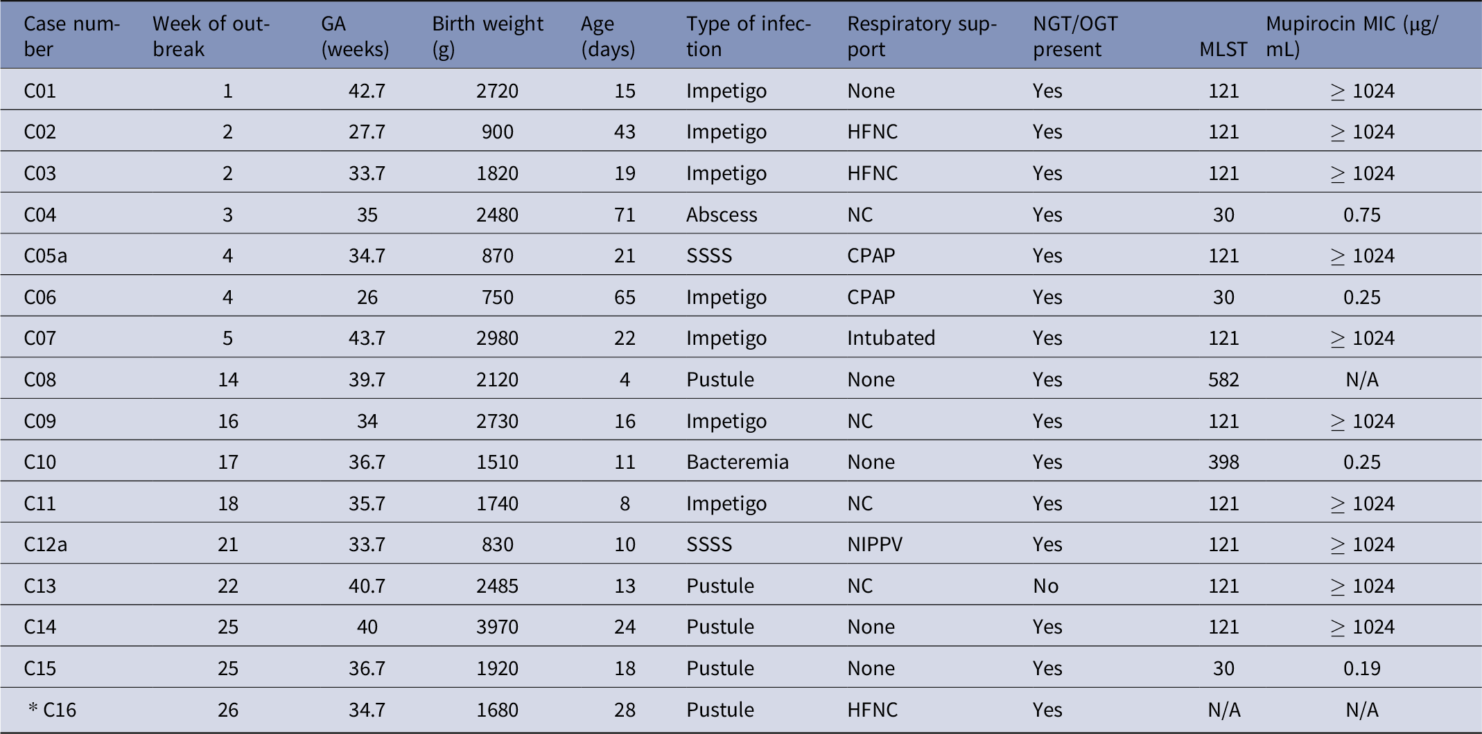
Case 1 was a 15-day-old full-term neonate in corridor B, who presented with perinasal erythema and skin erosion surrounding a nasogastric tube secured with medical tape. The lesions were consistent with impetigo. Case 2, a 6-week-old in corridor A, presented 10 days later with facial impetigo on the face where DuoDerm® secured the nasal cannula for respiratory support. Two more patients presented with SSTIs in corridor A including a 4-week-old with impetigo on the face and neck (12 days later) and an 8-week-old with an abscess at the site of their gastrostomy tube (16 days later). Case 5, a 3-week-old, developed facial impetigo during week 4, which progressed to a diffuse desquamated rash within 36 hours, consistent with staphylococcal scalded skin syndrome (SSSS). Two additional cases of impetigo occurred during weeks 4 and 5 (Table 1). No new MSSA infections occurred until week 14 (Figure 1, A) when 2 SSTIs in corridor A and 1 case of bacteremia in corridor B occurred. During the outbreak there were 16 infants who had MSSA infections including impetigo (n = 7), pustular rash (n = 5), SSSS (n = 2), abscess (n = 1), and bacteremia (n = 1). No deaths were associated with the outbreak.
Demographic and clinical characteristics of infants with MSSA infections

GA = gestational age, NGT = nasogastric tube, OGT = orogastric tube, MLST = multi-locus sequencing typing, MIC = minimum inhibitory concentration, SSSS = staphylococcal scalded skin syndrome, HFNC= high flow nasal cannula, CPAP = continuous positive airway pressure, NIPPV = non-invasive positive pressure ventilation, NC = nasal cannula, *WGS was not performed for the isolate of this infant.
Time line and geography of MSSA cases during the outbreak.
A. Epidemic curve showing the weekly case number of infection and colonization cases. ST indicated by color coding. Dates of surveillance cultures indicated with *. Positive cultures associated with infection indicated with X. One infant in week 22 had two isolates with different STs (ST-15 and ST-30) and both are included. Three infants that had two isolates with the same ST (ST-121) are included only once.
B. NICU map showing location of cases from weeks 1-7 (left) and weeks 14–32 (right). ST indicated by color coding. Numbers within boxes indicate week of outbreak. Cases of staphylococcal scalded skin syndrome (SSSS) are indicated with a black arrow. †Includes two cases (twins) with the same ST (ST-121). ‡Includes two cases (twins): twin 1 with ST-30, twin 2 with two isolates, ST-30 and ST-15.

Surveillance testing
Surveillance culture testing for MSSA was performed during weeks 4, 6, and 7 in patients in the rooms adjacent to and across from the first 4 cases (Figure 1), taking nursing assignments into consideration. Seven (33%) of 21 infants had positive surveillance cultures. In week 21, surveillance testing was expanded to the entire unit and continued monthly. In all, 147 patients were screened during 7 rounds of surveillance testing and 31 (21%) infants were colonized with MSSA.
Genomic testing
Genomic testing was performed on 46 isolates recovered from 42 patients [See Supplemental Figure 1 for MSSA isolates that underwent WGS]. Three colonized patients and 1 clinical case with pustular rash had MSSA detected by PCR instead of a culture and thus were not eligible to have WGS testing (Supplemental Figure 1). Analysis identified five main sequence clusters: ST-121 (n = 15), ST-398 (n = 10), ST-15 (n = 7), ST-30 (n = 6), and ST-582 (n = 2). Six additional isolates had other or unknown MLST: ST-2990 (n = 1), ST-672 (n = 1), ST-565 (n = 1), ST-8 (n = 1), and ST-unknown (n = 2). Three infants had two cultures with the same MLST (ST-121 resulting in 15 isolates for 12 infants). One infant had 2 isolates with different MLST (ST-15 and ST-30).
WGS analysis differentiated 40 isolates into five clusters and six isolates were singletons. Cluster assignment was consistent with MLST (Figure 2). The clusters corresponding to ST-15 and ST-398 included one isolate each (S16 and S09, respectively) that was of a different MLST type due to a single divergent allele, but which was similar enough by WGS analysis to warrant inclusion into the respective cluster.
Core-genome phylogenetic tree depicting the diversity of MSSA isolates recovered in this study. Isolates assigned to genomic clusters by the relatedness pipeline are denoted by color-coded boxes: blue = ST-398, yellow = ST-30, green = ST-15, teal = ST-582, pink = ST-121 and singleton isolates are labeled in gray. Genomic cluster assignment is consistent with MLST profiles.
*Isolates S09 and S16 differ by one allele each from the predominant MLST type but are otherwise very similar to the isolates in the corresponding cluster. Isolates are labeled based on source (C: clinical infection, S: surveillance).

All isolates in the ST-121 cluster differed by fewer than 10 genetic variants (Supplemental Figure 1), indicating that the isolates were all closely related and suggesting potential transmission between patients or acquisition from a common source. Both cases of SSSS were caused by ST-121. The clusters of ST-398, ST-15, and ST-30 each contained two or more closely related isolates that differed by fewer than 10 genetic variants (Supplemental Figures 2–4). However, a large number of isolates in these clusters differed by several hundred genetic variants, arguing against recent transmission events.
Mupirocin susceptibility
All ST-121 isolates had mupirocin MIC values ≥ 1,024 µg/mL and harbored mupA (Table 1). All other clusters were susceptible to mupirocin and did not harbor mupA.
Comparison of patients with ST-121 compared to patients with non-ST-121
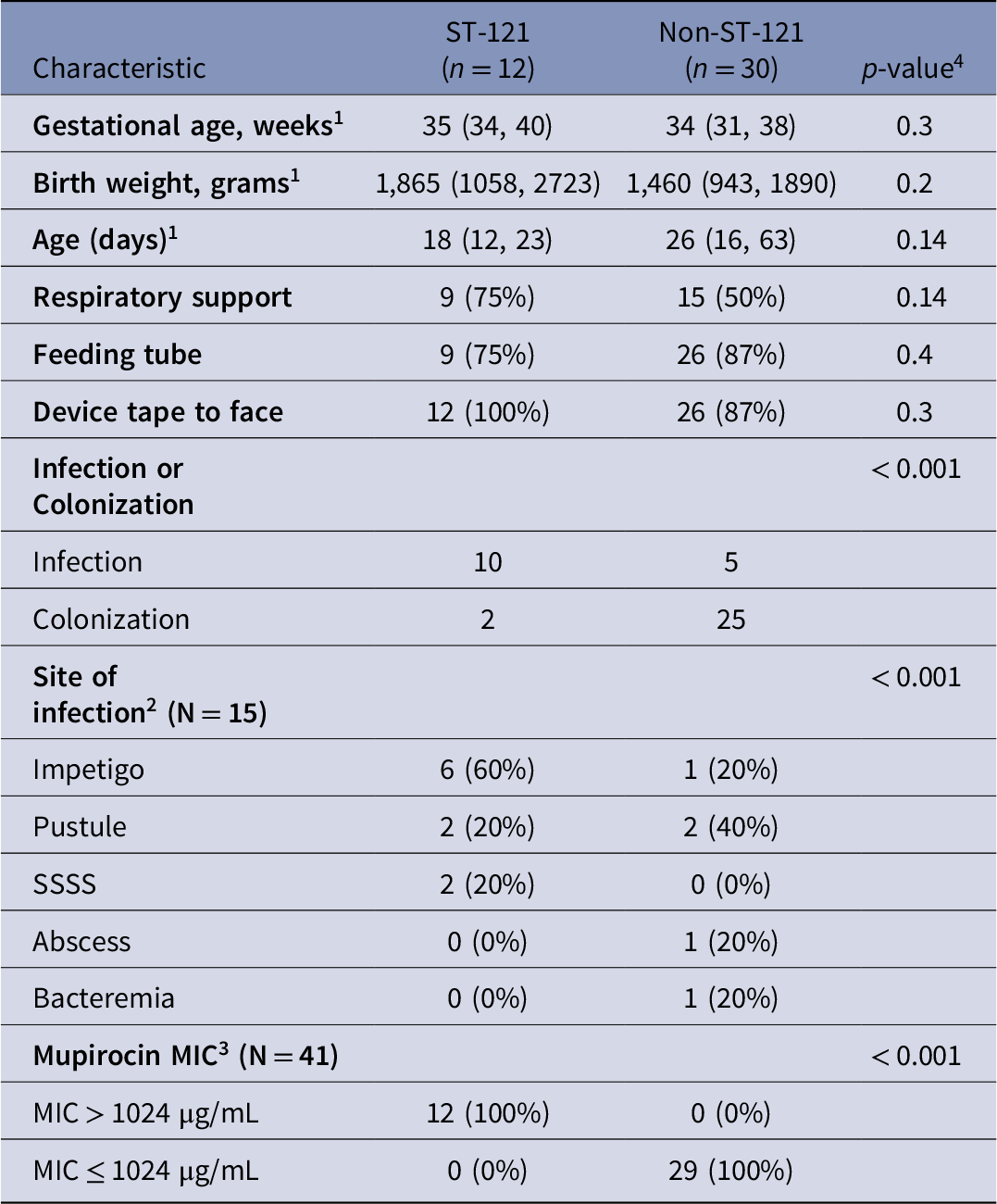
Gestational age, birthweight, and median age at the time of a positive culture were similar among infants with ST-121 isolates compared to infants with non-ST-121 isolates (Table 2). Those with ST-121 isolates were more likely to have infections compared to those with non-ST-121 isolates (83% vs 17%, < 0.001). The proportion of infants with the use of devices and/or tape applied to the face was similar in those infants with ST-121 versus those with non-ST-121 isolates.
Characteristics of infants with ST-121 isolates versus non-ST-121 isolates

1Median (IQR).
2Denominator includes only patients with infection: ST-121 (n=10), non-ST-121 (n=5)
3MIC = minimum inhibitory concentration
4Continuous variables compared using Wilcoxon rank sum test. Categorical variables compared using Pearson’s χ2 test or Fisher’s exact test.
Containment and investigation
In addition to the IP&C strategies described above, we convened a multidisciplinary quality improvement project to monitor compliance with our interventions. Skin checks of HCWs hands in week 7 did not identify potential sources of infection. We observed that respiratory therapists shared a workstation where the tape was stored and cut in preparation for securement of endotracheal tubes. Since many SSTIs were observed on neonates’ faces in association with securing a respiratory or feeding device, we implemented cleaning protocols for the shared respiratory workstation, educational initiatives for the respiratory therapy team, and revised their workflow whereby designated tape rolls and scissors were assigned to specific patient rooms. The NICU was mapped out to identify the teams responsible for cleaning patient rooms, and organizing and cleaning the respiratory and medical supply carts in patient rooms. Education regarding outbreak control measures was provided and decolonization protocols were shared with the team.
After week 26, no further cases of MSSA infections occurred. Monthly surveillance testing and WGS of recovered S. aureus isolates has continued in the NICU until present and no additional clusters of MSSA infections have occurred. During surveillance screening four months after the initial outbreak, one ST-121 isolate was identified that was not associated with infection.
Discussion
In this prolonged outbreak of SSTIs caused by MSSA, WGS refined our understanding of the outbreak by identifying a predominant cluster corresponding to ST-121 determined by MLST that was mupirocin resistant. Although the source of the outbreak was not identified, we suspect that respiratory devices and/or adhesives used to secure medical equipment to the face were involved given the frequency of facial lesions. Medical equipment, including contaminated surgical tape, has been suspected to be a reservoir or vector in healthcare-associated infections (HAIs). Reference Bernatchez and Schommer19 Multipronged interventions were successful in controlling the outbreak, including a BBE initiative and implementation of a multidisciplinary quality improvement effort.
Prior work has demonstrated a high degree of genetic diversity among MSSA isolates in the NICU, Reference Annavajhala, Kelly and Geng20 supporting the need for highly discriminatory molecular typing to differentiate outbreak from non-outbreak strains. Reference Carey, Duchon, Della-Latta and Saiman5,Reference Cho, Yang and Cunningham21 Recent studies have shown that WGS enables higher discrimination than prior methods used to determine strain relatedness in outbreak investigations. Reference Roach, Burton and Lee22–Reference Sundermann, Chen and Kumar24 WGS has become more readily available in clinical settings, allowing for timely identification of outbreak strains and differentiation of routes of acquisition allowing tailored interventions to mitigate transmission. In one neonatal MSSA bacteremia study, WGS distinguished outbreak-related isolates from non-outbreak isolates of the same MLST using a cut-off value of 12 allelic differences in the genome sequence. Reference Slingerland, Vos and Bras25 Similarly, we defined a cut-off of 10 genetic variants to categorize closely related isolates and found a high degree of relatedness within isolates of the outbreak cluster representing monoclonal expansion. Had we limited our investigation to conventional MLST, we may have erroneously concluded that the outbreak was caused by multiple clusters rather than one predominant clone. The isolates from the three other main clusters identified, ST-15, ST-30 and ST-398, differed generally by >100 genetic variants. This suggests a distant common ancestor among them, and these isolates diversified in the community or hospital and had repeated sporadic introductions to the NICU. Reference Uhlemann, McAdam and Sullivan26
Two neonates developed SSSS with ST-121. SSSS is caused by S. aureus strains that produce exfoliative toxin causing a severe diffuse skin reaction with fluid-filled blisters and skin desquamation. ST-121 is a recognized hypervirulent lineage with known production of exfoliative toxin and Panton-Valentine leukocidin and is frequently associated with SSTIs. Reference Rao, Shang, Hu and Rao27,Reference Giormezis, Doudoulakakis and Tsilipounidaki28
Application of mupirocin ointment to the nares is the standard approach for decolonization of S. aureus and can prevent MRSA infections in adult ICU patients when universal decolonization is instituted. Reference Huang, Septimus and Kleinman29 The emergence of mupirocin resistance among MSSA strains is a growing concern. Reference Dadashi, Hajikhani, Darban-Sarokhalil, van Belkum and Goudarzi30 Our investigation identified high-level mupirocin resistance with observed MIC values of ≥1,024 µg/mL within the ST-121 cluster that also harbored mupA. Reference Udo, Jacob and Mathew31 Over the past decade, reports from Europe have described an increase in mupirocin resistance among pediatric patients with MSSA SSTIs. Reference Giormezis, Doudoulakakis and Tsilipounidaki28,Reference Yin, Michel and Makki32
A central component of our interventions was adoption of BBE policy. Although the evidence regarding the impact of BBE on infections in hospital settings is limited, there is compelling rationale for BBE implementation. Sleeves and coats can harbor a high bacterial burden potentially facilitating transmission of organisms from HCW to patient. Reference Treakle, Thom, Furuno, Strauss, Harris and Perencevich33 Additionally, BBE practices can improve hand hygiene practices. A study of 7,544 HCWs from 123 hospitals found that HCWs who were compliant with BBE policy had better hand hygiene practices, disinfecting their hands correctly in 73% of cases, compared to 53% in the non-BBE group. Reference Szumska, Czajkowski, Zablocki and Rozkiewicz34 Nonetheless, further research is needed to determine if BBE practices reduce HAIs.
Limitations
This study has several limitations. This is a single center study; therefore, generalization of findings may be limited. Nasal surveillance testing of family members and HCWs was not performed nor was testing of medical surfaces and supplies. Success rates of decolonization were not investigated during this study.
Conclusions
This report highlights the ability of WGS to improve resolution of isolate relatedness compared to MLST and in this instance enabled identification of a predominant mupirocin-resistant strain associated with the outbreak. Successful control of this outbreak was achieved with several IP&C strategies, including BBE practice. Further guidelines are needed to determine the utility of MSSA surveillance in the NICU, and multi-center studies are needed to confirm sustained effect of mupirocin decolonization in neonates, especially with the risk of increasing mupirocin-resistant MSSA strains.
Supplementary material
To view supplementary material for this article, please visit https://doi.org/10.1017/ice.2025.10303.
Acknowledgments
We thank the New York-Presbyterian Hospital/Weill Cornell Medical Center Clinical Microbiology Laboratory and NICU staff, and the New York State Department of Health Wadsworth Center.
Competing interests
All authors have submitted the ICMJE Form for Disclosure of potential Conflicts of Interest.