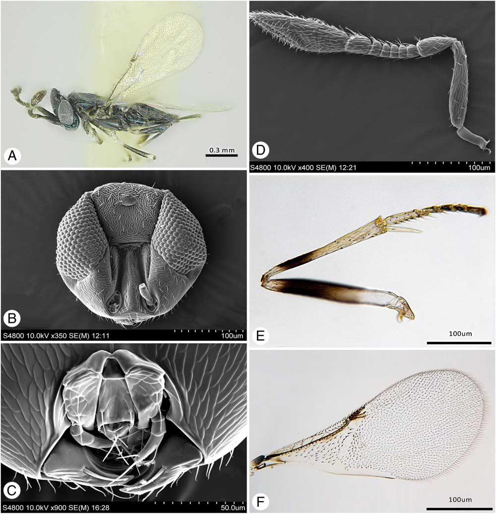
Introduction
The genus Oobius Trjapitzin, Reference Trjapitzin1963 (Hymenoptera: Encyrtidae) was not well studied until recently. Before 2010, only nine species were described (Trjapitzin and Volkovitsh Reference Trjapitzin and Volkovitsh2011), with six species from the Palaearctic Region (Nowicki Reference Nowicki1928; Trjapitzin Reference Trjapitzin1963; Myartseva Reference Myartseva1979; Guerrieri et al. Reference Guerrieri, Garonna and Viggiani1989; Zhang et al. Reference Zhang, Huang, Zhao, Liu and Bauer2005), and three species from the Afrotropical Region (Annecke Reference Annecke1967). Subsequently, Noyes (Reference Noyes2010) synonymised Avetianella Trjapitzin, 1968, Szelenyiola Trjapitzin, 1977, and Oophagus Liao, 1987 under Oobius and described 20 new species of Oobius from the Neotropical Region (Costa Rica). More recently, two new Oobius species were described from the Nearctic Region (United States of America) (Triapitsyn et al. Reference Triapitsyn, Petrice, Gates and Bauer2015) and one from the Palaearctic Region (Russia) (Yao et al. Reference Yao, Duan, Hopper, Mottern and Gates2016).
Most species of Oobius are solitary egg parasitoids of wood-boring Coleoptera in the families Buprestidae and Cerambycidae (Zhang et al. Reference Zhang, Huang, Zhao, Liu and Bauer2005; Triapitsyn et al. Reference Triapitsyn, Petrice, Gates and Bauer2015). Among those, O. longoi (Siscaro, 1992) was introduced from Australia to the United States of America (California) in the late 1990s, where it successfully established as a biocontrol agent of the invasive eucalyptus longhorned beetle, Phoracantha semipunctata (Fabricius, 1775) (Coleoptera: Cerambycidae), which feeds on ornamental Eucalyptus L’Héritier (Myrtaceae) trees (Hanks et al. Reference Hanks, Paine and Millar1996; Paine and Millar Reference Paine and Millar2002). Also, O. agrili Zhang and Huang, 2005 was introduced from China to the United States of America (Michigan) in 2007 for biocontrol of the invasive emerald ash borer, Agrilus planipennis Fairmaire, 1888 (Coleoptera: Buprestidae), which feeds on ash trees (Fraxinus Linnaeus; Oleaceae) and has become a devastating ash tree pest in North America (Federal Register 2007; Liu et al. Reference Liu, Bauer, Miller, Zhao, Gao and Song2007; Duan et al. Reference Duan, Bauer, Hansen, Abell and Van Driesche2012, Reference Duan, Jennings, Williams and Larson2014; Abell et al. Reference Abell, Bauer, Duan and Van Driesche2014; Bauer et al. Reference Bauer, Duan, Gould and Van Driesche2015). Most recently, O. primorskyensis Yao and Duan, 2016 from the Russian Far East, is being evaluated for possible use in biocontrol of the emerald ash borer in North America (Larson and Duan Reference Larson and Duan2016; Yao et al. Reference Yao, Duan, Hopper, Mottern and Gates2016).
Research in northeast China on Agrilus fleischeri Obenberger, 1925 (Coleoptera: Buprestidae), an emerging wood-boring pest of Populus Linnaeus (Salicaceae), led to the discovery of several species of insect natural enemies. These included two new species of Oobius that parasitise eggs of A. fleischeri, with combined egg parasitism rates on poplar trees ranging from 7% to 48% (Zang et al. Reference Zang, Wang, Yang, Wei and Duan2016). These two species are described here as O. saimaensis Yao and Mottern, new species and O. fleischeri Yao and Duan, new species. Herein, we describe the morphological, biological, and molecular differences between these two species, and contrast them to the morphologically similar congeners O. agrili (sympatric) and O. primorskyensis (allopatric), both of which parasitise emerald ash borer eggs on ash trees. In addition, we sequenced and analysed several ribosomal and mitochondrial genes to infer their phylogenetic relationships.
Materials and methods
Sample collection and rearing
Parasitised A. fleischeri eggs, which are darker in colour than healthy eggs, were first collected from European black poplar trees (Populus nigra Linnaeus; Salicaceae) infested with A. fleischeri on 18 September 2013 in Saima Village, Liaoning Province, China (40°57.473'N, 124°15.788'E). Additional parasitised A. fleischeri eggs were collected from other infested exotic poplar trees Populus nigra var. italica Münchhausen (Salicaceae) and native poplar trees P. davidiana Dode in Saima Village and nearby Nanmiao Village, Liaoning Province, China (40°54.658'N, 124°16.305'E) from May to September 2014, and 2–5 May 2016. The parasitised host eggs were placed in 1.5×7.5 cm glass vials and reared at the room temperature in the Key Laboratory of Forest Protection, China State Forestry Administration, at the Research Institute of Forest Ecology, Environment and Protection, Chinese Academy of Forestry (CAF), Beijing, China. Newly emerged adult egg parasitoids were either killed directly in 100% ethanol for molecular studies or killed with carbon dioxide and critical point dried for card or slide-mounting. Parasitoid wasps that emerged from the A. fleischeri eggs collected in 2013 were also reared individually on A. planipennis eggs freshly laid on coffee filter paper (Larson and Duan Reference Larson and Duan2016) for one generation in the quarantine laboratory at the United States Department of Agriculture, Agricultural Research Service, Beneficial Insects Introduction Research Unit, Newark, Delaware, United States of America. While some laboratory-reared (F1) parasitoids were designated as paratypes, most were used for DNA sequencing for molecular phylogenetic analysis at the Laboratories of Analytical Biology (Washington, District of Columbia, United States of America), Smithsonian Institution, National Museum of Natural History (NMNH).
Taxonomic studies
Selected parasitoid specimens were slide-mounted in Canada balsam (Platner et al. Reference Platner, Velten, Planoutene and Pinto1999) for detailed examination. All specimens were examined under a ZEISS Stemi 508 stereomicroscope (Bio-Research Scientific Cooperation, Agent Company, Beijing, China). Slide-mounted specimens were imaged using a Leica DM6000B microscope with a Leica DFC 300FX high sensitivity digital camera (Leica Microsystems, Wetzlar, Gemany) according to the methods described in Yao et al. (Reference Yao, Duan, Hopper, Mottern and Gates2016). Some specimens were also examined in ethanol to determine the number of multiporous plate sensillae on the antennae. Images of card-mounted specimens were generated using Helicon Focus software (Helicon Soft, Kharkiv, Ukraine) after generating an image stack with an Olympus CX31 microscope (Olympus, Tokyo, Japan) fitted with a Canon EOS 700D digital camera. Scanning electron microscope images were produced with a Hitachi/S4800 desktop unit (Hitachi, Tokyo, Japan). Some specimens were manually cleaned of external debris with forceps or brushes and affixed to 12.7×3.2 mm Leica/Cambridge aluminum scanning electron microscope stubs with carbon adhesive tabs (Electron Microscopy Sciences, #77825-12). Stub-mounted specimens were imaged after sputter coating using a Cressington Scientific 108 Auto with gold from all angles to ensure complete coverage (~10–20 nm coating). Morphological characters and measurements were obtained by examination of card-mounted specimens, slide-mounted specimens and scanning electron microscope photos. Morphological terms used in the species description follow Bouček (Reference Bouček1988) and Gibson (Reference Gibson1997). The holotypes and a portion of the paratypes are deposited in the Insect Collection Unit at the Chinese Academy of Forestry, Beijing, China. Additional paratypes are deposited in the Smithsonian Institution, Washington, District of Columbia, United States of America.
DNA extraction, amplification, and sequencing
DNA extraction, amplification, and sequencing were performed independently on different sets of specimens at two institutions: the Key Laboratory of Forest Protection at China State Forestry Administration, and the Laboratories of Analytical Biology at NMNH. At the China State Forestry Administration, a 610 base pair fragment of the mitochondrial cytochrome oxidase subunit I (COI) gene was amplified and sequenced from one female (F0) and one male (F0) specimen of O. saimaensis and one female (F0) O. fleischeri (GenBank, www.ncbi.nlm.nih.gov/genbank: MF568022–MF568026). In addition, we extracted DNA from two female (F0) O. agrili. Genomic DNA was extracted from the whole body of adult wasps using the Tiangen Blood and Animal Tissue Kit (Tiangen Biotech, Beijing, China). A region of COI of mtDNA was amplified using the following primer pair: New Par: 5'-TAAGWTTAATTATTCGRTTAGAATTARG-3' (forward), 5'-TAAACTTCWGGATGACCAAAAAATCA-3' (reverse) (Santos et al. Reference Santos, Besnard and Quicke2011). The amplification reactions were done in 0.5 μL of each primer (10 μmol), 12.5 μL 2×Taq PCR Master Mix (Tiangen Biotech), 10 μL ddH2O and 1.5 μL DNA template for a final volume of 25 μL. The amplification was performed in 0.2 mL tubes in Eppendorf B Mastercycler (Eppendorf, Hamburg, Germany) with the following profile: incubation for five minutes at 94 °C (initial denaturation step), then for 35 cycles consisting of 94 °C for 45 seconds, 46 °C for 45 seconds and 72 °C for one minute and a final extension for five minutes at 72 °C. The sequencing reaction was carried out using ABI BigDye Terminator version 3.1 cycle sequencing kit on an ABI 3730XL (SinoGenoMax, Beijing, China). Sequences were edited and aligned with Staden Package (Staden et al. Reference Staden, Beal and Bonfield1999) and DNAMAN version 6 (Lynnon Biosoft, San Ramon, California, United States of America).
At the National Museum of Natural History, Laboratories of Analytical Biology, DNA was nondestructively extracted from one female (F0) and four male (F1) O. saimaensis (USNMENT01231522, USNMENT01231524-26) and two male (F1) O. fleischeri (USNMENT01231521, USNMENT01231523). In addition to the two new species, we extracted DNA from one female O. agrili (USNMENT01119386) one female O. primorskyensis from Russia (USNMENT01119387), and one female O. primorskyensis from South Korea (new locality record, USNMENT01119503). To serve as outgroups for our molecular analysis, we included one female O. longoi (USNMENT01119464) and one female O. buprestidis (USNMENT01119332). These outgroups were chosen based upon a larger and more comprehensive molecular phylogeny of Oobius currently in developed by the co-authors J.L.M. and M.W.G. (unpublished data). Extraction, polymerase chain reaction amplification, purification, sequencing, and sequence verification/assembly followed the protocols outlined in Liu and Mottern (Reference Liu and Mottern2017). Amplified gene regions included the following, though not all genes were amplified for all specimens: a 766 base pair fragment of the small subunit 18S rDNA (GenBank: MF573302–MF573309), large subunit 28S rDNA expansion regions D2–5 (GenBank: MF573310–MF573322), a 1041 base pair fragment of COI (GenBank: MF568009–MF568025), and a 254 base pair fragment of cytochrome oxidase subunit II (COII) (GenBank MF567996–MF568008). All primer sequences are reported in Liu and Mottern (Reference Liu and Mottern2017) with the exception of the pair used for COII, which were as follows: 5'-ATTGGACATCAATGATATTGA-3' (forward) (Simon et al. Reference Simon, Frati, Beckenbach, Crespi, Liu and Flook1994) and 5'-CCACAAATTTCTGAACATTGACCA-3' (reverse) (Dowton and Austin Reference Dowton and Austin1997).
Sequence alignment and phylogenetic analysis
Ribosomal DNA sequences were aligned using the E-INS-i algorithm in MAFFT v6 (Katoh et al. Reference Katoh, Asimenos and Toh2009) with default settings. Ribosomal and mitochondrial genes were then concatenated using SequenceMatrix v100.0 (Vaidya et al. Reference Vaidya, Lohman and Meier2010) for a final alignment length of 3196 base pairs. Maximum likelihood analysis of the concatenated alignment was conducted using RAxML v8.2.10 (Stamatakis Reference Stamatakis2014) under a GTR+Γ substitution model as implemented through the CIPRES Web Portal (Miller et al. Reference Miller, Pfeiffer and Schwartz2010) accessed at www.phylo.org. The data were analysed with 1000 rapid bootstraps using five gene partitions: 18S, 28S-D2, 28 S D3–5, COI+COII codon positions 1 and 2, and COI+COII codon position 3. The resulting best tree (highest likelihood) was visualised and drawn using FigTree v1.3.1 (Rambaut Reference Rambaut2009).
Results
Molecular analyses
As the molecular data obtained at the National Museum of Natural History are more comprehensive in terms of gene and taxon sampling, the National Museum of Natural History data were used for phylogenetic reconstruction, and the COI sequences from Beijing were used for the barcoding gap analysis. Both laboratories found the same result regarding COI: O. saimaensis and O. fleischeri share identical COI sequences across the regions of the gene that we examined, with the exception of a single synonymous transition (C/T) in one O. saimaensis specimen. After trimming the low-quality bases from the edge, the data matrix consisted of 610 base pairs for the COI genes, and no insertions or deletions were found in the trimmed sequence. The average compositions of the nucleotides A, G, C, and T were 30.9%, 13.0%, 12.5%, and 43.6%, respectively. As expected, the sequences were rich in A+T, which accounted for 74.5% of the nucleotides.
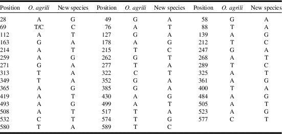
The genetic distance between O. saimaensis/fleischeri and O. agrili was computed using COI haplotype data based on the Kimura two-parameter and p-distance model. A barcode gap was discovered between the maximum intraspecific variation (0.17%) and the minimum interspecific divergence (K2P distance: 7.74%; p-distance: 7.32%). The application of character-based methods provides a combination of diagnostic nucleotides that can be used to distinguish the two new species from O. agrili.
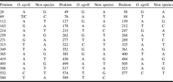
The species-specific nucleotide positions of COI (pure diagnostic barcode characters) for the two closely related species are listed in Table 1. There are differences in 44 selected nucleotide positions between the two distinct COI haplotypes (O. saimaensis versus O. agrili), and there is a switch between T and C at position 69 for O. agrili (Table 1).
Character-based DNA barcodes (COI) for Oobius saimaensis/Oobius fleischeri (new species) and Oobius agrili.
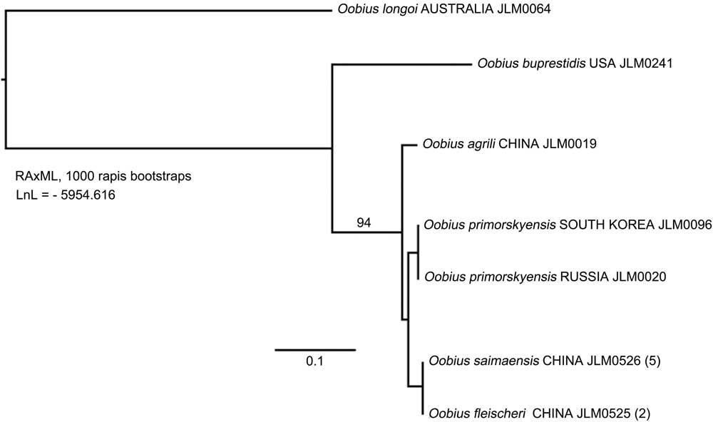
A phylogram (Fig. 1) demonstrates the close evolutionary relationship among O. agrili, O. primorskyensis, O. saimaensis, and O. fleischeri. The ribosomal genes are invariant among these four species, though there are likely multiple copies of 28S-D2 as evidenced by the presence of two double peaks (one C/G and the other C/T) in most sequence reads. Only COI and COII provide signal among the ingroup taxa, and none of our sampled genes could distinguish between O. saimaensis and O. fleischeri.
Maximum likelihood analysis of far eastern species of Oobius. Outgroups include Oobius longoi (Australia) and Oobius buprestidis (North America) base on COI. COII and 28S D2–5 ribosomal DNA. Support values are maximum likelihood bootstraps; values below 90% not shown.

During a visit to the Canadian National Collection of Insects, Nematodes, and Arachnids in Ottawa, Ontario, Canada, we found a single Oobius specimen in an ethanol lot collected in South Korea (USNMENT01119503: Gangwon-do: Chuncheon, Nam-myeon Hudong-li, 6–31.vii.2003, malaise trap in pastured area on trails close to forest edge). This specimen was both a morphological and molecular match with O. primorskyensis specimens collected in the Russian Far East.
Oobius saimaensis Yao and Mottern, new species
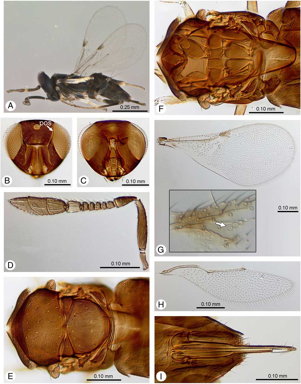
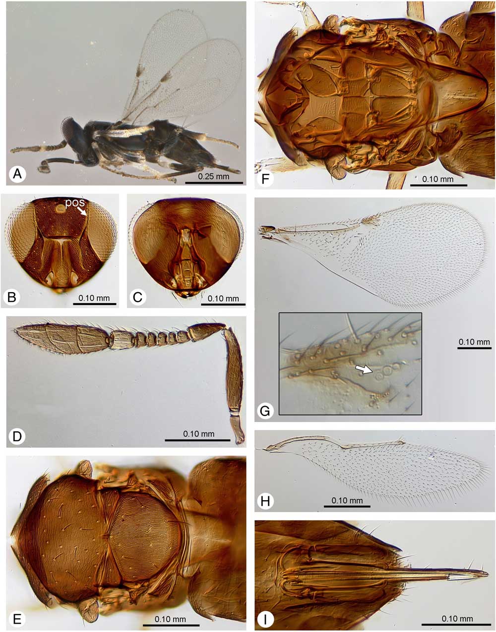
Oobius saimaensis Yao and Mottern, new species, female. A, Lateral habitus; B, anterior head, arrow indicating preorbital sensillum; C, posterior head; D, antenna; E, dorsal mesosoma; F, interior and ventral mesosoma; G, forewing, inset showing venation and annulated first stigmal sensillum; H, hind wing; I, ovipositor.
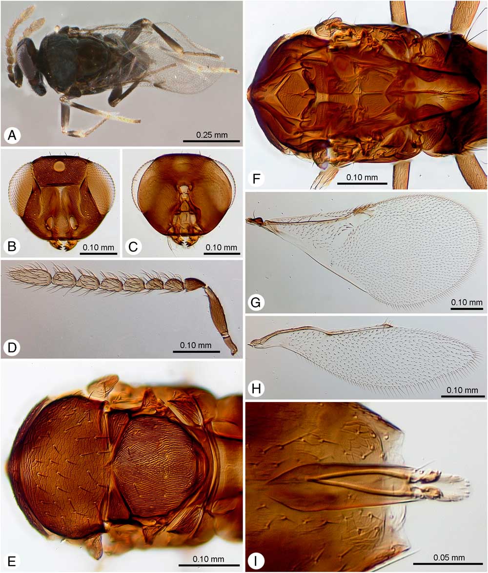
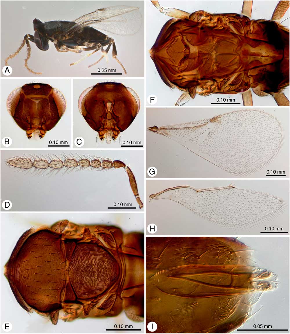
Oobius saimaensis Yao and Mottern, new species, male. A, Dorsal habitus; B, anterior head; C, posterior head; D, antenna; E, dorsal mesosoma; F, interior and ventral mesosoma; G, forewing; H, hind wing; I, aedeagus.

Type material. Holotype: ♀, China: Liaoning Province: Saima Village [40°57.473'N, 124°15.788'E], v–ix, 2014, Kai Zang, ex Agrilus fleischeri eggs, deposited in CAF. Paratypes: ♀: China: Liaoning Province, Saima Village (40°57.473'N, 124°15.788'E), 18.ix.2010, J.J. Duan, ex Agrilus fleischeri eggs, deposited in CAF; 3♀ and 2♂, China: Liaoning Province: Saima Village [40°57.473'N, 124°15.788'E], v–ix, 2014, Kai Zang, ex Agrilus fleischeri eggs, deposited in CAF; ♀: China: Liaoning Province: Saima Village (40°57.473'N, 124°15.788'E), 16.ix.2013, ex Agrilus fleischeri eggs collected from poplar trees, emerged 25–27.iv.2014 [USNMENT01231527, deposited USNM]. 4♂: progeny produced from parental generation collected from holotype locality, ex Agrilus planipennis eggs, emerged 24.xi–1.xii.2014 in quarantine at the Beneficial Insects Introduction Laboratory, Newark, Delaware [USNMENT01231522, USNMENT01231524-26, deposited in USNM].
Diagnosis. Oobius saimaensis is morphologically similar to O. agrili, O. primorskyensis, and O. fleischeri. Therefore, the following combination of characters is required to distinguish females from all other Oobius species: body length 0.63–0.74 mm; all tarsi with four tarsomeres; pedicel longer than the combined lengths of the first three funicular antennomeres; clava 2.81–3.25× as long as broad; exerted portion of ovipositor 0.33–0.42× as long as metasoma; ovipositor at least 1.3× the length of the mesotibia. Males can be distinguished from all other Oobius species by the combination of the tarsi with four tarsomeres and clava rounded apically, lacking transverse truncation.
Description. Female (Fig. 2). Body length 0.63–0.74 mm (including exerted ovipositor) (Fig. 2A).
Colour. Body dark brown to black with blue-green metallic sheen. Mandible yellowish brown with distal two teeth darker apically. Antenna dark brown except sixth funicular antennomere brownish yellow (in card-mounted specimens) or yellow (in slide-mounted specimens); multiporous plate sensilla pale yellow; clava brown. Scutellum dark brown with coppery sheen. Coxae brown, femora dark brown, lighter at apices; metatibia dark brown except distal third yellow; mesotibia mostly pale yellow with dark brown band in proximal quarter; protarsi and metatarsi pale brown, tarsomeres becoming darker distally; mesotarsus mostly pale yellow with fourth tarsomere dark brown. Wings hyaline except venation mostly brown, stigmal vein and middle third of submarginal vein yellowish brown. Metasoma dark brown; metasomal tergite 1 lighter than remaining terga. Visible portion of third valvula dark brown with coppery sheen.
Head (Fig. 2B, 2C). Frontovertex with coarse irregular reticulate sculpture and large setiferous punctures, its maximum width measured in anterior view about half that of the head. Ocelli form obtuse triangle, posterior ocellus separated by less than half its diameter from inner eye margin. Eye sparsely setose with each seta shorter than the width of an ommatidium; eye height 1.6–2.3×malar space. Frons with irregular rugulose sculpture; upper face with two lines of setae each along preorbital carina, interantennal area having two lines of setae each along inner edge of torulus; malar space with scattered setae. Torulus longer than broad, separated by greater than largest torular diameter; scrobal depression shaped like an inverted V, smooth, not reaching anterior edge of fronvertex. Preorbital sensillum (arrow, Fig. 2B, pos) present, directly adjacent to the compound eye, positioned on the frontovertex lower than the ventral margin of the anterior ocellus. Mandible tridentate, with basal tooth shorter than apical two tenth. Maxillary with three palpomeres, and labial with one palpomere. Antenna with scape subcylindrical, not reaching anterior ocellus, 4.32–5.00×as long as maximum width; pedicel 1.33–2.07×as long as the first three funicle antennomeres together, 1.60–2.67×as long as broad; funicular antennomeres 1–2 distinctly transverse with the following length width ratios: funicular antennomere 1, 0.70–0.85×, funicular antennomere 2, 0.52–0.64×, funicular antennomere 3, 0.45–0.50×, funicular antennomere 4, 0.50–0.57×, funicular antennomere 5, 0.60–0.64×, funicular antennomere 6, 1.18–1.53×as long as broad and with 1 or 2 multiporous plate sensilla. Clava with three antennomeres, 2.81–3.25×as long as broad, strongly obliquely truncate, truncation slightly more than a half the length of clava. Clava multiporous plate sensilla: claval antennomere 1: claval antennomere 2: claval antennomere 3=3:3:4.
Mesosoma (Fig. 2E–F). Mesosomal dorsum, 0.94–1.05×as long as broad, 0.68–0.89× as long as metasoma (including ovipositor). Pronotum short, with imbricate reticulate sculpture, posterior margin with a line of the longer bristles. Mesoscutum 0.39–0.53× as long as broad, with imbricate reticulate sculpture similar to pronotum but more superficial, with about 40 scattered setae, and posterior margin slightly produced medially. Axillae with superficial reticulation, not advanced and nearly meeting medially, but often covered by posterior margin of mesoscutum in dried specimens, making them appear well separated in dorsal view; usually with three setae each, forming a triangle with two setae laterally and one in about the center of the axilla. Mesoscutellum 0.74–1.14× as long as broad, with fine longitudinally striate sculpture, and with nine or 10 more-or-less paired setae, posterior-most pair the longest; mesoscutellar campaniform sensilla separated from each other by about 5× the diameter of a sensillum, and located ~0.33× length of the mesoscutellum from posterior margin. Forewing 1.94–2.51×as long as broad; marginal vein 0.87–0.90×as long as postmarginal vein, 0.81–0.90×as long as stigmal vein; stigmal vein with three sensilla, basal most sensillum with distinct annulation (arrow, Fig. 2G inset); linea calva closed posteriorly by several setae. Hind wing 3.76–4.40×as long as broad. All legs with four tarsomeres; mesotibial spur about 0.71–0.81×length of basitarsus, and basitarsus of mesothoracic leg with four stout lateral pegs and five ventral pegs.
Metasoma. Oval acuminate, 0.94×as long as broad excluding ovipositor. Cerci arise just posteriorly to the middle of the metasoma. Hypopygium nearly reaching apex of metasoma. Ovipositor about 2.04×as long as third valvula and 1.31–1.76×as long as mesotibia, ovipositor sheath exerted 0.41–0.50×metasomal length beyond apex of metasoma, apical half symmetrically lined with pairs of setae.
Male (Fig. 3). Body length 0.51–0.59 mm. Male similar to female except genitalia and the following: legs with coxae, femur, and tibiae dark brown with metallic tint, tarsi brown; antenna with scape 3.41–3.65× as long as broad, pedicel 1.58×as long as first funicular antennomere, 1.25×as long as broad; funicular antennomeres 1–2 distinctly smaller (both length and width) and darker in colour than the following antennomeres; all funicular antennomeres with long setae, the longest setae about as long as the width of the antennomere; length width ratios for funicular antennomeres as follows: funicular antennomere 1, 1.41–1.70×, funicular antennomere 2, 1.44–1.48×, funicular antennomere 3, 1.48–1.67×, funicular antennomere 4, 1.69–1.70×, funicular antennomere 5, 1.63–1.69×, funicular antennomere 6, 1.51–1.68×. Clava solid (segmented), without oblique truncation, 2.39–2.61× as long as broad; multiporous plate sensilla absent from funicular antennomeres 1–2; funicular antennomeres 3–6 each with two multiporous plate sensillae (one dorsal and one lateral) and clava with three multiporous plate sensillae. Forewing with marginal vein 0.70–0.86× the length of postmarginal vein. External male genitalia as in Figure 3I.
Etymology. The specific epithet is derived from the name of the region (Saima, Liaoning province, China) where the species was collected.
Distribution. China, Liaoning Province.
Natural history. Oobius saimaensis is a bisexual solitary egg parasitoid of A. fleischeri infesting Populus trees. Rearing experiments conducted at the United States Department of Agriculture, Agricultural Research Service, Beneficial Insects Introduction Research Unit quarantine laboratory confirmed that this species could successfully parasitise and develop in A. planipennis eggs, at least under laboratory-rearing conditions.
Oobius fleischeri Yao and Duan, new species
Oobius fleischeri Yao and Duan, new species, female. A, Lateral habitus; B, anterior head; C, posterior mouthparts; D, antenna; E, mesothoracic leg; F, forewing.

Oobius fleischeri Yao and Duan, new species, male. A, Lateral habitus; B, anterior head; C, posterior head; D, antenna; E, dorsal mesosoma; F, interior and ventral mesosoma; G, forewing; H, hind wing; I, aedeagus.
Type material. Holotype: ♀: China: Liaoning Province: Saima Village (40°57.473'N, 124°15.788'E), v–ix.2014, Kai Zang, ex Agrilus fleischeri eggs, deposited in CAF. Paratypes: 3♀2♂: China: Liaoning Province: Saima Village (40°57.473'N, 124°15.788'E), v–ix.2014, Kai Zang, ex Agrilus fleischeri eggs, deposited in CAF; 2♂: progeny produced from parental generation collected from holotype locality, ex Agrilus planipennis eggs, emerged 31.x–14.xi.2014 in quarantine at the Beneficial Insects Introduction Laboratory, Newark, Delaware [USNMENT01231521, USNMENT1231523, deposited in USNM].
Diagnosis. Oobius fleischeri is morphologically similar to O. agrili, O. primorskyensis, and O. saimaensis. Therefore, the following combination of characters is required to distinguish females from all other Oobius species: body length 0.69–0.76 mm; all tarsi with five tarsomeres; pedicel longer than the combined lengths of the first three funicular antennomeres; clava 2.97–3.33× as long as broad; exerted portion of ovipositor 0.23–0.38× as long as metasoma; ovipositor at least 1.2× the length of the mesotibia. Males can be distinguished from all other Oobius species by the combination of the tarsi with five tarsomeres and clava rounded apically, lacking transverse truncation.
Description. Note: because O. saimaensis and O. fleischeri can only be reliably distinguished based on tarsomere count, we include only measurements and some counts within the O. fleischeri description, as some of these may eventually prove to differ statistically between the two species.
Female: Body length 0.69–0.76 mm (including exerted ovipositor) (Fig. 4A) Colour. As in O. saimaensis.
Head (Fig. 4B). Eye height about 2.20–2.45×malar space. Scape 4.16–4.86× as long as maximum breadth; pedicel 1.11–1.47× as long as the first three funicular antennomeres together, 1.78–2.20× as long as broad; funicular antennomere 1 1.13–1.31× as broad as long, funicular antennomere 2–5 distinctly transverse with the following length: width ratios: funicular antennomere 2, 0.59–0.65×, funicular antennomere 3, 0.56–0.61×, funicular antennomere 4, 0.47–0.57×, funicular antennomere 5, 0.48–0.58×, funicular antennomere 6, 1.04–1.09× as long as broad and with one or two multiporous plate sensillae. Clava 2.97–3.33× as long as broad, strongly obliquely truncate, truncation about a half of the length of clava, multiporous plate sensilla: claval antennomere 1: claval antennomere 2: claval antennomere 3=3:3:4, but the number of multiporous plate sensillae in claval antennomere 2 and claval antennomere 3 variable.
Mesosoma. Mesosoma 0.79–0.85× as long as broad, 0.63–0.76× as long as metasoma (including ovipositor). Mesoscutum 0.50–0.59× as long as broad, scattered with about 40 setae. Scutellum about as long as or slightly longer than mesoscutum, 0.69–0.75× as long as broad, with 8–10 setae (four or five pairs). Forewing (Fig. 4G) 1.95–2.17× as long as broad, marginal vein 0.75–1.15× as long as postmarginal vein, 1.00–1.15× as long as stigmal vein. Hind wing 3.64–3.93× as long as broad (Fig. 4H). All legs with five tarsomeres; mesotibial spur about 0.67–0.83× length of basitarsus.
Metasoma. 1.29–1.46× as long as broad excluding ovipositor. Ovipositor 2.19–2.65× as long as outer plate of the ovipositor, 1.17–1.50× length of mesotibia, ovipositor sheath exerted 0.23–0.38× metasomal length beyond apex of metasoma.
Male (Fig. 5): Body length 0.53–0.57 mm. Similar to female except genitalia and the following: antenna with scape 2.65× as long as broad, pedicel just about or slightly shorter than first funicular antennomere, 1.38–1.48× as long as broad; all funicular antennomere longer than broad and with long setae, length: width ratios for funicular antennomeres as follows: funicular antennomere 1, 1.48–1.63×, funicular antennomere 2, 1.17–1.46×, funicular antennomere 3, 1.42–1.52×, funicular antennomere 4, 1.50–1.58×, funicular antennomere 5, 1.44–1.56×, funicular antennomere 6, 1.38–1.62×. Clava solid, without oblique truncation, 2.27–2.68× as long as broad; multiporous plate sensilla absent from funicular antennomeres 1–2; funicular antennomeres 3–6 each with two multiporous plate sensillae (one dorsal and one lateral) and clava with three multiporous plate sensillae. Forewing with marginal vein 0.55–0.60× as long as postmarginal vein, 0.86–1.05× as long as stigmal vein. External male genitalia as in Figure 5I.
Etymology. The specific epithet is derived from the scientific name of its host A. fleischeri.
Distribution. China, Liaoning Province.
Natural history. Oobius fleischeri is a bisexual solitary egg parasitoid of A. fleischeri infesting Populus trees. Rearing experiments conducted at the United States Department of Agriculture, Agricultural Research Service, Beneficial Insects Introduction Research Unit quarantine laboratory (Newark, Delaware) confirmed that this species could successfully parasitise and develop in A. planipennis eggs, at least under laboratory-rearing conditions.
Discussion
Most species of Oobius are primary egg parasitoids of buprestid and cerambycid beetles, and a total of 44 species were recorded worldwide before the present study (Noyes Reference Noyes2015; Triapitsyn et al. Reference Triapitsyn, Petrice, Gates and Bauer2015; Yao et al. Reference Yao, Duan, Hopper, Mottern and Gates2016). The two new species of Oobius described here are the primary and solitary egg parasitoids of A. fleischeri, a newly emerging pest of poplar trees in northeastern China. Both species appears to have co-existed on A. fleischeri hosts infesting poplar trees in the same geographic region (northeastern China). Our field collection records showed that both species appeared to be equally abundant in northeastern China (Y.X.Y., X.Y.W. unpublished data) and inflicted a combined parasitism of A. fleischeri as high as 48% (Zang et al. Reference Zang, Wang, Yang, Wei and Duan2016). More recently, a laboratory study at the quarantine facility of the United States Department of Agriculture, Agricultural Research Service, Beneficial Insects Introduction Research Unit showed that both species successfully parasitised eggs of the emerald ash borer A. planipennis. As A. planipennis also occurs in Liaoning Province (part of its native rage), where both O. fleischeri and O. saimaensis were collected from A. fleischeri eggs, we recommend future studies to determine if these two parasitoids species also attack A. planipennis in this region. If so, O. fleischeri and O. saimaensis may also be studied for biological control introduction against A. planipennis in North America.
We acknowledge that this may be an unusual polymorphism within a single species, especially in light of our inability to separate the two species using molecular data. However, as these wasps are part of a cryptic species complex that is being used for classical biological control, giving different names to these wasps that are morphologically diagnosable will allow biological control researchers to clearly communicate the identity of the agent to regulatory agencies during non-target risk assessment and host specificity testing. More importantly, we did not observe any variation in tarsomere numbers of the F1 males produced on A. planipennis eggs by the unmated female wasps collected in the field in each of the species. Although it is true that tarsomere counts vary within Chalcidoidea at various taxonomic levels, to our knowledge there are no cases where tarsomere counts vary intraspecifically.
The distribution of O. fleischeri and O. saimaensis overlaps with that of O. agrili, the egg parasitoid of A. planipennis in northeastern China. While O. fleischeri may be easily distinguished from O. agrili and O. saimaensis based on its five tarsomeres versus four tarsomeres in the latter two species, O. agrili and O. saimaensis are morphologically extremely similar. However, morphological results show that O. saimaensis is smaller than O. agrili and has a much longer ovipositor (at least 1.3× the length of the mesotibia) relative to overall body size than O. agrili. In addition, our molecular data show clear genetic divergence between these two species (Table 1).
As the three genera Avetianella, Szelenyiola, and Oophagus, were incorporated to Oobius (Noyes Reference Noyes2010), a few studies have examined the intraspecific and interspecific variation in morphological characteristics as well as the phylogenetic relationships among different species within Oobius as currently defined. In addition, morphological similarities among some species combined with the lack of molecular data and morphological detail in many of the early species descriptions has made the taxonomy more challenging. Further taxonomic and phylogenetic studies are clearly needed, particularly with the addition of more molecular markers through the use of next generation sequencing and biological crossing studies to further clarify species boundaries.
Acknowledgements
The authors thank the graduate students Kai Zang and Jun Yang (Chinese Academy of Forestry) for assisting in collection of parasitised A. fleischeri eggs. Dr. Yan-Zhou Zhang (Institute of Zoology, Chinese Academy of Science, Beijing, China) for checking the holotype of O. agrili and providing the specimens of the species. They also thank John S. Noyes (Natural History Museum, London, United Kingdom) for providing literature and helpful advice regarding character assessments, and Kim Hoelmer (United States Department of Agriculture, Agricultural Research Service, Beneficial Insects Introduction Research Unit) for helpful comments on the manuscript. The project was in part funded by Ministry of Human Resources and Social Security of China for Returned Overseas Chinese Scientific and Technological programme (#2016-176) and by United States Department of Agriculture Forest Service International Program Grant to J.J.D. and L.S.B. (United States Department of Agriculture, Agricultural Research Service agreement 60-1926-2-043). Mention of trade names or commercial products in this publication is solely for the purpose of providing specific information and does not imply recommendation or endorsement.