Case report
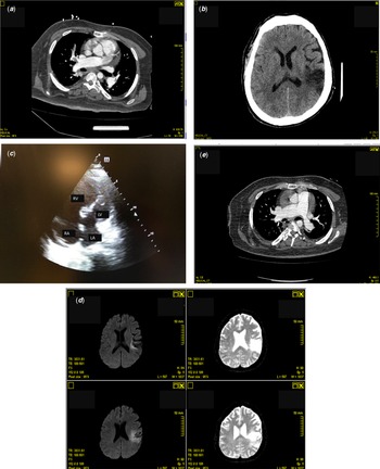
A 40-years-old male affected by Down syndrome and diabetes was admitted to our department for dyspnoea. Patients’ parents reported that the man was immobilised since 3 months because of muscular pains after COVID-19 vaccine administration. They also reported no other comorbidities or previous surgical therapies, with the exception of multiple pneumonia episodes during infancy and in adulthood. The ECG showed a complete right branch block and an anterior left hemiblock. SARS-CoV2 swab was negative and haematochemical examinations revealed a D-dimer of 13,593 ng/ml. Fast echocardiography showed a moderate enlargement of the right heart sections. CT scan revealed a bilateral pulmonary embolism (Fig 1a) mainly on the right pulmonary artery. The patient was assisted with CPAP ventilation in the ICU. Lower extremity vascular ultrasound showed left great saphenous vein thrombosis. One week later, the patient presented neurological right side signs and was obnubilated. Therefore, he underwent urgent CT scan (Fig 1b) evidencing a left parietal ischaemic lesion. The thrombophilic screening evidenced a heterozygosis MTHFR mutation. A new echocardiographic evaluation was then performed revealing a partial atrio-ventricular canal defect, typically associated to Down syndrome (Fig 1c). Reference Versacci, Di Carlo, Digilio and Marino1 A bidirectional shunt (left to right in systole and right to left in diastole) was found both on the ostium primum and ostium secundum atrial septal defects. Associated defect was a mild regurgitation of the mitral valve. A severe enlargement of right heart sections was also observed with severe tricuspid regurgitation allowing us to estimate a systolic pulmonary arterial pressure of 95 mmHg with a systemic arterial pressure of 100/60 mmHg. Left ventricular dimensions were severely decreased due to the leftward shifting of the interventricular septum with preserved left ventricular ejection fraction. Extra cardiac anatomy was normal with normal systemic and pulmonary veins return, as well as not obstructed left aortic arch. Patient parents’ reported that the diagnosis of CHD was never performed in advance. On this basis, we supposed that the concomitant pulmonary and neurological embolisation was due to paradoxical shunt at the atrioventricular septal defect level in apatient with other multiple risk factors such as the MHTFR mutation, the COVID-19 vaccine and the immobilisation. Fifteen days after the admission, the patient recovered well from the pulmonary embolism (confirmed by the chest CT scan, Fig 1d) and only partially from neurological point of view with a stable brain CT scan and MRI views (Fig 1e). Due to the neurological complication, the patient was collectively judged not eligible for cardiac surgery and he was referred to a centre specialised on adult CHDs and pulmonary arterial hypertension treatment.
(a) Thoracic CT scan showing pulmonary embolism, (b) Brain CT scan revealing the parietal neurological lesion, (c) Echocardiographic apical four chambers view showing the atrio-ventricular canal, (d) Brain MRI showing the evolution of the neurological lesion, (e) Thoracic CT scan immediately before hospital discharge showing the resolution of the pulmonary embolism.

To our knowledge, this is the first case reported in literature on concomitant left and right embolisation in a Down patient because of unknown atrio-ventricular septal defect. Previous paper in fact reported about pulmonary and neurological embolisation in non-syndromic patients presenting patent foramen ovalis. Reference Ieva, Magnesa, Gallo, Cassese, Di Biase and Brunetti2
Down patients affected by CHD are more prone to develop pulmonary vasculopathy than non-syndromic patients. Reference Kurokami, Takasawa and Takeda3 In this case, the pulmonary vasculopathy was further exacerbated by the pulmonary embolism and by the late diagnosis of CHD. Moreover, the atrioventricular septal defect would be surgically treated in childhood if timely discovered avoiding fatal or major complications such as, in this case, the stroke.
Conclusion
We report the case of a 40-years-old Down male admitted to our hospital for pulmonary embolism after COVID-19 vaccine and experiencing a stroke 1 week after the admission. Echocardiography revealed an unknown atrio-ventricular septal defect leading to the possible diagnosis of paradoxical embolisation. Atrio-ventricular septal defect is typically associated to Down patients which are also usually more prone to the general population to develop pulmonary vasculopathy. Therefore, Down patients should be routinely evaluated by a cardiologist during childhood and adulthood to avoid major and fatal complications. In our patient, the late cardiac diagnosis lead to a severe pulmonary vasculopathy with systemic right heart pressure further exacerbated by the pulmonary embolism and to a severe neurological empairment because of a stroke after COVID-19 vaccine probably due to a paradoxical embolisation on the atrioventricular septal defect.
Acknowledgment
None.
Financial support
None.
Conflict of interest
None.