The aqueous leaf extract of Dicliptera verticillata is used in some traditional societies of western Cameroon in association with Aloe buettneri, Hibiscus macranthus and Justicia insularis to regulate the menstrual cycle, and to cure dysmenorrhoea or infertility in women. The pharmacological effects of the mixture were demonstrated on some biochemical and reproduction parameters in female rats (Telefo et al., Reference Telefo, Moundipa, Tchana, Tchouanguep and Mbiapo1998, Reference Telefo, Moundipa and Tchouanguep2001). The mixture was also shown to be active on disturbed menstrual functions and functional sterility. It also showed a potentiating effect on puberty, follicular growth and maturation in immature rats pre-treated with pregnant mare serum gonadotrophin (PMSG) (Telefo et al., Reference Telefo, Moundipa and Tchouanguep2004; Goka et al., Reference Goka, Telefo, Mbemya, Awouafack, Lienou, Yemele, Njina, Donfack, Tagne and Fekam2016). When administered individually, AEDv has been shown to induce estradiol production by granulosa cells and, as the plant mixture, it could simulate steroidogenesis (Telefo et al., Reference Telefo, Guemo, Nguelefack, Watcho and Tchouanguep2005).
Traditional medicine practitioners of the western region of Cameroon advised over the toxicity of ADHJ (mixture of leaves extracts of Aloe buettneri, Dicliptera verticillata, Hibiscus macranthus and Justicia insularis) when administered through gestation and revealed the toxicity was dependent on the effect of D. verticillata. The toxicity of a plant extract can be expressed at the level of the liver, which is one of the principal organs implied in detoxification (Kieffer et al., Reference Kieffer, Martin and Adams2016). It can be manifested by the destruction of liver cells and release of many hepatic markers in the blood. The reproductive toxicity of medicinal plants can be expressed at the level of the reproductive organs an even in their hormonal release. Some additional studies done on AEDv showed that it induces the contraction of uterine smooth muscle (Guemo, Reference Guemo2002) and the principal hormone implied in uterine contractions is estradiol. Many reproductive parameters as well as the modifications of the hypothalamus–pituitary–gonad (HPG) axis are under the control of the same hormone. Conversely, estradiol, by inducing contraction of the uterine muscle, can be associated with abortion if administered during gestation. In the present study, the effects of AEDv on some reproductive and biochemical parameters of toxicity, its stimulating effect on folliculogenesis and its effect on different stages of gestation were investigated.
Materials and methods
Preparation of the extract
Fresh leaves of D. verticillata were harvested in October 2018 at the ethnobotanical garden of the University of Dschang. This plant material was washed, dried at room temperature and subsequently ground in a mortar. The powder obtained was infused for 1 h in distilled water. The solution was then filtered with a Whatman paper No. 1 and dried in a ventilated oven at 45°C. The powder obtained was further suspended in distilled water to prepare the extract at different concentrations for their daily administration to the animals at the needed doses, that is 0, 12.5, 50 and 100 mg/kg body weight (these dosages represent the control, the therapeutic dosage reconstituted from the traditional recipe and its multiples).
Animals
Female immature and mature Wistar rats, bred in the animal house of the Faculty of Agronomy and Agricultural Sciences (University of Dschang-Cameroon) were used. Immature animals were 21–23 d old while mature animals were 10–12 wk old. They were housed under uniform husbandry conditions of light (12-h cycle) and temperature (22 ± 2°C) and fed with standard laboratory diet and tap water ad libitum.
Treatment
Effect of AEDv on toxicity and fertility parameters
Sixty-four immature Wistar female rats were randomized into four groups of 16 animals each. All the groups were administered by the oral route with the AEDv at the dosages of 12.5, 50 and 100 mg/kg of body weight, except the control group, which received approximately the same quantity of distilled water. The animals were treated over 20 d and weighed throughout all the experimental period. At the end of this period, six animals per group were euthanized by intra-abdominal injection of thiopental sodium (6 mg/ml, 30 mg/kg). Their blood was collected by cardiac puncture; their liver, ovaries and uteri were also removed, blotted, weighed and conserved at −20°C until use.
The ovaries and uteri of the animals were homogenized in Tris–sucrose buffer (0.25 M sucrose, 1 mM EDTA and 10 mM Tris–HCl, pH 7.4) at 1% and 2% respectively. After centrifugation (4000 g, 15 min), the supernatants were collected and used for protein (Bradford, Reference Bradford1976) and cholesterol (Trinder, Reference Trinder1969; Richmond, Reference Richmond1973; Röschlau et al., Reference Röschlau, Bernt and Grube1974) assays. The liver was homogenized in phosphate buffer (0.1 M, pH 7.4) at 10%. After centrifugation (3000 g, 15 min), the supernatant was collected and used for protein assays (Gornall et al., Reference Gornall, Bardwill and David1949). The blood was centrifuged (2500 g, 15 min) and the serum collected was stored at −20°C for the dosage of proteins (Gornall et al., Reference Gornall, Bardwill and David1949), serum aspartate aminotransferase (ASAT) and alanine amino transferase (ALAT).
The remaining rats (10 per group) were mated, on the day following the last administration, with males of proven fertility over 2 wk. Vaginal smears were then collected the following day to look for the presence of spermatozoa, for which detection would be considered as the first day of gestation (Prakash and Roy, Reference Prakash and Roy1983). A laparoscopy was undertaken under diazepam (5 mg/ml, 5 mg/kg) and ketamine (50 mg/ml, 80 mg/kg), 10 d after the day of mating to count the number of implantation sites in uterine cords and the number of corpora lutea in ovaries. After delivery, the fetuses were weighed and their number recorded. From these data, the number of resorption sites (number of implantation site – number of live fetuses), implantation index [(total number of implantation sites/number corpora lutea) × 100], resorption index [(total number of resorption sites/total number of implantation sites) × 100], preimplantation loss [(number of corpora lutea – number of implantations/number of corpora lutea) × 100], postimplantation loss [(number of implantations × number of live fetuses/number of implantations) × 100], antifertility activity [(number of females without live fetuses/total number of females) × 100], anti-implantation activity [(number of females without implantation sites/total number of females) × 100], and gestation rate [(number of females with live fetuses at birth/total number of gestational females) × 100] were calculated (Costa-Silva et al., Reference Costa-Silva, Lyra, Lima, Arruda, Araújo, Ribeiro, Ribeiro, Arruda, Fraga, Lafayette and Wanderley2007).
Effect of AEDv on the stages of gestation
In total, 135 adult female rats (10–12 wk old) with a regular estrous cycle of 4 d were selected and mated with males of proven fertility on the evening of the proestrus phase of the cycle over 3 wk. Vaginal smears were collected on a daily basis to assess the presence of sperm. Among the positive animals, 90 were selected and shared into three sets (each for a considered period) of 30 rats each. Each set was divided into five groups of six animals and each administered distilled water (for control) or AEDv at the doses of 12.5, 50, 100 or 400 mg/kg, the last dosage. The experimental design was as follows:
1. Administration from the 1st to the 5th day of gestation to evaluate the contraceptive effect. The 10th day, the corpora lutea and implantation sites were counted through laparoscopy; these two parameters allowed calculating the implantation index and the anti-implantation activity (Badami et al., Reference Badami, Aniesh, Sankar, Sathishkumar, Sureh and Rajan2003).
2. Treatment from the 6th to the 9th day of gestation to evaluate the precocious abortive effect. On the 10th day, corpora lutea and implantation sites were counted through laparoscopy. At the delivery, the number of resorption sites, the number of in utero dead fetuses and the number of live ones were recorded. The implantation index, the anti-implantation and antifertility activities as well as the resorption and gestation rates (Badami et al., Reference Badami, Aniesh, Sankar, Sathishkumar, Sureh and Rajan2003) were evaluated.
3. From the 11th to the 20th day of gestation to evaluate the late abortive effect. A first laparoscopy was performed on the tenth day of gestation to ensure the effectivity of gestation. AEDv was administrated from day 11 to day 20 and the 21st day, a second laparoscopy allowed evaluation of the number of live and dead fetuses, the number of resorption sites, the gestation rate and the antifertility activity.
Statistical analysis
The data from biological assays were registered as mean ± standard error (SE) or percentages. The analysis of variance (ANOVA) test was used to evaluate the significance of the differences in mean values of parametric data (liver, ovarian and uterine weights). Student–Newman–Keuls and Fisher’s least significant difference (LSD) tests were used for comparison between means whenever experimental factors were significant for ANOVA. The percentages were compared using the chi-squared and Fisher’s exact probability tests. Non-parametric data were analyzed using the Kruskal–Wallis test (for treatment effects) and compared with the Mann–Whitney U-test when significant. The significance of the differences was established at the 5% level (P > 0.05).
Results
Effects of AEDv on some toxicity and fertility parameters
Oral administration of different doses of AEDv for 20 d to immature rats did not lead to any lethal effect. A significant increase (P < 0.001) was recorded in the body weight according to the treatment duration and independently of the administered doses (Fig. 1).
Effects of AEDv on body weight growth in immature rats. * and ** values indicate significant difference at P < 0.05 and P < 0.001, respectively, from those of the control group.

The administration of AEDv did not lead to any variation in the liver’s relative weight. However, a decrease was registered in hepatic proteins with the 50 and 100 mg/kg dosages. The same AEDv dosages were marked with a significant increase (P < 0.05) in serum proteins in addition, all reported to control animals. Although a gradual decrease in alanine aminotransferase (ALT) rate (according to AEDv doses) was observed, no significant variation in ASAT or in ALAT was recorded (Table 1).
Effects of AEDv on some toxicity parameters in immature rats

Each value represents the mean ± standard error of the mean (SEM) for five rats.
Asterisks indicate values significantly different from the control (distilled water) *P < 0.05, **P < 0.001 (Fisher’s LSD test).
Effects of AEDv on fertility parameters
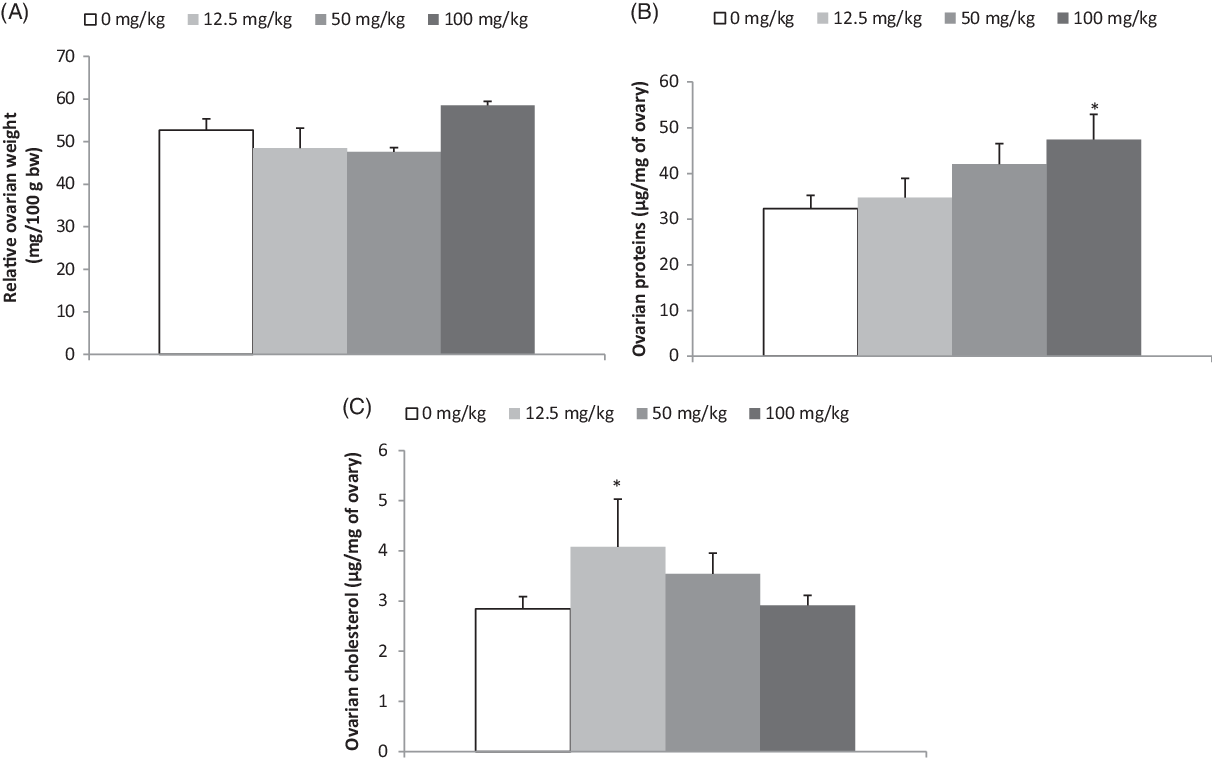
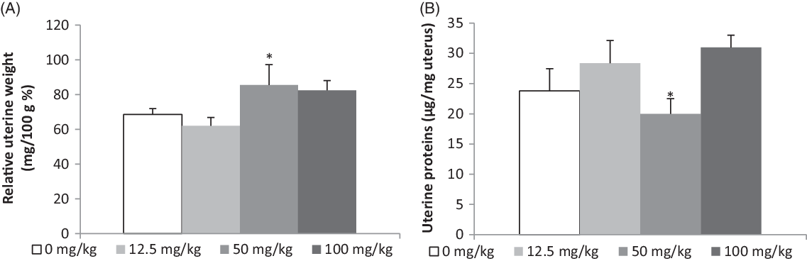
Different doses of AEDv administered along 20 d consecutively to immature rats did not lead to significant variation in ovarian relative weight. However, ovarian proteins experienced a dose-dependent increase that became significant (P < 0.05) with the 100 mg/kg dose. An increase was also recorded in ovarian cholesterol with the 12.5 mg/kg dose (Fig. 2). The 50 mg/kg dosage led to a 32% increase in uterine weight and a 28% decrease in uterine proteins (P < 0.05) (Fig. 3) compared with controls values.
Effects of AEDv on ovarian relative weight (A), proteins (B) and cholesterol (C) rates. Each histogram represents the mean ± standard error of the mean (SEM) for five rats. Values significantly different from the control (distilled water) *P < 0.05 (Fisher’s LSD test).

Effects of AEDv on uterine relative weight (A) and proteins rate (B). Each histogram represents the mean ± standard error of the mean (SEM) for five rats. Asterisks indicate values significantly different from the control (distilled water) *P < 0.05 (Fisher’s LSD test).

Effects of AEDv on fertility parameters in treated rats
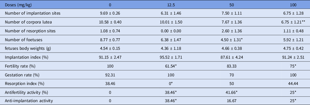
During and after the treatment, there was an increase in the body weight of treated animals. After mating, no significant effect was observed in the number of implantation sites, fetuses born body weight and implantation index. There was a 36% decrease in the number of corpora lutea (P < 0.01) with the 100 mg/kg dosage and at least a 25% increase in the antifertility and anti-implantation activities with all the AEDv doses (P < 0.05). The 12.5 mg/kg dose experienced a significant drop (P < 0.05) in resorption index and fertility rate, while this last parameter also dropped in 100 mg/kg dose-treated animals, all compared with controls (Table 2).
Effects of AEDv on fertility parameters in AEDv 20 d-treated immature rats mated with fertile males

Each value represents the mean ± standard error of the mean (SEM) for 13 to 14 rats.
In the same column, asterisks indicate values significantly different from the control (distilled water) *P < 0.05, **P < 0.01 (one-way ANOVA test for the number of implantation sites, corpora lutea, fertility rate, resorption sites, fetuses number and weights and Kruskal–Wallis test for implantation and resorption index, preimplantation and postimplantation losses, antifertility and anti-implantation activities).
Effects of AEDv on different stages of gestation
Contraceptive effect
AEDv administered from the first to the fifth day of gestation led, independently with the dose, to at least a 50% general increase tendency (P < 0.01) in the anti-implantation activity in animals treated with 50–400 mg/kg dosages and then, a decrease in the number of implantation sites and the implantation index in such animals (Table 3) all compared with the control group.
Effects of AEDv on gestation parameters from the 1st to the 5th day of pregnancy

Each value represents the mean ± standard error of the mean (SEM) for six rats.
In the same column, asterisks indicate values significantly different from the control (distilled water) *P < 0.05, **P < 0.01, ***P < 0.001 (one-way ANOVA test for the number of implantation sites, corpora lutea, and Kruskal–Wallis test for implantation index and anti-implantation activity).
Precocious abortive effect
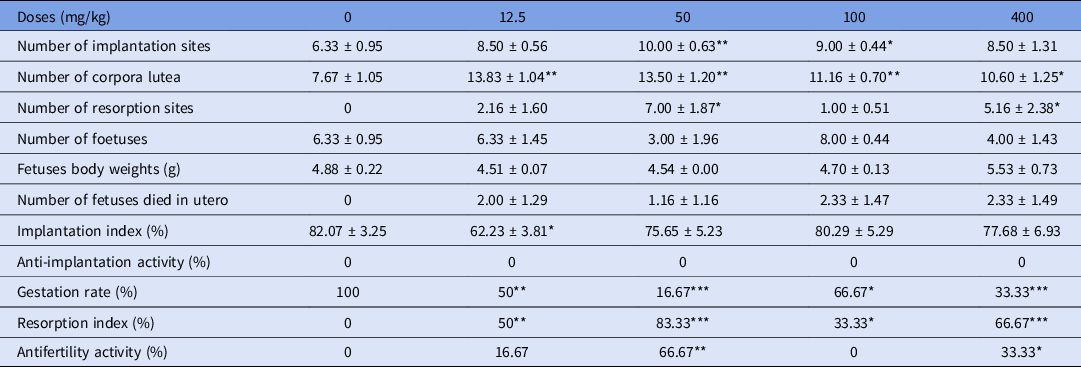
Table 4 reports a significant increase (P < 0.01) in the number of corpora lutea of all treated animals compared with the controls. An increase was also experienced in the number of resorption sites with the 50 and 400 mg/kg doses. The gestation rate significantly dropped (P < 0.01) in all treated animals, while a controversial tendency was shown in the resorption index as well as in the antifertility activity that rose (P < 0.05) in 50 and 400 mg/kg AEDv dosage-treated animals.
Effects of AEDv on gestation parameters from the 6th to the 9th day of pregnancy

Each value represents the mean ± standard error of the mean (SEM) for six rats.
In the same column, asterisks indicate values significantly different from the control (distilled water) *P < 0.05, **P < 0.01, ***P < 0.001 (one-way ANOVA test for the number of implantation sites, corpora lutea, fertility rate, resorption sites, fetuses number and weight and Kruskal–Wallis test for implantation and resorption indexes, preimplantation and postimplantation losses, antifertility and anti-implantation activities).
Late abortive effect
A significant decrease of 66.67% was observed in the gestation rate of the 50 mg/kg AEDv-treated animals (P < 0.05). A significant increase was therefore observed in the resorption index of the 12.5, 50 and 100 mg/kg dose-treated rats as well as in the 50 mg/kg for the antifertility activity, all compared with the control group (Table 5).
Effects of AEDv on gestation parameters from the 11th to the 20th day of pregnancy

Each value represents the mean ± standard error of the mean (SEM) for six rats.
In the same column, asterisks indicate values significantly different from the control (distilled water) *P < 0.05, **P < 0.01, ***P < 0.001 (one-way ANOVA test for the number of resorption sites, fetuses, and Kruskal–Wallis test for resorption index, fertility rate and antifertility activity).
Discussion
The present study evaluated the effects of AEDv on some biochemical and principally toxicological parameters of reproduction in female rats. The linear increase in the body weight of all treated animals through the treatment period showed that the growth of the animals was not negatively affected by AEDv and that instead led to a greater growth rate than the control. The significant decrease in liver proteins was correlated with an increase in serum proteins in high (50 and 100 mg/kg) dosage-treated animals; a similar result was obtained by Njindikouyou (Reference Njindikouyou2006). This result might show an inflammatory or necrosis effect at the level of the hepatocyte. In fact, the liver cell is responsible for the synthesis and release of the majority of serum proteins and an increase in proteins release can be provoked by an inflammatory process associated to cell necrosis (Brenner et al., Reference Brenner, Galluzzi, Kepp and Kroemer2013). Nevertheless, the rates of AST and ALT did not significantly change, showing that the adverse effect would be of lower extent. Indeed, ALT and AST rates increase in blood when the hepatic cellular permeability varies or when necrosis and cellular injury occur (Giboney, Reference Giboney2005). AEDv can then be classified as a relatively non-toxic substance according to the evaluated parameters. Pone et al. (Reference Pone, Telefo, Gouado and Tchouanguep2012) evaluated the acute and subacute toxicity of the aqueous extract of leaves mixture of Aloe buettneri, Justicia insularis, Hibiscus macranthus and Dicliptera verticillata on mice and female rats. Their results revealed that the mixture is devoid of toxicity; this confirms the harmlessness of AEDv at the administered doses.
Ovarian proteins (100 mg/kg) and uterine weight (50 mg/kg) significantly increased in treated immature rats. Indeed, estrogens, through activation of the synthesis of biomolecules (such as DNA, RNA, proteins, etc.) at the level of their target organs, induce cell proliferation that culminates in organ (uterus and ovary) weight increase (Jensen and Desombre, Reference Jensen and Desombre1972; Katzenellenbogen et al., Reference Katzenellenbogen, Bhakoo, Fergusson, Lan, Tatee, Tsia and Katzenellenbogen1979). This showed that the increase in organ weight and proteins would be the result of an oestrogenic effect of AEDv. The cholesterol rate was not affected through the study, meaning that the oestrogenic effect must be from an external source, as cholesterol is the main precursor of oestrogenic hormones in animals (Berg et al., Reference Berg, Tymoczko and Stryer2002). Many phytochemical trials have however been performed to evaluate secondary metabolites classes in D. verticillata. It has been revealed that this plant is rich in alkaloids, saponins, phlobatannins, steroids, terpenes, deoxy-sugar and cardiac glycoside (Sawadogo et al., Reference Sawadogo, Lompo, Guissou and Nacoulma2008; Akpakpan et al., Reference Akpakpan, Ukpong, Odiongenyi and Willie2017). Some of these compounds can act as structural analogues of endogenous hormones to express the oestrogenic effect observed. The oestrogenic effects of AEDv have already been described by Guemo (Reference Guemo2002) and Njindikouyou (Reference Njindikouyou2006) in previous studies. Mating the animals after treatment then allowed evaluation of whether that effect modified the fertility properties in female rats.
Corpora lutea were significantly reduced in the 100 mg/kg dose-treated animals. This could be due to a rapid regression of some corpora lutea in treated animals, probably linked to the rapid maturation of the follicles through folliculogenesis, as the parameters are evaluated 10 d after the mating. Also, regression can be due to rapid growth of the placenta that replaces the corpus luteum in the release of estradiol and progesterone for maintaining fetus growth. Another possibility is a possible downregulation effect induced by some plant compounds on the release of pituitary hormones that lead to low follicles recruitment and maturation (Driancourt et al., Reference Driancourt, Gougeon, Mouniaux, Royere, Thibault, Thibault and Levassseur2001; Saple et al., Reference Saple, Agrawal and Kawar2016). This last explanation seems to be the most relevant when considering the decrease in the fertility rate of treated animals. Therefore, the compounds could have acted through follicle growth inhibition or on early embryonic development phases, by reducing tubal transit duration of fertilized ova and then inhibiting all transformations the zygote should undergo before implantation in the uterus or the process itself. It was then interesting to evaluate the effects of AEDv on the phases of gestation.
After administration from the first to the fifth day of gestation, a significant decrease was reported in the number of implantation sites and the implantation index in all treated animals. A significant increase in anti-implantation activity was also reported. These results suggested that AEDv would have reduced the zygote tubal transit period or have destabilized the endometrial structure preventing implantation. These can be caused by high oestrogenic effect because oestrogenic compounds by linking to their endometrial receptor can provoke the contraction of the smooth muscle of the endometrium, leading to a resorption of the implant (Xu et al., Reference Xu, Chen, Wei, Brandon, Zava, Shi and Cao2017). This confirms the effect previously suggested. To evaluate the extent of that action, AEDv was administered from the 6th to the 9th day of gestation corresponding to the post implantation period. This led to a decrease in gestation rate, resorption, antifertility activity and, finally, there were at least two fetuses dead in utero in almost all the treated animals. This shows that the antifertility effect of AEDv also applies during the post implantation period, acting directly on placental unit and leading to fetal death or placental dislocation (Bhargava, Reference Bhargava1987). These results are similar to those obtained by Bhargava (Reference Bhargava1987) after administration of malvidin chloride isolated from Malvaviscus conzattii flowers to rats during gestation. In another study, Lau et al. (Reference Lau, Saksena and Chang1980) observed impairment in the normal luteal function, leading to an impaired feto-maternal relationship and a possible physical separation between fetal and maternal placentas after administration of trichosanthin, an active principle isolated from Trichosanthes kirilowi roots and they considered the barrier as the main cause of interruption of the gestation.
When administered from the 11th to the 20th day of gestation, high doses of AEDv led to an increase of antifertility activity and resorption index and to a decrease in the gestation rate. This effect can be explained by the oestrogenic action of the extract. At the endometrium, estrogens induce oxytocin receptors and stimulate prostaglandin synthesis. Both oxytocin and prostaglandins are highly implicated in parturition processes (Maltier et al., Reference Maltier, Germain, Breuiller, Thibault and Levassseur2001). The high resorption index can be an abortive effect provoked by the high content of AEDv in oestrogenic compounds. Furthermore, the high content in alkaloids (Akpakpan et al., Reference Akpakpan, Ukpong, Odiongenyi and Willie2017) can also be implied because these secondary metabolites can stimulate contractions of the smooth uterine muscle through prostaglandin biosynthesis (Gupta et al., Reference Gupta, Anang, Ghatak and Atal1978; Zutsi et al., Reference Zutsi, Rao, Saori, Gupta and Atal1980). The abortive effect of AEDv presented by the 50 mg/kg dosage was not experienced with high doses of the same extract (100 and 400 mg/kg). This observation should be because plant extracts are mixtures of hundreds or thousands of secondary metabolites with various pharmacological/toxicological effects and increasing the dosage could have led to increase in others metabolite concentrations that had anti-abortive effects. Of course, the plant is used at its therapeutic dosage in a mixture of four plants (ADHJ) and in that case these protective compounds are certainly provided by the other plants in the mixture (Telefo et al., Reference Telefo, Moundipa, Tchana, Tchouanguep and Mbiapo1998, Reference Telefo, Moundipa and Tchouanguep2001).
AEDv is then a non-toxic substance at the level of general metabolism, but presents weak toxic effects on the reproductive processes, and particularly for gestation, in which it leads to abortion and antifertility effects. Its use should be highly forbidden during gestation.
Acknowledgements
The authors’ special thanks go to the collaborators of respective institutions for technical assistance.
Financial support
This research received no specific grant from any funding agency, commercial or not-for-profit sectors.
Conflict of interest
None of the authors have any conflict of interest to declare.
Ethical standards
This study was performed according to the internationally accepted standard ethical guidelines for laboratory animal use and care as described in the European Community guidelines (Radzikowski, Reference Radzikowski2006).