
Introduction
As we age, physiological changes are observed in several body systems, including the immune system (Akbar et al., Reference Akbar, Henson and Lanna2016). The changes that occur within the immune system are termed immunosenescence and can be found in both the innate and adaptive immune compartments. Such alterations can be linked to different cellular processes such as loss of cellular diversity (Aiello et al., Reference Aiello, Farzaneh, Candore, Caruso, Davinelli, Gambino, Ligotti, Zareian and Accardi2019), impaired cell signalling (Larbi et al., Reference Larbi, Pawelec, Wong, Goldeck, Tai and Fulop2011; Solana et al., Reference Solana, Tarazona, Gayoso, Lesur, Dupuis and Fulop2012), telomeric erosion (Rufer et al., Reference Rufer, Brümmendorf, Kolvraa, Bischoff, Christensen, Wadsworth, Schulzer and Lansdorp1999), decreased proliferative capacity (Rebel et al., Reference Rebel, Miller, Eaves and Lansdorp1996; Mauch et al., Reference Mauch, Botnick, Hannon, Obbagy and Hellman1982) as well as a chronic inflammatory profile (Barbé-Tuana et al., Reference Barbé-Tuana, Funchal, Schmitz, Maurmann and Bauer2020), even in the absence of infection. Immunosenescence results in the impaired response to vaccines and increased susceptibility to pathogens and opportunistic infection in humans and animal models (Weinberger et al., Reference Weinberger, Herndler-Brandstetter, Schwanninger, Weiskopf and Grubeck-Loebenstein2008; Leng et al., Reference Leng, Stout-Delgado, Kavita, Jacobs, Tang, Du, Tussey and Goldstein2011; Chen et al., Reference Chen, Kelley and Goldstein2020).
The impact of infectious diseases on older individuals is far more significant in terms of morbidities and mortalities than on younger subjects (Norman, Reference Norman2016). Elderly individuals exhibit increased susceptibility to respiratory (Bender et al., Reference Bender, Johnson and Small1991; Krone et al., Reference Krone, Trzciński, Zborowski, Sanders and Bogaert2013; Williams et al., Reference Williams, José, Brown and Chambers2015), urinary (Pavlicek et al., Reference Pavlicek, Krebs, Capossela, Bertolo, Engelhardt, Pannek and Stoyanov2017) and intra-abdominal infections caused by viruses (Murasko and Jiang, Reference Murasko and Jiang2005), bacteria and protozoan (Krone et al., Reference Krone, Trzciński, Zborowski, Sanders and Bogaert2013; Sorci et al., Reference Sorci, Léchenault-Bergerot and Faivre2021).
Leishmaniasis is a complex of clinically unique diseases caused by obligate intracellular protozoa belonging to the genus Leishmania (Burza et al., Reference Burza, Croft and Boelaert2018). It is considered the fifth most important parasitic disease worldwide, affecting over 12 million individuals (Alvar et al., Reference Alvar, Vélez, Bern, Herrero, Desjeux, Cano, Jannin and de Boer2012). The disease manifestation ranges from self-healing cutaneous leishmaniasis (CL) to severe visceral leishmaniasis (VL), which is a systemic and fatal disease if left untreated (Torres-Guerrero et al., Reference Torres-Guerrero, Quintanilla-Cedillo, Ruiz-Esmenjaud and Arenas2017).
In leishmaniasis, the clinical outcome is dependent on a delicate balance between the parasite genetic background and host immunity features. Regarding the latter, a reasonable prognosis of the disease is related to the host's ability to develop a robust cellular immune response with the predominance of Th1 responses together with IFN-γ production, which is crucial for activating parasite-infected macrophages and controlling the infection (Scott et al., Reference Scott, Pearce, Cheever, Coffman and Sher1989; Reiner and Locksley, Reference Reiner and Locksley1995). Whereas, on the contrary, the exaggerated production of cytokines and inflammatory mediators such as TNF-α (Carvalho et al., Reference Carvalho, Passos, Bacellar, Lessa, Almeida, Magalhães, Dutra, Gollob, Machado and Ribeiro De Jesus2007; de Lima et al., Reference de Lima, Peiro and de Oliveira Vasconcelos2007) and nitric oxide (NO) (Horta et al., Reference Horta, Mendes, Roma, Noronha, Macêdo, Oliveira, Duarte and Vieira2012), the predominance of Th2-related or suppressive cytokines are linked to the host susceptibility and disease progression (Faria et al., Reference Faria, Gollob, Barbosa, Schriefer, Machado, Lessa, Carvalho, Romano-Silva, De Jesus, Carvalho and Dutra2005).
The overexpression of inhibitory checkpoint receptors has been potentially deleterious in many parasitic diseases (de Freitas e Silva and von Stebut, Reference de Freitas e Silva and von Stebut2021), including leishmaniasis. Of these, PD-1 and its ligands (PDL1 and PDL2) have been widely described during Leishmania infection and linked to the decreased proliferative ability and diminished Th1-related cytokines production by T cells (Liang et al., Reference Liang, Greenwald, Latchman, Rosas, Satoskar, Freeman and Sharpe2006; Covre et al., Reference Covre, Martins, Devine, Chambers, Vukmanovic-Stejic, Silva, Dietze, Rodrigues, de Matos Guedes, Falqueto, Akbar and Gomes2019; da Fonseca-Martins et al., Reference da Fonseca-Martins, Ramos, Pratti, Firmino-Cruz, Gomes, Soong, Saraiva and de Matos Guedes2019; de Moura et al., Reference de Moura, Covre, Fantecelle, Gajardo, Cunha, Stringari, Belew, Daniel, Von Zeidler, Tadokoro, de Matos Guedes, Zanotti, Mosser, Falqueto, Akbar and Gomes2021). Apart from leishmaniasis, the role of killer cell lectin-like receptor G1 (KLRG1) has been associated with T cell differentiation (Müller-Durovic et al., Reference Müller-Durovic, Lanna, Polaco, Mills, Henson and Akbar2016), decreased proliferative capacity (Plunkett et al., Reference Plunkett, Franzese, Finney, Fletcher, Belaramani, Salmon, Dokal, Webster, Lawson and Akbar2007) and poor T cells activation through TCR stimulation (Lanna et al., Reference Lanna, Gomes, Muller-Durovic, McDonnell, Escors, Gilroy, Lee, Karin and Akbar2017). Although little is known about its role during leishmaniasis so far, we have recently shown that KLRG1 is increased in both CD4+ and CD8+ circulating T cells (Covre et al., Reference Covre, Martins, Devine, Chambers, Vukmanovic-Stejic, Silva, Dietze, Rodrigues, de Matos Guedes, Falqueto, Akbar and Gomes2019) and its expression is found within lesional leucocytes of patients (de Moura et al., Reference de Moura, Covre, Fantecelle, Gajardo, Cunha, Stringari, Belew, Daniel, Von Zeidler, Tadokoro, de Matos Guedes, Zanotti, Mosser, Falqueto, Akbar and Gomes2021), suggesting that it may play an important role in the pathogenesis of the disease.
Although the impact of ageing on VL infection has not yet been explored, broad evidence suggests that ageing may represent a significant risk factor and affect the outcome of tegumentary CL infection (Jirmanus et al., Reference Jirmanus, Glesby, Guimarães, Lago, Rosa, Machado and Carvalho2012). In this scenario, elderly individuals have been reported to have higher frequencies of both mucosal and disseminated CL (Cincurá et al., Reference Cincurá, de Lima, Machado, Oliveira-Filho, Glesby, Lessa and Carvalho2017). Furthermore, both severity with higher numbers and size and longer healing times have been observed within this population (Araujo-Melo et al., Reference Araujo-Melo, Meneses, Schubach, Moreira, Conceição-Silva, Salgueiro, Pimentel, Araújo-Silva, Oliveira, Carmo and Valete-Rosalino2010; Oliveira et al., Reference Oliveira, Bafica, Rosato, Favali, Costa, Cafe, Barral-Netto and Barral2011, Reference Oliveira, Brito, Schubach, Oliveira, Saheki, Lyra, Salgueiro, Terceiro, Pimentel, Vasconcellos and Valete-Rosalino2013; Diniz et al., Reference Diniz, Costa and Escalda2012). Controversially, in an experimental model, aged mice were more efficient than young mice in generating a protective immune response and controlling the infection caused by L. major, suggesting the need for complementary studies to understand better the effect of ageing on Leishmania infection, particularly those caused by the different species (Ehrchen et al., Reference Ehrchen, Sindrilaru, Grabbe, Schönlau, Schlesiger, Sorg, Scharffetter-Kochanek and Sunderkötter2004).
Herein, we demonstrate that aged mice are more susceptible to L. infantum infection than younger mice, accompanied by a senescence-associated cytokine profile. Moreover, they have increased expression of inhibitory receptors that are associated with disease severity within the T cell populations. Overall, the data reported in this study suggest for the first time that ageing may negatively impact the VL outcome and provides a perspective for new therapeutic strategies involving manipulation of immunosenescence features against Leishmania infection.
Materials and methods
Animals
C57BL/6 mice were originally purchased from Jackson Laboratory (Bar Harbor, Maine, USA). They were bred and maintained at our facilities and given sterilized bedding, filtered water and pelleted food ad libitum. For each experiment, 5–8 female mice at 4–6 weeks old and at 72 weeks old were used for the young and aged groups, respectively. The Ethical Committee for Experimental Animal Use at the Universidade Federal do Espírito Santo approved all experimental protocols, registered under reference number: 014/2011.
Parasites and infection
L. infantum strain MHOM/BR/1975/PP75 promastigotes were cultured at 26 °C in Grace's medium (Gibco, USA), pH 7.2, supplemented with 10% heat-inactivated fetal bovine serum (FBS) (Gibco, USA), 2 mm l-glutamine, 25 mm HEPES and 20 μg mL−1 gentamicin (LGC, Brazil). The mice were infected by the intravenous route in the tail vein with 107 L. infantum promastigotes in the stationary growth phase.
Parasite burden
On day 30 of infection, the parasite burden in the liver and spleen of the mice were individually determined by limiting dilution assay, as described previously (Leal et al., Reference Leal, Mosquini, Covre, Stagmiller, Rodrigues, Christensen, Guedes, Rossi-Bergmann and Gomes2015). Briefly, each organ was weighed and homogenized in Grace's medium supplemented with 10% heat-inactivated FBS. The volume of the cell suspension was adjusted with supplemented Grace's medium according to tissue weight (100 mg of tissue mL−1) and plated in a 96-well plate (Corning, USA). Serial dilutions of the single-cell suspensions were cultured for 10 days at 26 °C. The original number of parasites in each organ was calculated from the reciprocal of the highest dilution containing promastigotes. The relative change values were obtained through parasite burden mg−1 tissue data normalization according to the following formula:

where μ A = aged group experimental average of parasite burden mg−1; and μ Y = young group experimental average of parasite burden mg−1.
Cytokines and nitrite production
Ex vivo cytokine quantification was performed in both liver and spleen of all groups. The organs were individually homogenized in 1 mL of phosphate-buffered saline (PBS), pH 7.2 with the addition of protease and phosphatase inhibitors (Sigma-Aldrich, USA) using a glass tissue grinder (Thomas, USA). The cells suspension volumes were adjusted with PBS according to tissue weight (100 mg of tissue mL−1), centrifuged at 20 000 g for 10 min at 4 °C, and the supernatants were collected for the subsequent tests. Cytokine levels were determined individually by ELISA following the manufacturer's instructions, considering the sensitivity of each test (R&D Systems, USA). Nitrite production was determined using the Griess method (Green et al., Reference Green, Wagner, Glogowski, Skipper, Wishnok and Tannenbaum1982). Briefly, 50 μL of the supernatants were mixed with 50 μL of Griess reagent (1% sulphanilamide and 0.1% N-1-naphthylethylenediamin dihydrochloride in 2.5% o-phosphoric acid) and incubated at room temperature for 10 min. The nitrite concentrations were determined at 540 nm against a standard sodium nitrite curve.
Flow cytometry
For phenotypic analysis, at least 106 splenocytes were stained with antibodies against cell surface markers for 30 min at 4 °C. The cells were then fixed using paraformaldehyde for 30 min at room temperature. Data from 50 000 events were acquired on a BD FACSCanto II flow cytometer to determine the following: CD4+ (Clone RM4-5/FITC; 1:100 dilution) or CD8+ (Clone 5H10-1/PerCP-cy5-5, 1:150 dilution) cells with KLRG1 (Clone 2F1/PE; 1:50 dilution) and PD-1 (Clone J43/APC-cy7; 1:50 dilution), all from biolegend, USA. For IFN-γ (Clone XMG1.2 /PE-cy7; 1:100 dilution) intracellular staining, cells were fixed and permeabilized with the Fix & Perm Kit (Invitrogen, Life Technologies, Carlsbad, CA) after the extracellular stainings, followed 20 min incubation with the antibody. To determine the intracellular expression of Ki67 (Clone B56/PE; 1:150 dilution), cells were fixed and permeabilized for 20 min using the kit from BD Biosciences after surface staining. Following this, the cells were marked with Ki67 for 1 h. After acquisition, data were analysed using FlowJo software (Version 10). All gates were based on pooled fluorescence minus one (FMO) control samples and applied identically across all samples.
Statistical analyses
Data were analysed using the GraphPad Prism software version 8.0 for Windows. Means of normally distributed variables were compared by an analysis of variance (ANOVA) simple factorial test, by one-way ANOVA-Tukey's honestly significant difference (HSD) posthoc test, and by the Spearman's rank correlation method. Data were considered significantly different when P < 0.05.
Results
Aged mice develop increased susceptibility to L. infantum infection
To compare the susceptibility of aged and young mice to L. infantum infection, we assessed the parasite burden in the liver and spleen at the parasitic peak, 30 days after L. infantum infection, as previously defined (Leal et al., Reference Leal, Mosquini, Covre, Stagmiller, Rodrigues, Christensen, Guedes, Rossi-Bergmann and Gomes2015). Our data demonstrate that aged mice exhibit a significant increase in the parasite burden in the liver and spleen (Fig. 1A); that was 18- and 23-fold greater than in the young mice, respectively (Fig. 1B). The parasitism was compatible with their weight loss and prostrate behaviour, in comparison to both the young-infected mice and the non-infected controls, which had a healthy appearance and dynamic behaviour (data not shown). Additionally, both the liver and spleen from aged-infected mice were significantly heavier and larger dimensions than young-infected mice (Fig. 1C and D), demonstrating a conspicuous hepatosplenomegaly clinical presentation.
Parasite burden and clinical features in infected aged mice. Young (4–6 weeks) and aged (72 weeks) mice were i.v.-infected with 107 L. infantum promastigotes. (A) Cumulative data of the parasite burden and (B) fold change of hepatic and splenic parasitism normalized with young controls. (C) Table with average organs size and weight and (D) representative image of spleens at the peak of parasitism. Results are represented as arithmetic means ± s.d. of pooled data (n = 16 mice/group). Statistical differences and P values were determined by t-test or ANOVA and indicated in the graphs.

Leishmania infection enhances the inflammatory profile in aged mice
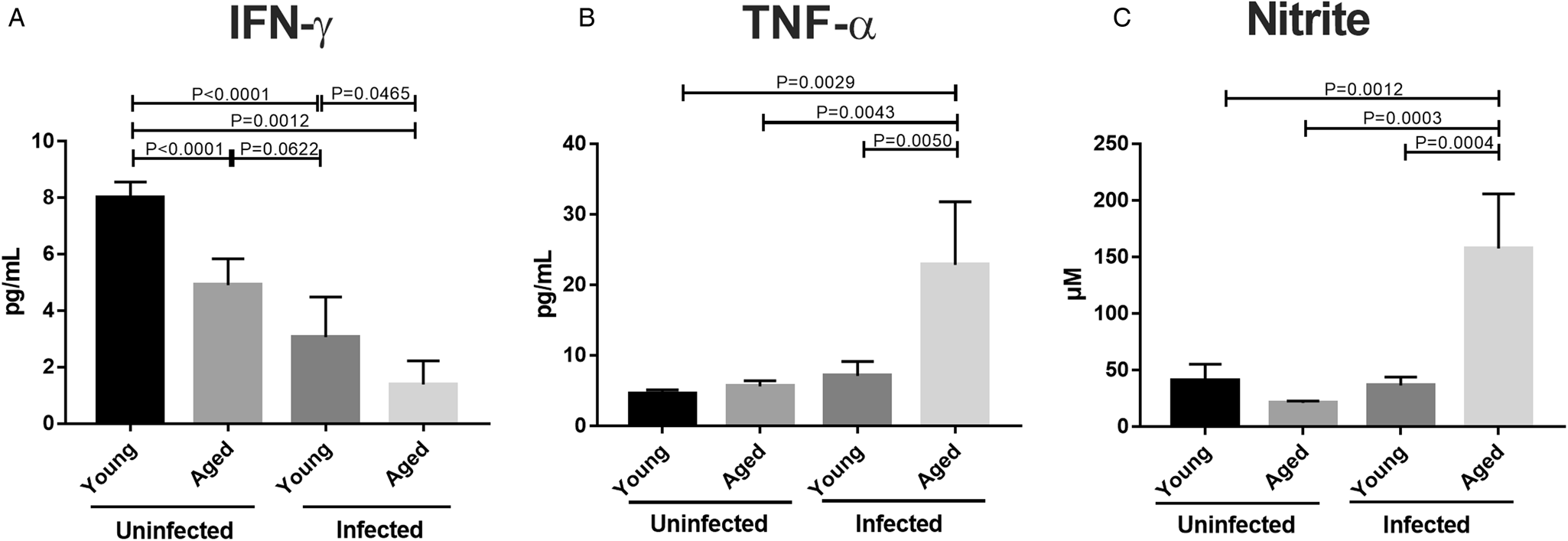
Ageing is a key factor affecting the inflammatory profile and the host proliferative ability (Ponnappan and Ponnappan, Reference Ponnappan and Ponnappan2011). Ex vivo analysis of the cytokine production in the spleen revealed increased amounts of both TNF-α (Fig. 2A) and nitrite (Fig. 2B), a breakdown product of nitric oxide (NO), in the aged-infected group that were not found in the respective non-infected control nor the young-infected group. Moreover, there were lower amounts of IFN-γ compared to all the other mouse groups (Fig. 2C), suggesting that aged mice acquire a conspicuous inflammatory profile following infection.
Cytokines and NO production in infected aged mice. Spleens were processed individually, and the in-situ production of IFN-γ (A) and TNF-α (B) were assessed by ELISA. Nitrite production was evaluated by the Griess assay (C). Results are represented as arithmetic means ± s.d. of pooled data (n = 16 mice/group). Statistical differences and P values were determined by ANOVA and indicated in the graphs

Leishmania infection promotes enhanced expression of inhibitory receptors, diminished frequencies of IFN-γ-producing-T cells and proliferative capacity impairments of aged mice
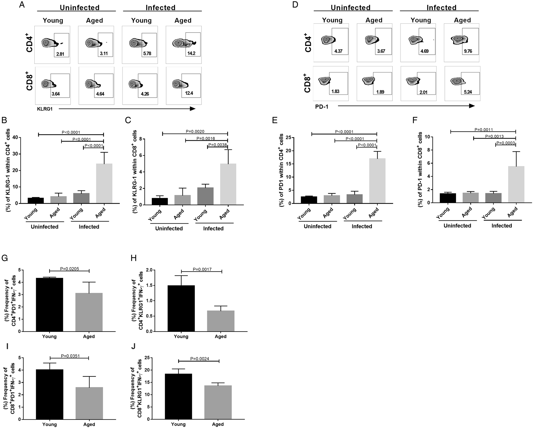
To further investigate the acquisition of an inhibitory profile, we next assessed the expression of surface differentiation-associated receptors such as KLRG1 and PD-1, as well as the proliferative capacity of both splenic CD4+ and CD8+ T cells. No differences in T cell frequencies were found between young and aged mice groups, pre- and post-infection (Supplementary Fig. 1). However, aged-infected mice displayed increased KLRG1 (Fig. 3A–C) and PD-1 (Fig. 3D–F) frequencies within both T cell populations compared to the young-infected mice and the non-infected groups.
Increased expression of inhibitory KLRG1 and PD-1 receptors in aged mice following Leishmania infantum infection. Representative histograms and pooled data of ex vivo (A) KLRG1 expression on (B) CD4+ and (C) CD8+ T cells; (D) PD-1 expression on (E) CD4+ and (F) CD8+ T cells from young and aged mice. Pooled data of the ex vivo frequencies of IFN-γ-producing cells within PD-1+ by (G) CD4+ and (I) CD8+ T cells; and KLRG-1+ by (H) CD4+ and (J) CD8+ T cells from young and aged mice. Results are represented as arithmetic means ± s.d. of pooled data (n = 16 mice/group). Statistical differences and P values were determined by t-test and ANOVA and indicated in the graphs.

During leishmania infection, the increased frequencies of PD-1 and KLRG1 receptors are linked to impairments of leishmanicidal mechanisms (Covre et al., Reference Covre, Martins, Devine, Chambers, Vukmanovic-Stejic, Silva, Dietze, Rodrigues, de Matos Guedes, Falqueto, Akbar and Gomes2019; da Fonseca-Martins et al., Reference da Fonseca-Martins, Ramos, Pratti, Firmino-Cruz, Gomes, Soong, Saraiva and de Matos Guedes2019; Oliveira Silva et al., Reference Oliveira Silva, Marin Chiku, Luvizotto Venturin, Correa Leal, de Almeida, De Rezende Eugenio, Dos Santos, Fabrino Machado and De Lima2019; de Moura et al., Reference de Moura, Covre, Fantecelle, Gajardo, Cunha, Stringari, Belew, Daniel, Von Zeidler, Tadokoro, de Matos Guedes, Zanotti, Mosser, Falqueto, Akbar and Gomes2021). To evaluate this on aged- and young infected mice, we next accessed the frequencies of IFN-γ – producing T cells within PD-1+, and KLRG-1+ subsets. Compared to young-infected control, aged-infected mice had lower frequencies of CD4+ T cells expressing IFN-γ within subsets of PD-1+ (Fig. 3G and I) and KLRG1+ cells (Fig. 3H and J). These decreased frequencies were also observed within the CD8+ T cell compartment, suggesting that aged mice may have more significant impairments in activating leishmanicidal mechanisms, and supporting our previous result demonstrating a greater susceptibility of these animals to infection.
In complement to this, analysis of their proliferative capacities conducted by staining the cell cycle-related nuclear antigen Ki67 demonstrated that aged-infected mice had decreased T cells proliferative capacity. It was a 0.5- and 0.6-fold change in the CD4+ and CD8+ T cell compartments, respectively, than we found in the young-infected controls (Supplementary Fig. 2). No difference in the Ki67 staining was found between the non-infected groups (data not shown), suggesting that the infection may affect the T cell compartments.
Acquisition of senescence features correlates with parasite burden of visceral leishmaniasis
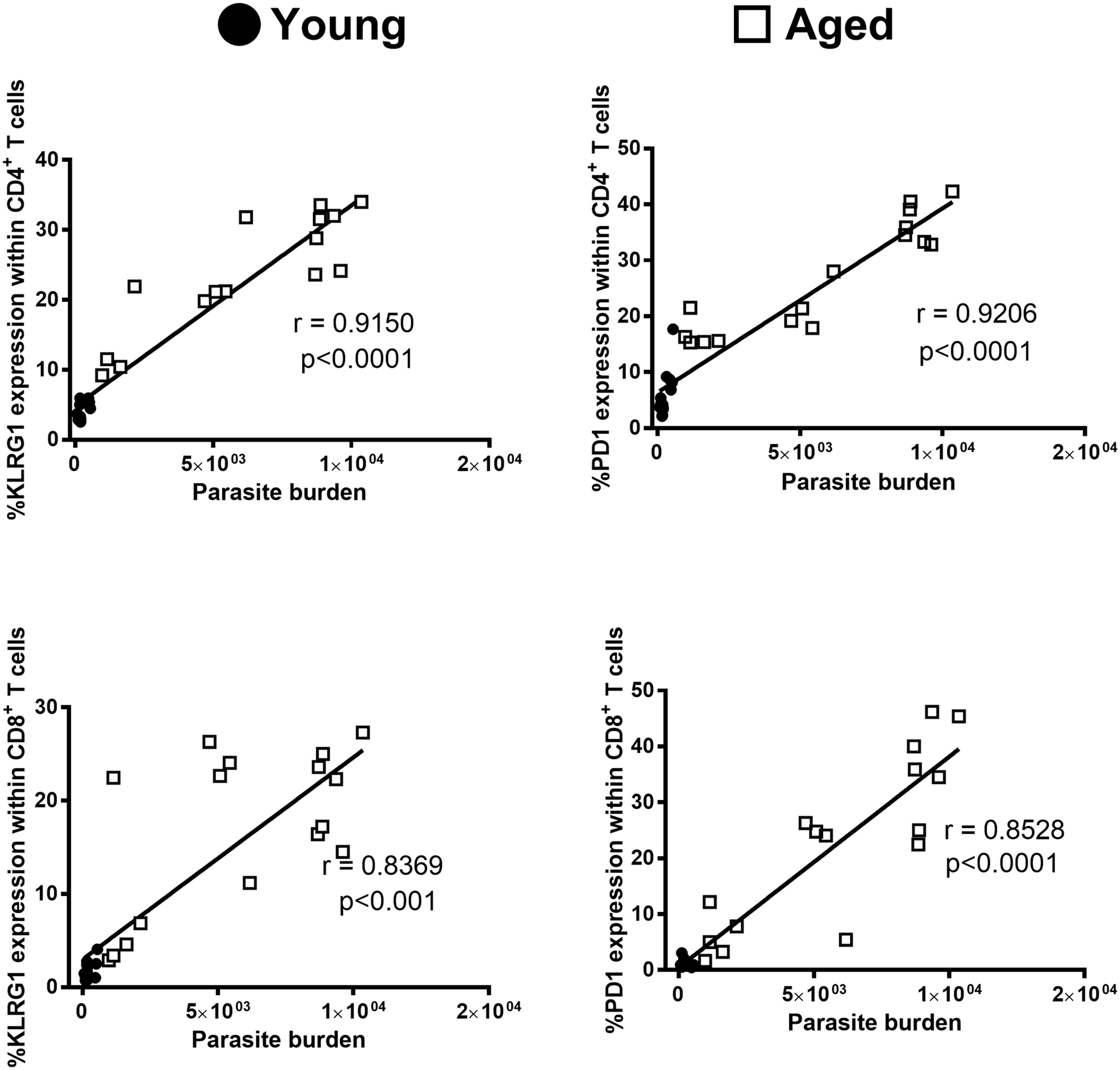
As T cells with senescence features appeared to accumulate, especially in the aged-infected mice, we next investigated if these cells were associated with the pathology during VL. To achieve this, we correlated the frequencies of KLRG1 and PD-1 expressing-CD4+ and CD8+ T cells with the parasite burdens observed in the liver and spleen. KLRG1-expressing CD4+ and CD8+ T cells and the parasite burden in liver and spleen were strongly correlated for aged, but not young, mice (Fig. 4A and B). The same correlation was also found for the expression of PD-1 for both T cell populations (Fig. 4C and D), suggesting that the expression of both inhibitory receptors by T cells may contribute to the pathology of VL.
Inhibitory KLRG1 and PD-1 receptors correlate with the severity of VL in aged mice. Pearson's correlation test between frequencies of CD4+ and CD8+ T cells expressing KLRG1 (A and C, respectively) and PD-1 (B and E, respectively) and the splenic parasite burden of young (black dots) and aged (white dots) mice. Results are represented as arithmetic means ± s.d. of pooled data (n = 16 mice/group). Statistical differences and P values were determined linear regression and indicated in the graphs.

Discussion
The immunosenescence induced by physiological processes or chronic infection has been associated with a significant increase in host susceptibility to infections (Gardner, Reference Gardner1980; Marston et al., Reference Marston, Plouffe, File, Hackman, Salstrom, Lipman, Kolczak and Breiman1997; Barbé-Tuana et al., Reference Barbé-Tuana, Funchal, Schmitz, Maurmann and Bauer2020; Tsuji et al., Reference Tsuji, Minami, Hashimoto, Konishi, Suzuki, Kondo, Sasai, Torii, Ono, Shichinohe, Sato, Wakita, Okumura, Nakano, Matsudaira, Matsumoto, Kawamoto, Yamamoto, Watanabe, Matsuura, Takayama, Kobayashi, Okamoto and Hara2022). Here we extended this concept by showing for the first time that senescent C57BL/6 mice are prone to L. infantum infection compared to young-infected mice. In our experiments, aged mice presented increased parasitism in the liver and spleen and a propensity for developing clinical symptoms of VL such as splenomegaly. These data support the greater susceptibility of elderly mice to infections as previously reported (Toapanta and Ross, Reference Toapanta and Ross2009; Fang et al., Reference Fang, Roscoe and Sigal2010; Speziali et al., Reference Speziali, Aranha, Teixeira-Carvalho, Santiago, Oliveira, Rezende, Carneiro, Negrão-Corrêa, Coelho and Faria2010; Krone et al., Reference Krone, Trzciński, Zborowski, Sanders and Bogaert2013) and seemingly contradict previous findings that reported an association between ageing and resistance to L. major infection (Ehrchen et al., Reference Ehrchen, Sindrilaru, Grabbe, Schönlau, Schlesiger, Sorg, Scharffetter-Kochanek and Sunderkötter2004). Differences between the experimental models that used distinct mice strains, age analysis and the natural evolution of visceral vs cutaneous infections could account for the observed discrepancies, thus highlighting the need for further complementary studies.
In the present study, the next series of experiments tested whether the susceptibility observed in aged mice was associated with impaired anti-Leishmania immune mechanisms. The development of VL caused by L. infantum is not defined by a constant Th1/Th2 pattern of cytokines as observed in CL caused by L. major (Wilson et al., Reference Wilson, Jeronimo and Pearson2005; Tripathi et al., Reference Tripathi, Singh and Naik2007). However, in both CL and VL, a good prognosis is associated with a robust IFN-γ production that mediates the clearing of intracellular Leishmania by activated macrophages and disease control (Squires et al., Reference Squires, Schreiber, McElrath, Rubin, Anderson and Murray1989; Scott, Reference Scott1991). In our experiments, aged-infected mice were less able to produce IFN-γ compared to young-infected mice and non-infected controls but these mice presented an increased ability to make TNF-α and NO. Although the last two are essential to mediate macrophage activation and Leishmania clearance (Liew et al., Reference Liew, Li and Millott1990), their chronic production is usually associated with more extensive and unspecific inflammatory processes, defective T-cell response and severity of VL (Rodrigues et al., Reference Rodrigues, Laforge, Campillo-Gimenez, Soundaramourty, Correia-de-Oliveira, Dinis-Oliveira, Ouaissi, Cordeiro-da-Silva, Silvestre and Estaquier2014, Reference Rodrigues, Cordeiro-Da-Silva, Laforge, Silvestre and Estaquier2016; Bogdan, Reference Bogdan2020). Moreover, both TNF-α and NO are components of the senescence-associated secretory phenotype (SASP) (Coppé et al., Reference Coppé, Desprez, Krtolica and Campisi2010). An array of cytokines, chemokines and matrix metalloproteinases are associated with a pronounced pro-inflammatory secretome found in chronic stimulatory processes, which is linked with disease pathology (Childs et al., Reference Childs, Durik, Baker and van Deursen2015).
The pronounced production of inflammatory mediators, like TNF-α, NO may exacerbate the inflammatory effects on tissues (Freund et al., Reference Freund, Orjalo, Desprez and Campisi2010; Prata et al., Reference Prata, Ovsyannikova, Tchkonia and Kirkland2018; Cerqueira et al., Reference Cerqueira, Marinho, Neiva and Lourenço2020). These can directly modulate the expression of stress ligands, including inhibitory receptors and ligands by immune and non-immune cells (Groh et al., Reference Groh, Rhinehart, Randolph-Habecker, Topp, Riddell and Spies2001; Gasser et al., Reference Gasser, Orsulic, Brown and Raulet2005). In our experiments, the marked pro-inflammatory profile was found in association with the increased expression of KLRG1 and PD-1 within T cell populations. Further to this, the expression of these markers positively correlated with the spleen parasitism. Both KLRG1 and PD-1 inhibit T cell function, impairing immunity and promoting disease progression in mice (da Fonseca-Martins et al., Reference da Fonseca-Martins, Ramos, Pratti, Firmino-Cruz, Gomes, Soong, Saraiva and de Matos Guedes2019) and humans (Covre et al., Reference Covre, Martins, Devine, Chambers, Vukmanovic-Stejic, Silva, Dietze, Rodrigues, de Matos Guedes, Falqueto, Akbar and Gomes2019; de Moura et al., Reference de Moura, Covre, Fantecelle, Gajardo, Cunha, Stringari, Belew, Daniel, Von Zeidler, Tadokoro, de Matos Guedes, Zanotti, Mosser, Falqueto, Akbar and Gomes2021). Moreover, further evidence has demonstrated their deleterious role in impairing microbicidal function (Wang et al., Reference Wang, Cheng, Shi, Ying, Wu, Li, Moorman and Yao2013; Müller-Durovic et al., Reference Müller-Durovic, Lanna, Polaco, Mills, Henson and Akbar2016), which could contribute to the increased susceptibility to infection observed in the aged mice.
The current study provides an overview of the global effects that ageing has on the immune system following L. infantum infection and demonstrates how the acquisition of a pronounced inflammatory cytokine profile and the expression of inhibitory receptors may impact the disease severity. The data presented here have important implications for understanding the differences in the immune response between young and aged individuals during infection, which could pave the way for vaccine development and immunotherapies specifically for the elderly.
Supplementary material
The supplementary material for this article can be found at https://doi.org/10.1017/S0031182022000828
Acknowledgements
The authors would like to thank MSc. Glênia Daros (Núcleo de Doenças Infecciosas – UFES), MSc. Divan Barcelos (Núcleo de Doenças Biotecnologia – UFES) and MSc. Renan Garcia (Núcleo de Doenças Infecciosas – UFES) for their scientific discussion and technical support.
Author contributions
C. L. S., A. F. M., L. P. C. and H. L. M. G. performed experiments. C. L. S., A. F. M., L. P. C. analysed data. D. G., L. P. C., H. L. M. G. and A. F. designed the project and discussed the data. D. G., C. L. S. and A. F. wrote the manuscript.
Financial support
This work was supported by Fundação de Amparo a Pesquisa do Espírito Santo-FAPES/Newton Fund and Medical Research and Coordination for the Improvement of Higher Education Personnel – CAPES-Brazil.
Conflict of interest
The authors have no conflicts of interest.
Ethical standards
The Ethical Committee for Experimental Animal Use at the Universidade Federal do Espírito Santo approved all experimental protocols, registered under reference number: 014/2011.