Introduction
The use of light rather than electricity to probe and manipulate neural networks is a relatively new approach. Given the wide variety of interactions between tissue and various parts of the electromagnetic spectrum, from the infrared to the ultraviolet, photonic technologies are quickly finding applications in both clinical and experimental neuroscience. Applications span a wide range and include imaging of electrical activity in single cells and neuronal networks, neuronal stimulation, as well as alteration of molecular pathways and gene expression.Reference Callaway and Yuste1–Reference Hillman3 In 2010, the journal Nature Methods declared one technique of optical neuronal stimulation, optogenetics, the method of the year, proclaiming that: “with the capacity to control cellular behaviors using light and genetically encoded light-sensitive proteins, optogenetics has opened new doors for experimentation across biological fields.”Reference Anon4
Apart from satisfying our scientific curiosity, a key motivation for the development of better neural interfaces is their potential for treatment of neurological disorders, particularly for restoration of lost function in vision, hearing, and limb movement, and for treating disorders such as Parkinson’s disease and epilepsy. In order to be used clinically, however, optical devices must be safe, reliable, and robust. Here we review optical methods for probing neural networks and address the materials challenges that must be overcome before these interfaces are fully realized for therapeutic use.
Benefits of optical neural interfaces
It is known that neuronal communication is electrochemical in nature, thus it is perhaps not intuitive why we should consider light as an alternative to electrophysiological methods, the gold standard for interfacing with the nervous system for over a century.Reference Galvani5, Reference Fritsch and Hitzig6 We postulate that optical techniques may overcome some of the fundamental limitations of electrophysiology. The first problem is spatial selectivity. Electrodes cannot discriminate between different current sources, and therefore detect either point sources such as single cells or the averaged activity of neuronal populations. Similarly, fine control over the stimulated area is limited due to passive spread of current along the path of least resistance, which is dictated by tissue anisotropy. These problems can be solved by using a microelectrode array, which effectively discretizes the tissue into small compartments, but practical considerations, such as the invasive nature of the device and the large parallel hardware architecture needed, impose limitations on this method. Another problem encountered in electrophysiology is the difficulty in pairing stimulation with recording, since the stimulating current masks endogenous neural signals immediately following stimulation. The final drawback of electrical recording techniques is that they only measure changes in membrane potential, which are often secondary to a variety of physiological processes, such as neurotransmitter concentrations or intracellular ion signaling. In fact, the information-processing role of astrocytes, a non-neuronal cell type found throughout the nervous system, has been mostly overlooked until recently, precisely because these cells are largely electrically silent. Optical techniques can overcome some or all of these problems and even combine with or complement traditional electrophysiological techniques. Finally, optical probes can be used for imaging tissue micro-morphology as well as neural functions other than electrical activity.
Light-tissue interactions
The design of optical neural interfaces is ultimately constrained by the optical properties of tissue, which depend on wavelength, temperature, and tissue hydration. The two governing parameters are scattering and absorption. A detailed discussion of tissue optics is beyond the scope of this article, but we refer the reader to the recently published second edition of the book on laser-tissue interactions by Welch and van Gemert.Reference Welch7 Most scattering is by organelles rather than whole cells, and brain tissue has a high scattering coefficient due to high mitochondrial content.Reference Mourant, Freyer, Hielscher, Eick, Shen and Johnston8 Light scattering is largely anisotropic and mostly forward directed. Myelin, an insulating sheath surrounding some axons, also scatters strongly, accounting for the difference in scattering between white brain matter comprised largely of axonal tracts, and cortical tissue, composed of a looser network of neurons and other cell types. Myelin is anisotropic, and optical properties of nerve tracts depend on their orientation to the light beam.Reference Bevilacqua, Piguet, Marquet, Gross, Tromberg and Depeursinge9 The light scattering probability scales loosely as λ–n with n = 0.5–0.8 and thus decreases with increasing wavelength (λ). Scattering is strongly affected by the index of refraction mismatch between microscopic tissue structures and the inter- and intracellular fluid. Refractive index matching techniques can make tissue virtually transparent, but they are currently not suitable for use in live specimens.Reference Hama, Kurokawa, Kawano, Ando, Shimogori, Noda, Fukami, Sakaue-Sawano and Miyawaki10 The practical significance of scattering is that it limits the penetration depth of light into tissue as well as the ability to create sharply delineated regions of illumination within the brain.
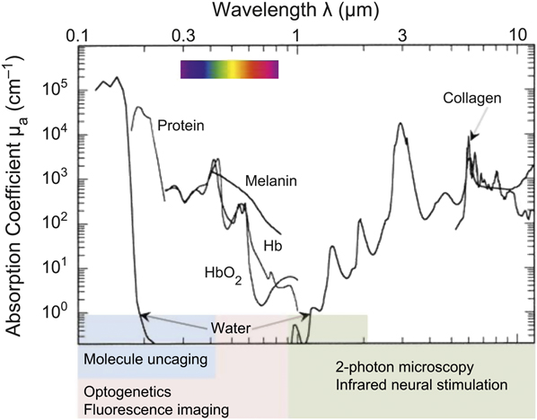
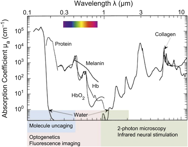
Absorption is the other key phenomenon in light-tissue interaction. In the brain, the strongest absorber in the visible range is hemoglobin, with a peak at about 580 nm, while absorption by water dominates in the infrared, with notable peaks at 1450, 1935, and 2940 nm, as shown in Figure 1. Absorption imparts energy to the tissue, which is manifested by ensuing physical, chemical, or mechanical phenomena, the precise combination of which are a function of the wavelength, the laser irradiance (W/cm2), and the duration of the light pulse. Absorption is key in determining what wavelengths of light can be used to activate or detect neural activation. Thermal confinement and stress confinement conditions, which lead to either highly localized heating or a mechanical pressure wave, establish the type of interaction that is expected between the laser energy and the tissue, and one or both may be used to activate neural tissue.Reference Jacques11 Whether the laser is operating in either of these regimes is determined by wavelength-dependent optical penetration depth and pulse duration. Additionally, stress confinement has been used in photoacoustic applications to perform imaging of neural tissue.Reference Zhang, Hong and Cai12
The use of optical methods in biological tissues is fundamentally dependent on the optical properties of the tissue itself. This figure shows the absorption curves of several important biological compounds as well as the wavelengths used for the most common imaging and stimulation modalities. Hb, hemoglobin.
Absorption can also be used to target specific chromophores, molecules that strongly absorb light at a specific wavelength, to produce an effect. These molecules can either be engineered dyes introduced into the body or naturally occurring molecules, such as some proteins. Photoactivation of neurotransmitters and optogenetics both rely on non-native chromophores to sensitize neurons to light.Reference Callaway and Yuste1 Various optical sources may be tailored to produce a variety of effects in biological and, specifically, neural tissue. However, despite the so called “therapeutic window” in the near infrared (700–900 nm) between the absorption peaks of water and hemoglobin, scattering and absorption make targeting of specific brain and nerve regions, especially below the surface, challenging.Reference Tromberg, Shah, Lanning, Cerussi, Espinoza, Pham, Svaasand and Butler13 Therefore, spatial selectivity must be achieved either by channeling light to the right location through optical conduits such as optical fibers, miniaturized light sources, or by engineering selectivity into the tissue itself.
Material considerations for implantable fiber optics
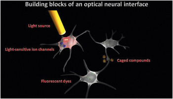
Optical fibers allow for delivery of light to specific locations within the brain. As shown in Figure 2, in many neural interfaces, most components can be externalized, and fibers are the sole synthetic materials coming into direct contact with subdermal tissues. Initially developed for the telecommunications industry, optical fibers consist of two concentric layers of silica glass, known as the core and the cladding, and a protective coating.Reference Crisp14 The glass is biologically inert and can be made to have excellent transmission properties in different parts of the spectrum (0.3–2.5 μm), depending on the concentration of hydroxyl ions, with low OH glasses favoring the infrared and quartz fibers being most useful in the UV. The difference in the refractive indices of the cladding and the core dictate the maximal angle off the fiber axis at which light can enter the optical conduit, known as the acceptance angle or numerical aperture (NA), and the width of the light cone at the exit. Low NA fibers allow for lower beam divergence but require tighter focusing of light into the fiber. Tapering or beveling the fiber tip provides additional control over the illumination spot.
An optical neural interface in its basic form consists of an appropriate targeted light source. Pulsed, infrared light channeled via an optical fiber stimulates neuronal tissue directly by a thermally mediated process. Electrical currents generated in neurons are the result of ion flow across the cells’ membranes. Introduction of genetically engineered light-sensitive ion channels allows one to control neuronal behavior by turning these channels on and off with flashes of visible light. Such transient, highly localized variations in neurotransmitter concentrations can be modulated artificially with the addition of caged neurotransmitters. These synthetic analogs remain inactive until they are “uncaged” by a pulse of UV light.

The design and fabrication of implantable fiber optics must address several issues to ensure effective performance within a physiological setting. Short distances between source and target mean that fiber transmission performance can be partially sacrificed to satisfy biomechanical considerations: stretching, bending, and twisting of the fiber and biocompatibility with the surrounding tissue. An implanted fiber must be able to deform with the surrounding tissue, yet due to optical considerations, the majority of fibers are brittle and stiff. While such properties can be tolerated in rigid brain implants, they are unacceptable in neural prosthetics, where the fibers would be subjected to continuous deformation.
The design of optical fibers can borrow much from recent advances in implantable electrodes. By layering metals and polymers, several groups report that electric signal quality can be maintained in flexible, bendable, and stretchable materials, reducing scar formation by minimizing interactions with the surrounding tissuesReference Lacour, Benmerah, Tarte, FitzGerald, Serra, McMahon, Fawcett, Graudejus, Yu and Morrison15–Reference Cheung, Renaud, Tanila and Djupsund18 (see the article by Capadona et al.). However, insertion of such flexible materials poses a new problem as penetration into the tissue becomes difficult. New methods for the insertion of flexible structures have been developed, including magnetic systems, chemical coatings, and ultrafast resorbable polymers.Reference Jaroch, Ward, Chow, Rickus and Irazoqui19–Reference Lewitus, Smith, Shain and Kohn21 Such composite materials with tunable parameters might provide the best characteristics to meet the requirements for implantable fiber optics. An example of an optical system using some of these laminate techniques is the BeamPath by OmniGuide, which utilizes a hollow waveguide fiber composed of alternating submicrometer-thick layers of chalcogenide glass and either polystyrene or poly(ether sulfone).Reference Fink, Winn, Fan, Chen, Michel, Joannopoulos and Thomas22, Reference Temelkuran, Hart, Benoit, Joannopoulos and Fink23 The transmission windows of these fibers range from 0.75 to 10.6 μm and can be altered by changing the layer dimensions. Additionally, because the fibers are composed of ∼98% polymer by volume, they exhibit excellent mechanical properties that allow for stretching, bending, and twisting.Reference Temelkuran, Hart, Benoit, Joannopoulos and Fink23
Biocompatibility of implanted materials is crucial in chronic optical implant design. Poly(methyl methacrylate) (PMMA) fibers are available, although they are generally opaque to infrared and, therefore, unsuitable for applications at these wavelengths. Fibers can also be treated to either promote or inhibit cell adhesion to the surface using specialized coatings originally developed for electrical implants. For example, Lee and Schmidt used electrically conductive polypyrrole conjugated with hyaluronic acid, a common extracellular matrix component, to coat the surfaces of metal electrodes to improve biocompatibility while minimizing cell adhesion and migration in vitro. Reference Lee and Schmidt24 In contrast, Kang and co-workers used polydopamine to uniformly and reproducibly coat neural interface materials to promote neuronal adhesion in vitro. Reference Kang, Choi and Nam25 Peripheral neural applications may benefit from the design by Liao et al., which involves coating a standard polyimide optical fiber jacket with collagen to promote cell adhesion, anchoring it within the skin layer, and preventing it from becoming explanted.Reference Liao, Hogen-Esch, Richmond, Marcu, Clifton and Loeb26 Ultrathin, microporous polyimide films demonstrated excellent biocompatibility in vivo when implanted subcutaneously, and cell adhesion and survival when implanted in the eye.Reference Julien, Peters, Ziemssen, Arango-Gonzalez, Beck, Thielecke, Büth, Van Vlierberghe, Sirova, Rossman, Rihova, Schacht, Dubruel, Zrenner and Schraermeyer27 Most types of polyimide films are optically transparent over the entire visible spectrum, and their refractive indices can be altered by changing the chemistry of the side groups or macromolecular structure.
Intracortical implants fabricated from the synthetic polymer benzocyclobutene (BCB), with an underlying stiff silicon backbone, have been shown to exhibit reliable biocompatibility, flexibility, facile surface modification, and long-term stability in vitro. Reference Lee, He, Clement, Massia and Kim28 Finally, it is important to point out that unlike electrical stimulation, where the tissue response consisting of inflammation or scarring can dramatically change the current flow in the tissue, in optical stimulation, if such a response were to occur, optical interaction is unlikely to be significant, particularly in the infrared.
Use of light for neuronal stimulation/modulation
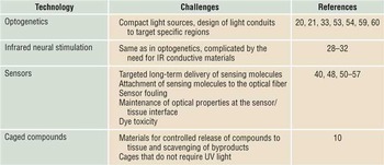
The ability to modulate neural activity with light has been of interest since the work of Arvanitaki and Fork, who discovered that neurons of invertebrates altered their firing rates when exposed to light of different wavelengths.Reference Fork29 The tight beam spot of small optical fibers, combined with the ability to localize light offers excellent spatial selectivity in applications requiring focal stimulation of excitable tissue. Current methods used to stimulate tissue with light include optical uncaging, infrared neural stimulation, and optogenetics. Each technique presents unique materials science challenges, which need to be overcome before it can be used in a clinical setting. Some of these technological barriers are listed in Table I.
Material challenges.

This table identifies some of the material challenges designers have faced in incorporating them into a robust design aimed at clinical applications.
Optical uncaging is one of the oldest methods of photostimulation, where UV light is used to activate or “uncage” an externally applied neurotransmitter, which is then free to exert its action on neurons. The rapid and localized changes in neurotransmitter levels achieved by optical uncaging allow for precise modulation of brain circuits without directly interfering with their electrical activity. UV light is toxic to tissue, and efforts are under way to replace UV-based excitation with two-photon techniques with light sources in the NIR or visible range.Reference Callaway and Yuste1 Two-photon excitation may also be used to activate small focal volumes of neural tissue in the absence of caged compounds. These techniques require sophisticated optical systems that produce pico- or femtosecond laser pulses and laser sources that are difficult to miniaturize.
Infrared neural stimulation (INS) utilizes infrared light to stimulate or modulate neural activity in both peripheral and central nervous tissues, with applications ranging from localization and monitoring of specific nerves during neurological surgery to small groups of auditory neurons in cochlear implants.Reference Teudt, Nevel, Izzo, Walsh and Richter30–Reference Cayce, Friedman, Jansen, Mahadevan-Jansen and Roe33 INS takes advantage of the high absorption of infrared light by water, which is abundant in neural tissues. A short pulse of infrared light creates a transient rise in temperature in a small region immediately below the optical fiber coupled to the light source. The temperature rise alters the electrophysiological properties of neural membrane and leads to generation of action potentials. Unlike electrical stimulation, INS does not suffer from current spread, allowing for stimulation of a single nerve (or even individual fascicles within it) without affecting neighboring structures. Although it offers better spatial selectivity compared to traditional electrical methods, to our knowledge, INS does not discriminate between neuronal types, and the recruited volume is determined primarily by the beam parameters and wavelength-dependent optical properties. However, significant engineering and materials challenges exist for chronic applications, such as the cochlear implant, where light must be delivered to multiple locations within a small and highly convoluted structure, necessitating the use of thin, highly flexible optical fibers.Reference Moreno, Rajguru, Matic, Yerram, Robinson, Hwang, Stock and Richter34 INS, in general, is highly amenable to miniaturization because it does not require additional optics or exogenous chemical agents and uses compact diode lasers.
Precise stimulation selectivity may be accomplished by genetically introducing light-sensitive molecules into tissue, a method known as optogenetics. It is now possible to target specific neuronal populations with viral vectors and introduce alga-derived, light-activated ion channels such as the excitatory channel-rhodopsins and inhibitory halo-rhodopsins, which, upon activation, either generate or inhibit neuronal action potentials. The challenge for developing neural interfaces to take advantage of these optogenetic techniques is the delivery of the light to the modified tissue.Reference Aravanis, Wang, Zhang, Meltzer, Mogri, Schneider and Deisseroth35 The light-activated channels respond to visible wavelengths of light, which are highly scattering in biological tissue. Optical fibers are used to deliver light subcortically, and high power miniature LEDs can be used as light sources in implantable devices, although heat dissipation by them is a concern that should be carefully addressed. Perhaps the bigger challenge is the more ubiquitous issue of being able to efficiently, reliably, and safely perform gene therapy in human patients.
Optical sensors
The electrical nature of neural impulses has made the measurement of electrical activity an obvious choice for a feedback signal to drive neural stimulators. While electrical activity cannot be directly visualized, the propagation of an electrical impulse changes the optical properties of the nerve, such as the scattering coefficient and the refractive index due to a combination of effects.Reference von Muralt36 These effects are small, and the signal to noise ratio is generally poor, with detection being limited to excised nerve preparations, where movement artifacts from respiration and heartbeat are not an issue, with signal averaging of multiple responses.Reference Cohen, Keynes and Hille37
Recently, indirect measures of optical changes in neural tissue have emerged. Optical coherence tomography is an interferometric technique for detecting changes in light scattering and has been used to detect changes in neural activity in different layers of the cerebral cortex of a cat and in marine sea slug Aplysia neurons.Reference Maheswari, Takaoka, Kadono, Homma and Tanifuji38, Reference Graf, Ralston, Ko and Boppart39 Another method is surface plasmon resonance, which relies on optical properties of an interface between a nerve and a conductive film (usually gold) rather than those of the nerve itself.Reference Ae Kim, Min Byun, Lee, Kim, Kim, Baac, Schuler and Kim40 This design enables detection of single action potential-related optical events, and specialized coatings of the sensor, including deposition of nanotubes and nanowires, may significantly improve performance.Reference Byun, Yoon, Kim and Kim41 However, these techniques suffer from extreme sensitivity to motion, including heartbeat and respiration. This problem may be reduced by improving sensor packaging that promotes consistent contact between the sensor and the tissue. Another potential approach uses metal-impregnated polydimethylsiloxane (PDMS) microbeads attached to fiber optics in order to measure the electric fields caused by conduction of action potentials. The electric field modulates the evanescent field of light using the whispering gallery mode. While this approach lends itself well to multiplexing, sensitivity in the in vivo setting is a challenge, and work is ongoing to determine optimal materials for these sensors.Reference Ioppolo and Otügen42 Optical detection of neuronal activity in this manner is appealing because it requires no modification of neuronal tissue or external contrast agents and the signals are, in some cases, well correlated to electrical impulses.Reference Wininger, Schei and Rector43
Engineering optical properties into the tissue can improve the specificity of the signal and gives the designer a wider choice in selecting a feedback signal for the device, including intra- and extracellular concentration of ions, neurotransmitters, or other molecules. Voltage sensitive fluorescent dyes (VSDs) are molecules that intercalate into the lipid membrane of neurons and respond to neuronal electrical activity by changes in their fluorescence, which can be imaged with a high speed camera. They have been used as early as 1968, and the latest variants are bright enough to provide the millisecond temporal resolution necessary for capturing neural dynamics.Reference Tasaki, Watanabe and Hallett44, Reference Chemla and Chavane45 Unlike microelectrode arrays, whose spatial resolution is limited by the number of electrodes and which emphasize action potentials rather than subthreshold activity, VSDs emphasize communication between neurons via release of neurotransmitters at synapses, providing a different but complementary picture of neural activity.Reference Petersen, Grinvald and Sakmann46 There are several problems that currently preclude the use of voltage, ion, and pH sensitive dyes: dye delivery, dye toxicity, and maintenance of a stable optical window for imaging. Surface-loaded dyes penetrate less than half way into the cortex (∼400 μm), which bars them from being used in imaging of deeper structures.Reference Chemla and Chavane45 In some cases, dyes can be injected into the cortex, but this requires a more invasive approach. Moreover, dyes are degraded (sometimes into toxic byproducts) under continuous exposure to light and need to be replenished.Reference Phillips, Zhou and Baltz47–Reference Thaler, Haritoglou, Choragiewicz, Messias, Baryluk, May, Rejdak, Fiedorowicz, Zrenner and Schuettauf49
Periodic VSD recordings have been performed by Slovin et al. using an optically transparent silicone window placed over the brain, with a sterile port at its side used to replenish the dye.Reference Slovin50 Filling the port with dye is a manual process that takes several hours, and the technique would greatly benefit from controlled delivery of the dye either through a pump or through timed release. A material for sequestering the dye byproducts would also be useful. The long-term viability of the technique is limited by the stability of the optical window, since it gradually becomes opaque, due to a slow growth of pial and arachnoid membranes below the silicone membrane.Reference Arieli, Grinvald and Slovin51 In this case, having a bio-inert material is insufficient. What is needed is a material that slows down or prevents the normal physiological response.
Sensing applications generally do not require the high irradiance levels needed for neural stimulation. Extremely thin glass optical fibers, between 20 and 500 nm in diameter, permit investigation of not only single cells, but the intracellular environment as well.Reference Cullum and Vo-Dinh52–Reference LeChasseur, Dufour, Lavertu, Bories, Deschênes, Vallée and De Koninck54 Fabrication techniques include pulling laser-heated fibers or chemical etching, followed by coating with reflective materials, such as aluminum.Reference Hoffmann55, Reference Stöckle, Fokas, Deckert, Zenobi, Sick, Hecht and Wild56 Because biologically active compounds, such as neurotransmitters, are often confined to highly specialized compartments, the small sensing space of the probe enables reflection of physiologically relevant changes in their concentration rather than measuring an average concentration in the brain. For example, Tan et al. used fibers 100–1000 nm in diameter that were silanized and cross-linked to a photopolymerizable polycarbonate membrane doped with a pH-sensitive dye.Reference Tan, Shi, Smith, Birnbaum and Kopelman57 The fiber was small enough to be used in measurement of the pH of the extra-embryonic space of rat fetuses, and its response time (300 ms) was about 100 times better than that of conventional electrodes.
The process of attaching optical probes to the fiber is challenging, because glass is non-reactive, while entrapment of dyes in the sensor matrix results either in gradual leaching of the dye or poor access of analytes to the sensing space and long response times. Recent advances have included the use of plastic fibers and a hybrid organic silicate matrix that offers good ion mobility and excellent adhesion to the fiber itself.Reference Fabbri, Pilati, Rovati, McKenzie and Mijovic58 In some cases, the probe can be attached to the fiber directly. Cordek and colleagues developed a sensor for glutamate, the major excitatory neurotransmitter found in the brain, by silanizing silica fibers with trimethoxysilylpropyldiethylenetriamine (a DETA functionalized methoxy silane) and conjugating them with glutaraldehyde, to which the enzyme glutamate dehydrogenase was attached.Reference Cordek, Wang and Tan59
Advances in optical probes have greatly improved our understanding of the nervous system.Reference Hillman3 Not only do VSDs provide a valuable tool for monitoring neural network activity, but fluorescent sensors for calcium, pH, and various neurotransmitters are being used to investigate biological signaling at an incredible level of detail in such phenomena as synaptic plasticity and neuronal-astrocyte interactions. The materials science contribution and challenge to the field lies largely in creating an appropriate interface between the dye and the body that is compatible with both. This involves creation of appropriate fibers to deliver the light and send the information back, packaging of the dye either on the surface of the fiber or in separate vehicles, targeting the probes to the appropriate neuroanatomical structures, and ensuring long-term biocompatibility of the entire assembly.
Hybrid electrical-optical interfaces
Both optical and electrophysiological techniques have their advantages and drawbacks and can complement each other when used in hybrid electrical/optical devices. These devices fall into three general categories: (1) microprobes for interrogation of single cells, (2) multi-electrode/optical fiber arrays, and (3) non-traditional photoactivation technologies.
Micro-optrodes, essentially micropipettes amalgamated with tapered optical fibers, can be used to locate specific neuron types using optical methods and then record electrophysiological signals from them.Reference Dufour, Dufour, Chever, Vallée and Amzica53, Reference LeChasseur, Dufour, Lavertu, Bories, Deschênes, Vallée and De Koninck54 These interfaces can provide both field potential recordings and intracellular fluorescence measurements. Recently a method of constructing a hybrid microprobe using a single optical fiber was reported.Reference LeChasseur, Dufour, Lavertu, Bories, Deschênes, Vallée and De Koninck54 This design comprises a hollow glass pipette (filled during use with an electrolyte solution for electrophysiological measurements) and a graded-index optical fiber. The fiber and pipette are heated and pulled to a final tip diameter less than that of the target neuron cell body to allow for single cell identification.
The study of neuronal networks can benefit from hybrid micro-optrode/multi-electrode recording arrays (MEAs). An MEA provides high temporal and spatial resolution electrophysiological recording over the dimensions of the array, while an integrated optrode is able to stimulate and/or inhibit neuronal activity without generating electrical artifacts. Hybrid MEA-optrode arrays generally have one of the silicon shanks of a commercially available MEA replaced with a tapered multimode glass optrode. However, a recent design incorporated a gold film at the tip of the optrode to allow for simultaneous stimulation and recording.Reference Wang, Borton, Zhang, Burwell and Nurmikko60 In addition to modifications of commercially available MEA devices, a hybrid complementary metal-oxide-semiconductor (CMOS) sensor device arranged along a penetrating shank was developed as a tool for recording fluorescence and electrical activity from deep brain structures.Reference Tagawa, Mitani, Minami, Noda, Sasagawa, Tokuda and Ohta61 The device includes an LED and image sensors for exciting and detecting fluorescence, respectively, as well as electrodes for recording electrical potentials. For the hybrid CMOS sensor device, biocompatible materials such as gold, platinum, and tungsten were deposited on the electrodes, and the entire device was packaged in a parylene layer to add water resistance and biocompatibility. Parylene is transparent in the visible region and is a good packaging material for optical devices.
Novel hybrid electro-optical techniques make use of semiconductor nanoparticle films, carbon nanotubes, and even hydrogels as a means of interfacing optical and electrical signals to/from neural tissue.Reference Pappas, Wickramanyake, Jan, Motamedi, Brodwick and Kotov62–Reference Mohy-Ud-Din, Woo, Kim and Cho64 These materials offer exciting possibilities for optoelectrical stimulation due to material properties that may be tuned to offer efficient conversion from optical to electrical energy. Recently, a bio-organic semiconductor operating as a photovoltaic cell was developed for neuronal stimulation.Reference Ghezzi, Antognazza, Dal Maschio, Lanzarini, Benfenati and Lanzani65 The bio-organic interface presented, poly-L-lysine treated indium-tin oxide/region-regular poly(3-hexylthiophene-2,5-diyl) with phenyl-C61-butyric-acid-methyl ester (rr-P3HT:PCBM), was shown to have no toxic effects. This new interface may motivate the development of retinal implants using bio-organic polymers.
Conclusion
A number of exciting optical techniques for interacting with neural tissue have been developed. These include methods for both stimulation and sensing of neural activity. Optical methods can be used alone or combined with electrophysiological methods. While chronically implanted electrical devices have a well-proven track record of providing sustained clinical benefits, optical neural interfaces remain in the earlier stages of development, mostly limited to acute or experimental applications. Some of the key barriers that must be overcome for the clinical use of optical neural implants to become a reality are an incomplete understanding of chronic effects of light on tissue, and the difficulty in maintaining an optimal interface between the sensor or stimulator and its biological environment. The light guides and sources used in the current generation of optical neural devices have been borrowed from the fields of telecommunications, which initially guided the development of these components. We hope that with the help of the materials science community, a new generation of optical interfaces can be developed that specifically address the issues of this application: long-term biocompatibility, small size, improved mechanical qualities, and so on. We believe that if these optical devices can be made at least as robust as the current electrical implants, they will revolutionize the treatment of neurological disorders.