After surgery for complex congenital cardiac defects in newborns, immediate closure of the chest is not always feasible. In patients with a high risk of low cardiac output due to pronounced cardiomegaly, bleeding or tissue swelling, delayed sternal closure may enable circulatory stabilisation in the early postoperative period. Delayed sternal closure is used in two-fifths of newborns in our institutionReference Vojtovič, Tláskal and Selko1. Closure of the chest often causes haemodynamic and ventilatory deterioration that may be missed at the early stages by sole clinical assessment and standard monitoringReference McElhinney, Reddy, Parry, Johnson, Fineman and Hanley2, Reference Egan, Festa, Cole, Nunn, Gillis and Winlaw3. On the other hand, close monitoring of the cardiac output may assist timely therapeutic interventions in order to prevent deleterious consequences of unapparent low cardiac output state. In adults, the transpulmonary thermodilution technique using pulmonary arterial catheters is a standard procedureReference Schwann, Zacharias, Riordan, Durham, Engoren and Habib4–Reference Hollenberg and Hozt7. The method, however, poses risks in childrenReference Introna, Martin, Pruett, Philpot and Johnston8. It is possible, however, to achieve transpulmonary thermodilution in small children via the femoral artery by injecting a defined amount of cold saline into the central venous catheter and monitoring the temperature of the blood within the femoral arteryReference Fakler, Pauli, Balling and Lorenz9–Reference Pauli, Fakler, Genz, Henning, Lorenz and Hess11. The aim of our prospective study, therefore, was to analyze haemodynamic changes caused by the chest closure by means of the standard monitoring methods along with transpulmonary thermodilution. At the same time, we evaluated the changes in ventilatory parameters, the indexes of tissue perfusion, and the frequency and character of any necessary therapeutic interventions.
Patients
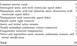
The study was done at a single nation–wide tertiary referral centre with an annual average of 330 open-heart and 110 closed-heart paediatric cardiac surgeries. At the time span of the study, 139 newborns underwent surgery using cardiopulmonary bypass, and in 50 of them (36%), it was necessary to delay sternal closure. Between April, 2006, and December. 2008, 25 newborns with delayed sternal closure after a biventricular correction of a complex congenital cardiac defect were included in a prospective study. Proper measurements were prevented in 2 patients, in one by a malfunction of the arterial thermodilution catheter, and in the other by a false detection of significant right-to left shunt that was excluded by contrast echocardiography. Data of remaining 23 newborns, aged from 1 to 30 days, with a median of 7 days, and with body weights from 1.9 to 4.2 kilograms, with a median of 3.25 kilograms, were subjected to the statistical analyses. The defects corrected by the surgery are listed in Table 1. The study was approved by the University Hospital Ethics Committee. The parents of all the children were familiarized with the principle of the thermodilution measurement and the need for the insertion of the specialised arterial catheter. They all signed the informed consent form.
Cardiac lesions surgically corrected.

Methods
Perioperative management
The patients were prepared for the surgery in a usual manner except for a special 3F arterial catheter with a thermistor at the tip (Pulsiocath PV 2013L07, Pulsion Medical Systems, Munich, Germany). The catheter was capable of simultaneous determination of the arterial blood pressure and the blood temperature changes. After the surgery, all the patients were subject to a standard haemodynamic and laboratory monitoring, time cycle pressure limited ventilation, and total or supplemental parenteral nutrition. The delayed sternal closure was indicated according to the surgeon’s decision. The indications were significant cardiomegaly in 7 patients, nonsurgical bleeding in 6, mediastinal tissue oedema in 5, and more than 10 mmHg systemic arterial pressure drop or global respiratory insufficiency due to attempted chest closure in 5 patients. When the delayed sternal closure was indicated, the sternum was stabilized using one or two splinter bars between sternal edges and the skin defect was covered with silastic patch (Perthese®, Perouse Plastie, Bornel, France). A continuous intravenous infusion of Fentanyl 8 micrograms/kilogram/hour and Midazolam 4–6 micrograms/kilogram/minute was used for analgosedation. At least 24 hours after the surgery and whenever haemodynamic instability or bleeding was encountered, myorelaxation was maintained with iv. Pipecuronium 0.1 milligrams/kilogram every three hours. There was no strict protocol introduced for the delayed sternal closure timing at the intensive care unit. Prerequisites for the closure attempt were as follows: stable hemodynamics, favourable blood gases without a need for an aggressive ventilatory support, negative fluid balance for at least 24 hours, and no signs of active wound infection or sepsis. The closure was performed under general anaesthesia (Ketamin 3 milligrams/kilogram with Fentanyl 20 micrograms/kilogram) and myorelaxation (Pipecuronium 0.1 milligram/kilogram) at the cardiac intensive care unit. Whenever a cardiopulmonary deterioration occurred due to the chest closure, vigorous therapeutic interventions were immediately employed: Dobutamine and Dopamine was increased step by step up to double initial dose and if this was insufficient Epinephrine and Milrinone were added. According to the filling pressures and haematocrit, colloid volume of 5–15 ml/kg over the 10–30 minutes interval was administered. In case of respiratory problems, oxygen fraction or inspiratory pressures were increased as appropriate. In only one patient the medical treatment was unsuccessful and the chest had to be re-opened for another 48 hours.
Monitoring
Heart rate (HR) and invasive pressures – systolic arterial (APs), diastolic arterial (APd), mean arterial (APm), and mean central venous pressure (CVP) – was recorded by the monitoring system Siemens SC 9000XL, Infinity Modular Monitoring Series (Siemens Medical Systems, Inc, Danvers, USA). Mean airway pressure (MAWP) and dynamic lung compliance (DynC) were recorded by servo-ventilator Avea™ (VIASYS Healthcare, Care Division, California, USA). Blood gases and lactate levels were examined from arterial blood using Roche OMNI® S analyser (F. Hoffmann-La Roche Ltd., Basel, Switzerland). Oxygenation index (OI) was determined from the equation OI = MAWP × FiO2/PaO2 (MAWP- mean airway pressure in cmH2O; FiO2- oxygen fraction in the inspired gas in %; PaO2- partial oxygen pressure in arterial blood in mmHg).
Transpulmonary thermodilution
Central venous lines routinely inserted in the jugular, subclavian, or femoral vein were used for the administration of 3 ml cold (below 8°C) saline bolus. For this purpose the distal lumen of the catheter was connected to the injectate temperature sensor housing (PV 4046, Pulsion Medical Systems, Munich, Germany). Arterial blood temperature was senzored by the femoral arterial catheter (see above). Signals from both the catheters were used by PiCCO Plus system (Pulsion Medical Systems, Munich, Germany) for construction of thermodilution curves, pulse contour analysis and computation of the following parameters: cardiac index (CI), stroke volume index (SVI), stroke volume variation (SVV), systemic vascular resistance index (SVRI), global end-diastolic volume index (GEDVI), intrathoracic blood volume index (ITBVI), and extravascular lung water index (ELWI). For details on the PiCCO measurements and calculations see the manual website.12 Residual intracardiac shunts were excluded by echocardiography in all the included patients. No complication in connection with the thermodilution catheter was encountered.
Data collection
All the above parameters were recorded before and immediately after the chest closure and then at 0.5, 1, 2, 6, 12, 24, and 48 hours after the closure. A single set of the monitored data was recorded at each time-point. For the thermodilution parameters, the average of two to three measurements was calculated for each time-point. Five patients are not included in the heart rate statistics because they required a temporary epicardial pacing.
Statistics
Statistical analyses were performed using SigmaStat 3.5 (SPSS Inc.). The data sets were tested for normal distribution and for equal variance when they were compared. In case of normal distribution and equal variance, comparison of all the post-closure values to the pre-closure one was done by One way RM ANNOVA with the Bonferroni post-hoc analysis and the results are displayed as mean (standard deviation). Otherwise the data were compared by RM ANNOVA on ranks with Dunn’s post-hoc analysis and the results are displayed as median (25th centile/75th centile). All the tests were two-sided. Linear regression analysis was performed to evaluate correlations between the pulse pressure and the cardiac index and the pulse pressure and the stroke volume index values for individual patients at each time stage. Results with a probability value p < 0.05 were considered significant.
Results
Hospital mortality in our cohort was 8.7%, with 2 of the 23 patients dying, one of sepsis and the other of a chronic heart failure caused by significant residual heart defects not amenable to a surgical re-intervention. The median interval from surgery to chest closure was 48 hours, with a range from 24 hours to 7 days. In the time of the closure, or shortly afterwards, 35 therapeutic interventions were needed in 18 of the 23 (78.3%) patients, giving an average of 1.9 interventions per a patient. In 15 patients, an increase was required in the inotropic support, 11 colloid volume administration, and 9 an increase in ventilatory support. In 1 patient, the chest had to be re-opened 2 hours after the closure because of untreatable low cardiac output syndrome and 48 hours later, an uneventful delayed sternal closure was performed.
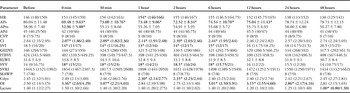
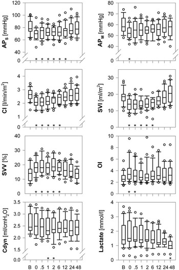
The changes in the studied parameters due to the chest closure and throughout the following two days are depicted in Table 2 and Figure 1. The closure caused a statistically significant decrease in systolic and mean arterial blood pressure, cardiac index, stroke volume index and increase in stroke volume variation. Diastolic arterial pressure remained unchanged. The increase of the heart rate was only significant since one hour after the chest closure in the coincidence with an increased inotropic support in 15 of the 23 patients. The central venous pressure as well as the volume indexes - global end-diastolic volume index, intrathoracic blood volume index, and extravascular lung water index – did not change significantly. Significant decrease in the blood lactate level occurred later after the chest closure. In presence of unchanged mean airway pressure, the dynamic lung compliance decreased significantly one hour after the closure and the oxygenation index temporarily significantly increased reflecting the deterioration in the gas exchange. No significant correlations were found between the pulse pressure and the cardiac index and the pulse pressure and the stroke volume index at any time before and after the delayed sternal closure, all having p values of greater than 0.05.
Changes in the studied parameters examined.

In case of normal distribution, the values are presented as mean (standard deviation), otherwise as median (25th centile/75th centile). Significant changes as compared to the baseline value (p < 0.05) are marked by bold font and asterisk.
Abbreviations: APd – diastolic arterial pressure (mmHg), APm – mean arterial pressure (mmHg), APs – systolic arterial pressure (mmHg), CI – cardiac index (l/min/m2), CVP – central venous pressure (mmHg), Cdyn – dynamic lung compliance (ml/cmH2O), ELWI – extravascular lung water index (ml/kg), GEDVI – global end-diastolic volume index (ml/m2), HR – heart rate (p/min), ITBVI – intrathoracic blood volume index (ml/m2), Lactate – serum lactate (mmol/l), MAWP – mean airway pressure (cmH2O), OI – oxygenation index, SVRI – systemic vascular resistance index (dyn.s.cm−5.m2), SVV – stroke volume variation (%), SVI – stroke volume index (ml/m2).
Parameters that shoved significant change from the baseline value due to the chest closure: Values before (B) and immediately after the closure (0) and then at 0.5, 1, 2, 6, 12, 24, and 48 hours after the closure are shown. Boxes indicate 25th and 75th centiles, error bars 10th and 90th centiles, and circles cases outside this interval. The lines within the boxes indicate medians. Significant changes (p < 0.05) are marked by asterisk. Abbreviations: APm – mean arterial pressure, APs – systolic arterial pressure, CI – cardiac index, Cdyn – dynamic lung compliance, Lactate – serum lactate, OI – oxygenation index, SVV – stroke volume variation, SVI – stroke volume index.

Discussion
Due to the size of their chest, newborns are at the risk of cardiopulmonary compromise caused by the chest closure after a cardiac surgery. It has been reported that the circulatory deterioration due to the chest closure is caused by a pseudo-tamponade, a significant increase in the pericardial and right atrial pressure along with a decrease in the end-diastolic ventricular volumeReference Jogi and Werner13, Reference Kay, Brass and Lincoln14. According to our search, our study is the first to use the femoral artery transpulmonary thermodilution in order to measure precisely the haemodynamic changes caused by the chest closure in newborns. We proved a significant decrease in cardiac output caused by a significant decrease in stroke volume. Also the variation in stroke volume significantly increased after the closure, presumably due to the enhanced preload changes caused by the positive-pressure ventilation. The fact that the pulse pressure did not correlate either with the cardiac index or with the stroke volume index shows that the pressure, which is routinely used as the principal measure of haemodynamics, does not purely reflect changes in the cardiac output. Indeed, the pressure is influenced also by other variables such as the heart rate and the systemic vascular resistance.
Interestingly, we did not prove a significant increase in central venous pressure in connection to the chest closure. Also intrathoracic fluid volumes, specifically the global end-diastolic volume index, intrathoracic blood volume index, and extravascular lung water index, did not change significantly due to the closure in our patients despite of volume administration in half of them. This probably reflects the contrary effects of the chest volume restriction and the plasma expansion. As expected with the time cycle pressure limited ventilation, we did not prove a significant change in the mean airway pressure due to the closure. Mean airway pressure increased on average by 1 cmH2O due to the increase in the ventilatory support, albeit that the change did not reach statistical significance. Dynamic lung compliance, however, significantly decreased, presumably due to decreased chest compliance after its closure. In consequence with the cardiopulmonary compromise, also gas exchange worsened, as reflected by significantly increased blood oxygenation index.
Lactate and mixed venous saturations are easily available in the paediatric cardiac intensive care units and serve to direct management in the absence of measurements of cardiac output. Although absolute levels of lactate in the serum over the postoperative course vary according to numerous factors, the lactate trend has been proved as a prognostic predictorReference Siegel, Dalton, Hertzog, Hopkins, Hannan and Hauser15. Serum lactate gradually decreased in all our patients in connection with the cardiopulmonary stabilisation. Interestingly, chest closure was not connected with any increase in lactate, despite the haemodynamic deterioration. We believe that this was caused by the fact, that decrease in cardiac output was quickly counteracted by appropriate therapeutic interventions. The usual way of assessing the mixed venous blood saturation is sampling in the superior caval vein. We did not attempt to evaluate the mixed venous blood data, as three-quarters of our patients had their central venous line inserted in the femoral vein.
Femoral artery transpulmonary thermodilution has been proved a reliable method for cardiac output measurement in childrenReference Pauli, Fakler, Genz, Henning, Lorenz and Hess11, Reference McLuckie, Murdoch and Marsh16, Reference Rupérez, López-Herce, García, Sánchez, García and Vigil17. In presence of intracardiac shunts, significant valvar incompetence, and arrhythmias, the method does not provide reliable resultsReference Tibby, Hatherill, Marsh, Morrison, Anderson and Murdoch10. Some of our thermodilution measurements indicated a presence of a significant right-to-left shunt, which was then reliably excluded by contrast echocardiography. In all but one of these patients a modification of injectate temperature enabled proper measurements. In one patient, however, the false-detection of the right-to-left shunt could not be prevented, and the patient was excluded from our analyses. The artefact occurred only when the cold bolus was injected into the femoral venous catheter. We believe that the false detection of the shunt was caused by the cold irradiation from the inferior caval vein to the descending aorta. Although normal values of the cardiac index in children are well recognizedReference Tibby, Hatherill, Marsh, Morrison, Anderson and Murdoch10, the normal values of the volume indexes are controversialReference Lopéz-Herce, Ruperéz, Sanchéz, García and García18, Reference Schiffmann, Erdlenbruch and Singer19. Therefore, unlike in adultsReference Sakka, Ruhl and Pfeiffer20, the volume indexes are seldom used to guide intravenous volume administration. In newborns the method may be limited by the fluid load necessary for calibration and measurements. The volume load may be prevented by using the thermodilution for a calibration of the arterial pressure tracing which is then used for a subsequent calculation of the cardiac output by the pulse contour cardiac index method. It has been reported that the results of the femoral artery transpulmonary thermodilution correlated very well with the pulse contour cardiac indexReference Zollner, Haller and Weis6, Reference Fakler, Pauli, Balling and Lorenz9, Reference Godje, Hoke and Lichtwark-Aschoff21, Reference Mahajan, Shabanie, Turner, Sopher and Marijic22. Published data shows that the pulse contour method correlates well with thermodilution, and therefore may be optimal for a beat-to-beat cardiac output monitoring in newborns because it is not limited by the necessity of volume administration.
Conclusions
Chest closure causes a significant transitory decrease in stroke volume with a consequent decrease of cardiac output and arterial blood pressure. Also lung compliance and blood oxygenation are temporarily significantly compromised. With aggressive therapeutic interventions that are necessary in more than three-quarters of patients, the cardiopulmonary instability gradually subsides.
Acknowledgement
Supported by the grant NR 9046-3, Internal Grant Agency of the Ministry of Health, Czech Republic.