Echocardiography has long been the non-invasive method of choice for diagnosis and follow-up of pulmonary arterial hypertension in general and that related to congenital cardiac disease in particular. This is despite the fact that echocardiographic parameters have not been found valuable as part of management algorithms in clinical practice or as endpoints in clinical trials due to marginal correlation with invasive measurements and also due to poor reproducibility.
The use of computed tomography and magnetic resonance imaging is becoming more commonplace in the evaluation, management and follow-up of all forms of pulmonary hypertension. As the quality and quantity of information provided by magnetic resonance increases, its potential for use in clinical decision making, especially in this patient population, becomes ever more attractive.
To date, large clinical studies and/or randomized clinical trials examining computed tomography or magnetic resonance imaging endpoints in pulmonary arterial hypertension associated with congenital cardiac disease are in essence, non-existent. Recommendations, therefore are based on extrapolation of existing data related to either congenital cardiac disease or pulmonary hypertension and expert consensus.
Assessment of anatomy and function
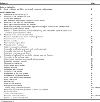
Magnetic resonance imaging has been shown to deliver improved reproducibility for both volumetric and functional parameters when compared with echocardiography, and is currently considered to be the gold standard for evaluating many forms of congenital cardiac disease in general, and those involving the right ventricle in particular, especially in the adult population (Table 1). Magnetic resonance imaging allows for measurements of right ventricular volumes, systolic function, and severity of pulmonary valvar regurgitation with good reproducibility.Reference Davlouros, Kilner and Hornung1, Reference Tulevski, Dodge-Khatami, Groenink, van der Wall, Romkes and Mulder2
Indications for cardiac magnetic resonance in congenital cardiac disease (Footnote *).

(*) from reference 8.
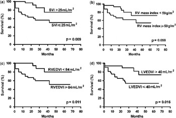
Using magnetic resonance imaging in 64 patients with idiopathic pulmonary arterial hypertension, van Wolferen et al. demonstrated a stroke volume ⩽25 ml/m2, a right ventricular end diastolic volume ⩾84 ml/m2, and a left ventricular end diastolic volume ⩽40 ml/m2 to be strong independent predictors of mortality (Fig. 1).Reference van Wolferen, Marcus and Boonstra3 Interval changes in these parameters at one year of follow-up were strong predictors of mortality, while right ventricular mass and wall thickness were not. Furthermore, van Wolferen et al. also demonstrated, in a separate study (including idiopathic and congenital cardiac disease-associated pulmonary arterial hypertension), significant improvement in right ventricular performance and cardiac index after three months of sildenafil added to baseline bosentan therapy.Reference van Wolferen, Boonstra and Marcus4 This study, however, is small and data should be interpreted with caution.
Survival in patients with idiopathic pulmonary arterial hypertension as a function of cardiac magnetic resonance parameters. LVEDVI, left ventricular end-diastolic volume index; RV, right ventricle; RVEDVI, right ventricular end-diastolic volume index; SVI, systolic volume index. Reproduced with permission from van Wolferen SA et al., Reference Reference van Wolferen, Marcus and Boonstra3.

Gan et al. examined right ventricular diastolic function and the acute effects of sildenafil in 25 pulmonary hypertensive patients (mainly idiopathic pulmonary arterial hypertension) using magnetic resonance imaging, brain natriuretic peptide and invasive measurements.Reference Gan, Holverda and Marcus5 Interventricular relaxation time correlated positively with brain natriuretic peptide, (r = 0.70), and inversely with cardiac index (r = −0.70) and right ventricular ejection fraction (r = −0.69). Both interventricular relaxation time and right ventricular ejection fraction improved after sildenafil and this may be a result of afterload reduction or direct effects of sildenafil on the right ventricle.
Flattening of the interventricular septum as an indicator of right ventricular hypertension is routinely applied in the echocardiographic evaluation of congenital cardiac disease and pulmonary arterial hypertension. Roeleveld et al. reported on 37 patients (including idiopathic and congenital cardiac disease-related pulmonary arterial hypertension) and demonstrated a linear relationship (r = 0.68) between the shape of the septum determined by magnetic resonance imaging and catheterization derived systolic pulmonary arterial pressure.Reference Roeleveld, Marcus and Faes6 This correlation is not as good as that reported in the original series involving echocardiography (r = 0.86), and is likely due, in part, to the fact that septal curvature is more closely related to the ratio of right-to-left ventricular pressures than an absolute number.
Numerous other studies have attempted to find echocardiographic correlates with magnetic resonance measurements with limited success. Parameters such as right ventricular fractional area change, tricuspid annular velocity, transverse fractional shortening and others have all been proposed. Due to either poor correlation with magnetic resonance imaging, poor reproducibility or the inherent time involved for acquisition and data analysis, none have gained wide acceptance. Recent studies using three-dimensional echocardiography have demonstrated excellent correlation with magnetic resonance and radionuclide estimates of right ventricular function.Reference Nesser, Tkalec and Patel7 This and some of the newer echocardiographic indices such as tissue Doppler imaging and myocardial performance index (the so-called Tei index), may challenge some of the current advantages of cardiac magnetic resonance in the future. The current availability and use of three-dimensional echocardiography is much less than that of magnetic resonance however, and indices require further evaluation, particularly in adult patients, where transthoracic views of the right ventricle can be problematic.
In some forms of left heart disease, magnetic resonance imaging exceeds echocardiography in the quality and type of information it can provide.Reference Pennell, Sechtem and Higgins8 Magnetic resonance imaging can define the pulmonary venous anatomy with improved accuracy as compared to echocardiography or cardiac catheterization.Reference Masui, Seelos, Kersting-Sommerhoff and Higgins9 In Shone’s complex, delayed enhancement has been used to document endocardial fibroelastosisReference Tworetzky, del Nido, Powell, Marshall, Lock and Geva10 and may offer a surgical option to reverse pulmonary hypertension due to left atrial hypertension.
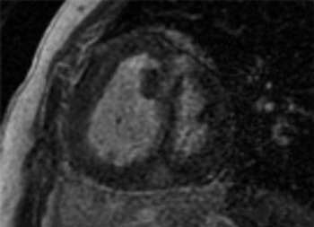
The application of delayed enhancement to examine the right ventricle has been far more limited than for the left ventricle. In patients with corrected tetralogy of Fallot, delayed enhancement due to presumed fibrosis has been demonstrated in the right ventricular outflow tract and in the area of the ventricular septal defect patch.Reference Babu-Narayan, Kilner and Li11 Multiple studies in patients with systemic right ventricles found areas of abnormal myocardium as demonstrated by delayed enhancement.Reference Babu-Narayan, Goktekin and Moon12 The presence of abnormal myocardial regions was associated with known markers of adverse outcome, such as right ventricular dysfunction, poor exercise tolerance, arrhythmia, and clinical deterioration over time. In adult patients with pulmonary hypertension, a small number of studies have suggested that the presence of delayed enhancement at the right ventricular insertion points is common, regardless of etiology, and is inversely related to right ventricular function and pulmonary hemodynamics, with systolic pulmonary arterial pressure correlating most closely (Fig. 2).Reference Blyth, Groenning and Martin13, Reference Sanz, Dellegrottaglie and Kariisa14 In patients with pulmonary arterial hypertension associated with congenital cardiac disease, the relative contribution of pulmonary hypertension versus the cardiac defect itself to delayed enhancement maybe difficult to differentiate.
Delayed enhancement is a common finding in patients with pulmonary hypertension of all etiologies. Most commonly seen in the interventricular septum at the “hinge points,” enhancement has also been demonstrated at the site of surgical fibrosis, papillary muscles and right ventricular outflow tract.

Computed tomography scanning is a reasonable alternative to magnetic resonance imaging for the acquisition of anatomic and functional data when magnetic resonance is not feasible, unavailable or contraindicated.Reference Nicol, Gatzoulis, Padley and Rubens15 Current resolution of computed tomography presents a good alternative to pulmonary angiography in the evaluation of clot or pulmonary arterial stenoses in all but the smallest vessels. Furthermore, computed tomography acquisition times are very short (a few minutes) which is an advantage for patients with claustrophobia or those with other disabilities such as Down’s syndrome.
Computed tomography evaluation of coronary artery compression
A particular area where computed tomography, for the time being, is still felt to be the non-invasive imaging modality of choice is the coronary arteries. Magnetic resonance imaging resolution, while improving and useful, has still not reached the sub-millimeter resolution offered by cardiac gated computed tomography often required for coronary investigations.
In both the idiopathic form and congenital cardiac disease-related pulmonary arterial hypertension, particularly atrial septal defects with or without Eisenmenger’s syndrome, numerous reports in adults and children as young as 6 months have documented coronary artery compression by an enlarged and pulsatile main pulmonary artery. In the largest study, using echocardiographic and catheterization based angiography in 36 patients with pulmonary arterial hypertension either idiopathic or associated with congenital cardiac disease, Mesquita et al. identified 7 patients with left main coronary artery compression.Reference Mesquita, Castro, Ikari, Oliveira and Lopes16 Angina was reported in 26 of 36 patients, and all 7 with compression. Compression was related to both main pulmonary arterial diameter and the pulmonary artery to aorta diameter ratio. Dynamic compression (⩾50% obstruction) was seen in 37% of patients with pulmonary arterial diameters ⩾40 mm, but in none <40 mm. Only patients with pulmonary artery to aorta diameter ratios of >1.21 showed compression and in this group, the rate was 26%. If catheterization is not planned for other reasons, and functional data is not required, computed tomography could be the diagnostic modality of choice. Untreated patients can develop ischemic left ventricular dysfunction that precludes bilateral transplantation of the lungs, requiring heart-lung transplant with considerably worse prognosis.
A not uncommon cause of death in pulmonary arterial hypertension is rupture of a markedly enlarged pulmonary artery (with aneurysm formation). Serial recording of the pulmonary arterial diameter as well as the thickness of the main pulmonary arterial wall is useful, since some of these patients must be considered for transplantation.
The Eisenmenger population
Because of the commonplace inability adequately to image adolescents and adults with congenital cardiac disease, magnetic resonance imaging is increasingly used in this population.Reference Broberg, Ujita and Prasad17 In addition to assisting in providing complete anatomic definition, cardiac magnetic resonance can replace echocardiography and catheterization partially (e.g. yearly) in the follow up of patients with the Eisenmenger’s syndrome. The known sequelae such as pulmonary arterial thrombosis and pulmonary arterial dissection have all been recently documented by magnetic resonance screening in the literature. However, it should be emphasized that neither magnetic resonance imaging nor computed tomography can replace confirmatory right heart catheterization for diagnosis of pulmonary arterial hypertension. Although these studies are excellent additional tools, right (and eventually left) heart catheterization remains the gold standard for confirmation of diagnosis.

Assessment of hemodynamic and mechanical properties
One of the most important hemodynamic parameters obtained during cardiac catheterization is the determination of cardiac output (cardiac index). As a strong predictor of mortality, cardiac index is often used in clinical decision making and therapeutic choices. Excellent correlation between catheterization and cardiac magnetic resonance velocity mapping methods have been demonstrated on many occasions in terms of both absolute and relative systemic and pulmonary flows in the setting of shunts.Reference Brenner, Caputo and Mostbeck18, Reference Hundley, Li and Hillis19 A distinct advantage of magnetic resonance measurements over catheterization based techniques is the indifference of the former to pulmonary and tricuspid valvar regurgitation in measuring cardiac output.
One further advantage of magnetic resonance over cardiac catheterization is its ability to measure flow in essentially any reasonably sized structure whether arterial or venous. This is particularly important in cases of multiple sources of pulmonary blood flow or anatomic abnormalities which prevent accurate thermodilution or Fick determination of flow; in some of these instances, measurement of total pulmonary vein flow may be used to provide the most accurate measurement of total pulmonary flow. Additionally, the ability of magnetic resonance to measure relative right and left lung perfusion as part of a comprehensive evaluation may obviate the need for radionuclide studies. In contradistinction to measurement of cardiac output, magnetic resonance estimation of pulmonary arterial pressures has not been successful to date.
Biomechanical properties of the pulmonary arteries have also been examined as markers of the disease process. Specifically, pulmonary arterial stiffness (capacitance, compliance, distensibility) which is felt normally to comprise approximately 30% of the total workload of the right ventricle, has been investigated as a measure of right ventricular work in pulmonary hypertension. The ratio of stroke volume to pulse pressure (expressed in ml per mmHg) as an indirect measure of arterial compliance, and ventricular work, was originally investigated in the systemic circulation in patients with hypertension and was found to be predictive of cardiovascular death.Reference de Simone, Roman, Koren, Mensah, Ganau and Devereux20 This concept was then evaluated in the setting of pulmonary hypertension by Muthurangu et al. who described a method of measuring pulmonary arterial capacitance utilizing magnetic resonance flow data and invasive pressure measurements.Reference Muthurangu, Atkinson and Sermesant21 An inverse relationship between capacitance and both resistance (r = 0.89) and mean pulmonary artery pressure (r = 0.72) was found.
Capacitance was subsequently found to be a predictor of mortality as well. Mahapatra et al. studied 104 patients with idiopathic pulmonary arterial hypertension and calculated the capacitance based on invasive measurements of pulse pressure and echocardiographic calculations of stroke volume.Reference Mahapatra, Nishimura, Sorajja, Cha and McGoon22 During the 4-year follow-up period, the capacitance index was the strongest univariate predictor of mortality (hazard ratio 17.0 per ml·mmHg−1 decrease) when compared to conventional markers. Patients in the lowest capacitance index quartile had a mortality of 61% while those in the highest had no deaths. Other investigators observed good correlations between echocardiographic and catheterization derived measures of capacitance and that impedance was better at predicting patient outcomes than pulmonary vascular resistance alone.
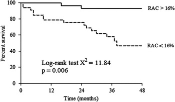
Adapting these methods, Gan et al., in 70 patients consisting primarily of idiopathic pulmonary arterial hypertension or systemic sclerosis used magnetic resonance imaging to examine proximal pulmonary artery stiffness.Reference Gan, Lankhaar and Westerhof23 The relative cross-sectional area change in pulmonary hypertensive patients was one-third that of normal controls and showed an inverse relationship with mean pulmonary arterial pressure. Patients with a relative cross-sectional area change ⩽16% had worse prognosis than those with values >16% (Fig. 3).
Kaplan Meier survival curves demonstrating improved survival with relative area change (RAC, i.e. pulmonary artery compliance measured by magnetic resonance) > 16%. Reproduced with permission from Gan et al., Reference Reference Gan, Lankhaar and Westerhof23.


Heart-lung transplant assignment
Right ventricular changes in response to chronic pressure overload regress after lung transplantation. Neither magnetic resonance imaging nor computed tomography have been used routinely to decide between lung or heart-lung transplant in patients with pulmonary arterial hypertension with or without associated congenital cardiac defects, and no studies have been published examining this approach. The choice of heart-lung transplant is usually based on the severity of tricuspid regurgitation (quantitative measurements are not felt to be clinically important), and in the case of unrepaired congenital cardiac disease, the complexity of the underlying cardiac anatomy and the extent of intracardiac repair required (personal communication, Dr. Bruce Reitz).

Acknowledgements
The authors would like to thank Michelle Ogawa, RN, CPNP-AC and Shannon Blalock, MD for their assistance in the preparation of this manuscript.