Enterococci are the most numerous gram-positive cocci colonizing the intestine, and Enterococcus faecalis is the most common species, followed by E. faecium. These opportunistic healthcare-associated pathogens can be transmitted between patients or acquired indirectly from the environment, contaminated equipment, or healthcare worker (HCW)s hands.Reference Arias and Murray1 Published outbreaks in neonatal units are usually associated with vancomycin-resistant E. faecium, although a few outbreaks have been attributed to vancomycin-sensitive E. faecalis (VSEfe).Reference Softic, Tahirovic, Di Ciommo and Auriti2, Reference Strabelli, Cais and Zeigler3
The neonatal infection surveillance network (neonIN; www.neonin.org.uk) has collected data on invasive neonatal infections from 60 participating units across Europe and Australia (including Monash Newborn (MNB), Monash Children’s Hospital) since 2004. Enterococcus spp were the second most common cause of infections from blood, cerebrospinal fluid, or urine in neonates after 48 hours post delivery. Enterococcal infection was reported from 383 cultures with most speciated isolates identified as E. faecalis (72%), followed by E. faecium (7%). Among blood cultures alone, 324 (84.6%) were enterococcal-positive culture, and 54.6% of these neonates had a central venous catheter (CVC) line.Reference Wang, Kortsalioudaki and Heath4
From 2011 to 2014, our neonatal unit detected >30 cases of VSEfe bacteremia. A significant increase was noted from 2013 through the end of 2014, with 22 cases detected compared to 10 cases the previous 2 years. The aim of this study was to establish the cause of this outbreak and to determine whether this cluster was from a single, common, point source. We conducted a case control study, reviewed previously described risk factors, and employed whole-genome sequencing (WGS) as a molecular typing tool to determine whether the isolate genomes were suggestive of recent transmission from a point source.
Methods
Study population and outbreak setting
The Monash Newborn Unit is a large neonatal intensive care (NICU) nursery (Victoria, Australia) consisting of 24 intensive care beds and 30 special care beds distributed over 8 separate bays. A significant increase in the number of VSEfe bacteremia was noted in 2014. This outbreak was predominantly bacteremia specific, with few other clinical sites (eg, urine, swabs, and respiratory samples) growing VSEfe. Inpatient neonatal data such as bed movements, bay locations, and durations and types of venous catheters were prospectively collected on a daily basis by the infection control team.
Microbiological methods
Positive blood cultures were plated onto standard agar with enterococcal isolates identified using a matrix-assisted laser desorption and ionization time of flight (MALDI-TOF) mass spectrometer (Bruker, Billerica MA). Sensitivities were performed using the Vitek 2 P612 card (bioMèrieux, Marcy-l’Étoile, France) according to standard microbiological methods.
Environmental samples were collected; they targeted shared equipment throughout the unit: weighing scales, baths, trolleys, stethoscopes, breast pumps, refrigerators, and computer keyboards. Swabs were enriched in trypticase soya broth for 48 hours and subcultured onto chromID UTI (bioMèrieux) plates to grow a typically blue colony color of Enterococcus spp. Identification and sensitivities were performed as described in the previous section.
Case control study
A case control study of 30 consecutive VSEfe bacteremia cases was conducted to investigate risk factors. A case was defined as a neonate admitted to MNB that had a positive blood culture for VSEfe between June 2011 and December 2014. Each case was matched to 2 controls for gestational age (±1 week), birth weight (±15%), and year of birth. According to published risk factors from the literature, data were extracted from electronic medical records for gender, antibiotics, ventilation, and type of venous catheter.Reference Samanta, Farrer, Breathnach and Heath5
Statistical analysis
Continuous variables were described as median and interquartile range (IQR). Inferences about categorical data were analyzed using the χ2 or Fisher exact test as required. For the case control study, univariate analyses were performed using Stata version 14 software (StataCorp, College Station, TX).
Molecular typing
In total, 30 VSEfe blood culture isolates from MNB case neonates (June 2011–December 2014) were sent to the reference laboratory, Microbiological Diagnostic Unit, Public Health Laboratory (University of Melbourne, Parkville, Victoria) for WGS. Multilocus sequence type (MLST) was determined in silico from WGS data using a previously described scheme for E. faecalis, publicly available through the PubMLST database (https://pubmlst.org/).Reference Ruiz-Garbajosa, Bonten and Robinson6 A phylogeny was inferred from core-genome, single-nucleotide polymorphisms for comparison with 5 maternity-ward neonatal isolates (nonoutbreak) from our hospital and 13 neonatal isolates from a national AGAR 2014 collection of bloodstream isolates (www.agargroup.org.au/). The project was approved by the Monash Health Human Research Ethics Committee.
Multimodal interventions
In December 2013, a change was made to the antibiotic regimen for late onset bacteremia (>48 hours postnatally) in neonates due to a large colonization outbreak with vancomycin-resistant Enterococcus faecium (VREfm) within the unit.Reference Lister, Kotsanas and Ballard7, Reference Gillespie, Tabbara, Scott, Lovegrove, Kotsanas and Stuart8 The standard empiric sepsis regimen of vancomycin and imipenem was replaced with flucloxacillin and gentamicin in 2013. In 2015 after the VSEfe bacteremia outbreak was recognized, benzyl penicillin (BenPen 600-mg vial) was included in the regimen to cover enterococci.Reference Lister, Kotsanas and Ballard7, Reference Gillespie, Tabbara, Scott, Lovegrove, Kotsanas and Stuart8 Beginning in February 2015, probiotics (Infloran oral, containing Lactobacillus acidophilus and Bifidobacterium bifidum) were given to neonates <1,500 g and <32 weeks gestation to reduce the occurrence of necrotizing enterocolitis. Reference Taylor, McDonald and Earnest9
Because the VSEfe cluster was primarily in blood cultures, interventions focused on education and quality improvement systems for insertion, management, and changing of lines. A line-related infection was defined as a VSEfe isolated from blood culture and not related to an infection at another site. Each episode was further investigated and deemed to be vascular-device–related after consultation with the neonatologist. Furthermore, 3 CVC checklists were designed and implemented in May 2015; they covered insertion, daily maintenance, and procedural access following the aseptic nontouch technique (ANTT). The key elements for improvement were feedback, education, and engagement. The insertion and daily maintenance checklists followed those produced by The Joint Commission (USA) and were modified for use in the neonatal unit.Reference Taylor, McDonald and Earnest9
After the checklist quality improvement initiative in 2016, additional interventions were introduced to further reduce central-line infection: Larger alcohol swabs (70% v/v isopropyl alcohol, 2% v/v chlorhexidine: Briemarpak, Victoria, Australia) were introduced. Routine CVC line changes previously conducted overnight were performed by the dayshift. A neutral-displacement, needle-free set with inbuilt filter (Vygon Bionector, UK) was introduced. Also, line-related infections were graphed as a running sheet and were posted on the unit noticeboard for educational purposes.
The introduction of diaper bags in January 2014 was a simple intervention implemented to reduce fecal contamination of the internal bed surfaces when changing soiled diapers. Plastic bags were placed inside the bed during diaper changes so nursing staff could directly seal and dispose of the diaper.
Results
A cluster of VSEfe bacteremia was first observed in November 2013. A retrospective look at cultures from June 2011 through December 2014 identified 30 neonates with VSEfe-positive blood cultures. During this same period, 24 additional neonates had VSEfe cultured from other sites: urines (n = 9), swabs (n = 8), respiratory samples (n = 5), peritoneal fluid (n = 1) and cerebrospinal fluid (n = 1). Only 1 vancomycin-sensitive Enterococcus faecium bacteremia had been detected over the same period.
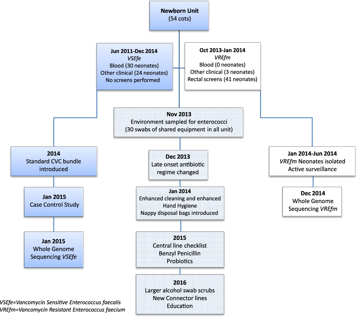
Figure 1 describes both VREfm and VSEfe overlapping outbreaks and the development of multimodal interventions that were applied to reduce enterococcal colonization and infection in the neonatal unit.
Multimodal interventions used during both enterococcal outbreaks are shown in the center of the flowchart. Vancomycin-sensitive Enterococcus faecalis and vancomycin-resistant Enterococcus faecium–specific interventions are shown on either side.

No VSEfe were isolated from the 40 environmental samples collected from shared unit equipment in November 2014, whereas 3 VREfm samples proved to be positive from the same collection.Reference Lister, Kotsanas and Ballard7 No epidemiological pattern was observed for bed or bay location as a possible point source in the unit nor rostered staff associated for caring of case neonates for the VSEfe outbreak described here.
Case control study
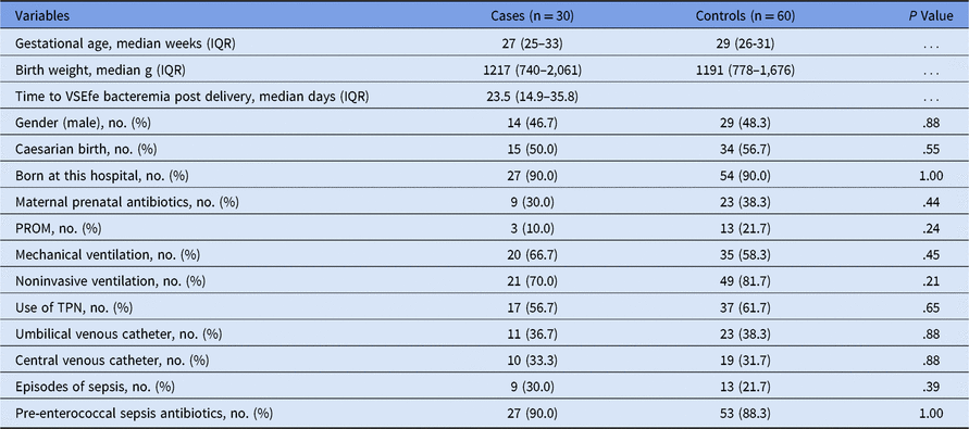
The cases had a gestational median age of 27 weeks (interquartile range [IQR], 25–33) and a median birth weight of 1,217 g (IQR range, 740–2,061). The time taken to detect VSEfe bacteremia was a median of 23.5 days (IQR, 14.9–35.8). Table 1 describes the basic demographics and assessed risk factors of cases and matched controls.
Demographics and Assessed Risk Factors of Vancomycin-Sensitive Enterococcus faecalis Bacteremia Cases and Matched Controls
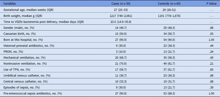
Note. IQR, interquartile range; PROM, prolonged rupture of membranes >18 h; TPN, total parenteral nutrition.
No difference was detected for gender, caesarean birth, born at this hospital, ventilation, parental nutrition, presence of line, or antibiotic use between cases and controls. However, VSEfe bacteremia was significantly associated with infection from an intra-abdominal source (confirmed necrotizing enterocolitis, gastroschisis, or bowel surgery) for 7 cases (23.3%) compared to 5 controls (8.3%; P = .03).
Molecular typing
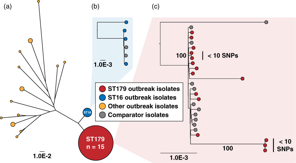
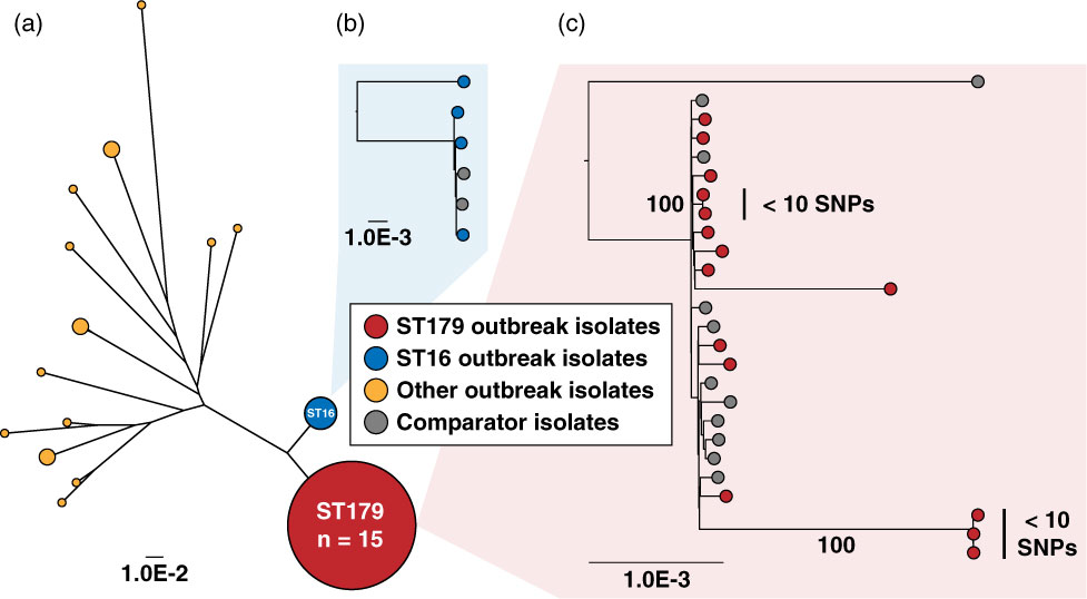
Of our 30 outbreak isolates and extra 5 maternity ward isolates from our hospital that underwent WGS, 15 of 35 (43%) were ST179 by in silico MLST. An additional 4 isolates (11%) were ST16, differing from the ST179 isolates by a single-locus allele (xpt gene). Also, 2 isolates each of ST30, ST34, and ST40 were identified, and the remainder of the isolates were unique sequence types within the study.
Among the ST179 and ST16 isolates, phylogenetic comparison with similar neonatal VSEfe from the AGAR national collection showed that most were unlikely to represent direct cross transmission in the outbreak. We identified 2 small clusters of ST179 outbreak isolates with <10 single-nucleotide polymorphisms (SNPs). Most likely, ST179 is a main circulating Australian clone because 11 of 13 comparator isolates (85%) were represented by this clone. Figure 2 illustrates the phylogenetic comparisons of all the VSEfe isolates implicated in the outbreak (Fig. 2A). The ST179 outbreak isolates can be seen in context with the 11 comparator isolates from the national collection in Fig. 2C, whereas Fig. 2B shows the relationship to ST16.
Phylogenetic comparisons of vancomycin-sensitive Enterococcus faecalis isolates implicated in the outbreak. (A) Comparison of all sequenced outbreak isolates. Size of the nodes indicates relative number of isolates. (B) ST16 outbreak isolates in context with comparator (n = 2) isolates from a national collection. (C) ST179 outbreak isolates in context with comparator (n = 11) isolates from a national collection. Note that 2 small clusters (<10 SNPs in core genome separating isolates) are highlighted, each comprising a small number of outbreak isolates. Bootstrap values from 1,000 replicates supporting these clusters are shown along the corresponding branches.
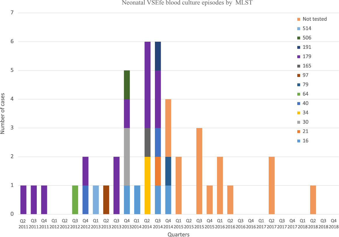
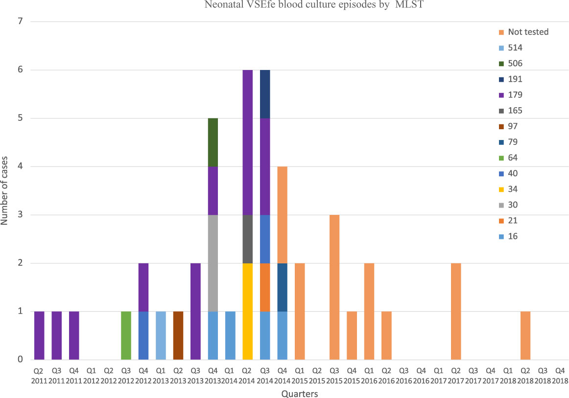
No dominant sequence type was observed in any quarter period to indicate a clonal outbreak, and most STs appeared randomly distributed (Fig. 3). A clear decrease in cases was observed by the beginning of 2015 as many of the multimodal interventions were implemented. The epidemic curve shows only 3 cases of VSEfe over 2017 and 2018. There was no difference in ST179 for those born by normal vaginal delivery (4 of 12, 33.3%) compared to those by caesarean section (5 of 15, 33.3%).
Vancomycin-sensitive Enterococcus faecalis bacteremia isolates, June 2011 through December 2018. Multilocus sequence type results for the 30 outbreak isolates tested from the Monash Newborn Unit, 2011–2014.
Line infection rate
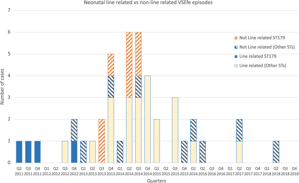
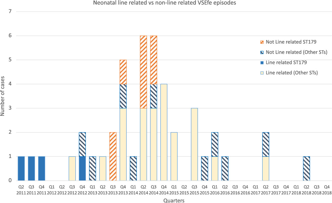
A decrease in the number of total line-related VSEfe from 12 in 2014, 5 in 2015, and 1 each for 2016 and 2017 can be seen in Fig. 4. Episodes due to the dominant ST179 represented 8/12 (67%) of non–line-related cases. This significant reduction in CVC-related infections was reported by the unit as 13.8 to 7.8 per 1,000 central-line days after quality improvement initiatives. The initiative bundle included the 3 checklists and was supported by education and feedback. All medical and nursing staff were educated on the checklists with compliance of 90% for insertion and 75% for daily maintenance by June 2016.Reference Taylor, McDonald and Earnest9 The 3 line-related infections detected in the third quarter of 2015 coincided with a loss of diaper bag supply and purchasing delays.
Vancomycin-sensitive Enterococcus faecalis isolates from line-related and non–line-related cases from the Monash Newborn Unit June 2011 through December 2018. ST179 isolates are displayed from the outbreak period, 2011–2014.
Bacteremia microbial review
A retrospective microbiology review was undertaken of all neonatal bacteremia in our unit to ensure that no other increase in bacteremia was seen with any other microorganism. This review showed no line-related infections due to Enterobacterales (eg, E. coli, Serratia spp, Klebsiella spp, Enterobacter spp, or Citrobacter spp) since the second quarter of 2017. For Staphylococcus spp (S. aureus, S. epidermidis, and S. capitis), line-related infections consistently ranged from 1 to 3 cases per quarter up to December 2018.
Discussion
We identified a VSEfe bacteremia outbreak that was resolved by a multifaceted intervention. The components included changes to antibiotic regimen to cover for late onset enterococcal sepsis, the introduction of diaper disposal bags to prevent environmental contamination, the introduction of new lines with CVC checklist bundles as well as probiotics for low-birth-weight premature neonates.
The neonatal microbiome is initially colonized during delivery by maternal vaginal and fecal bacteria followed by skin flora from close contact and breast feeding. Neonates are frequently exposed to and often colonized with healthcare-associated pathogens in the NICU, either by contact with the inanimate environment or by contaminated HCW hands. Low birth weight, prematurity, use of invasive devices and broad-spectrum antibiotics are contributing factors to the onset of infection and spread of outbreak events.Reference Gritz and Bhandari10–Reference Younge, Araujo-Perez, Brandon and Seed12
Enterococci can survive on inanimate surfaces for long periods of time and can tolerate a wide range of temperatures. A recent paper by Brooks et alReference Brooks, Olm and Firek13 describes how rooms can be reservoirs for microbiome colonization of hospitalized neonates and how neonates in the NICU can often be colonized by the same strains. In this study, E. faecalis was the most commonly detected species found in the neonatal feces samples as well as the room environment, which led us to conclude that colonization is part of the cycle of microbial exchange between the two.Reference Brooks, Olm and Firek13
In the NICU setting, the room environment may be representative of the interior space of the neonatal bed.Reference Babu, Tandur, Sharma and Murki14 Staff change diapers inside the confined space of the bed, and soiled diapers are often pushed to the bottom or placed on top of the bed before disposal. This procedure has the potential to directly contaminate the internal and external immediate bed environment, leading to colonization of the neonate and surrounding area with fecal microbes. Contamination of intravenous catheters from the colonized neonatal skin could then provide access for microbes to the blood system, especially if line procedures are not carefully undertaken.Reference Babu, Tandur, Sharma and Murki14
VSEfe outbreaks are difficult to recognize because there is no resistance marker, such as vancomycin, to alert infection control. Outbreaks may be detected when there is a clustering of cases from sterile sites such as blood cultures. A search of the literature references revealed only 2 outbreak articles with VSEfe in a neonatal setting, and both outbreaks were discovered from blood cultures.Reference Softic, Tahirovic, Di Ciommo and Auriti2, Reference Strabelli, Cais and Zeigler3 In total, 4 neonates were identified with bacteremia in 2004 during a 1-month active surveillance in a 10-bed NICU cardiology service in Brazil.Reference Strabelli, Cais and Zeigler3 All neonates had a CVC that breached the skin barrier, which was believed to be the portal of entry of VSEfe from the skin environment, especially because episodes of Rotavirus diarrhea had been reported in the NICU. A combination of infection control measures were implemented to reinforce measures to prevent further infections: enhanced environmental cleaning, unit layout modifications, neonatal bathing with chlorhexidine-gluconate soap, and increased nursing ratios.Reference Strabelli, Cais and Zeigler3
The second article was set in an 18-bed NICU in Bosnia and Herzegovina. This was a prospective observational study of neonatal sepsis over a 12-month period in 2012–2013 and involved 200 neonatal admissions. Culture-positive sepsis occurred in 43.4% of neonates, with Klebsiella pneumoniae (n = 10) and VSEfe (n = 10) predominating. In this setting, the high incidence was thought to be attributable to overcrowding, inappropriate use of antibiotics, and low HCW staffing. The risks included the presence of CVCs and surgical intervention.Reference Softic, Tahirovic, Di Ciommo and Auriti2 No molecular typing was performed in either outbreak.
In Victoria, sepsis is monitored in 4 high-level-care neonatal units by VICNISS Coordinating center (www.vicniss.org.au) for line-related infections since 2002. Recently, Worth et alReference Worth, Daley, Spelman, Bull, Brett and Richards15 reported 203 CVC events from 2008 to 2016 with vancomycin-sensitive Enterococcus spp contributing to 26 cases (9.1%). Overall CVC rates showed a significant reduction over this period from as high as 5 per 1,000 CVC days in 2009 to 2 per 1,000 CVC days in 2016, mostly due to prevention programs such as insertion practices and line management.Reference Worth, Daley, Spelman, Bull, Brett and Richards15 In a recent study in Taiwan, 3 catheter-related bundles were introduced in a newly built NICU when the unit was moved to a new facility on the same floor. Surveillance of the old unit in the previous 2 years, compared with 2 years in the new unit, showed a reduction in bloodstream infection from 8.3% to 3.7% (P = .03) attributed to the new central-line bundle. The elements of this bundle included aseptic techniques for insertion, changing tubing, maintaining a closed system, as well as daily evaluation of the catheter site.Reference Chen, Lin and Rehn16
In 2014, our NICU introduced checklists for insertion, daily maintenance, and procedural access for CVC lines. Over 24 months, we observed a 41% decrease in CVC infection rates. This quality improvement was supported with monthly education sessions and feedback using run charts with the date of last infection clearly displayed.Reference Taylor, McDonald and Earnest9 Other changes to staffing availability and times of CVC insertion and changes were implemented to improve adherence. These interventions correlate with the absence of line infections attributed to VSEfe since the second quarter of 2017.
Molecular tools such as WGS are advancing at a rapid speed and provide robust and high-resolution methods that are superior to MLST for epidemiological typing of outbreak microorganisms.Reference Johnson and Quach17 To be useful for intervention, WGS data need to be assembled, analyzed, and interpreted in a timely fashion during an outbreak, which was possible in our study.Reference Kwong, McCallum, Sintchenko and Howden18 Comparison of our VSEfe isolates using WGS assisted in determining that this was a polyclonal outbreak with ST179 comprising 43% of cases. In addition, WGS showed that 85% of the comparator neonatal bacteremia isolates were also predominantly ST179, indicating that this is the main circulating clone in Australia.
Our outbreak investigations had several limitations. Environmental samples were not collected from the interior of the beds to establish fecal contamination, nor were samples collected from the neonates’ mothers to determine whether VSEfe was maternally acquired and therefore part of an endogenous infection. VSEfe bacteremia was significantly associated with cases having gastrointestinal complications such as NEC, gastroschisis, or bowel surgery. This finding is consistent with the 2018 neonIN data by Wang et al.Reference Wang, Kortsalioudaki and Heath4 Although a retrospective analysis cannot assess causation, it is biologically plausible that enterococci may translocate from the gut with any infection or abdominal surgery. Furthermore, no WGS was undertaken for the 24 clinical nonbacteremic isolates nor from the remaining 14 VSEfe bacteremia samples to 2018.
We also observed considerable overlap of multimodal interventions: both enterococcal outbreaks (VREfm and VSEfe) had been concurrently detected between 2013 and 2014 (Fig. 1). Environmental cleaning, hand hygiene, diaper bags, antibiotic regimen changes, and probiotics would apply as interventions to both outbreaks. Initiatives around line maintenance and improved access would have benefitted the VSEfe bacteremia-specific outbreak.
In this study, WGS evidence directed investigators to focus on unit procedures rather than search only for a single point-source outbreak. The multimodal intervention with the primary focus on the 3 CVC checklists, which commenced in 2015, was a valuable element of the infection prevention effort. The possible contamination of HCW gloved hands from the bed environment as well as the surface colonization of neonates may have increased the risk of direct inoculation of VSEfe through the skin barrier during insertion of lines. Although we were unable to decipher which factors made the greatest contribution, the simple intervention of removal of soiled diapers directly into plastic waste bags inside the bed may have prevented environmental contamination and assisted in successful ending of the outbreak.Reference Babu, Tandur, Sharma and Murki14
Acknowledgments
We thank the microbiology laboratory for carrying out cultures and sensitivity testing.
Financial support
No financial support was provided relevant to this article.
Conflicts of interest
All authors report no conflict of interest relevant to this article.
Supplementary material
To view supplementary material for this article, please visit https://doi.org/10.1017/ice.2019.202