Introduction
Trichinellosis is a zoonotic disease in humans caused by multiple species of the Trichinella genus, with Trichinella spiralis being one of the most prevalent and pathogenic species. The primary transmission mode is consuming undercooked pork products containing infective larvae (Dyab et al., Reference Dyab, Ahmed and Abdelazeem2019; Abuelenain et al., Reference Abuelenain, Fahmy, Elshennawy, Selim, Elhakeem, Hassanein and Awad2022). Trichinella spiralis infection in humans occurs in two stages: an intestinal phase (enteral infection); and a muscle phase (parenteral infection) (Elgendy et al., Reference Elgendy, Othman, Saad, Soliman and Mwafy2020). During the initial stage of infection, the disease may manifest itself with symptoms such as diarrhoea, vomiting and abdominal pain. In contrast, the muscle phase, which begins one week after infection, can lead to facial swelling, rash, fever and muscle pain due to the migration of larvae to muscles (Gottstein et al., Reference Gottstein, Pozio and Nöckler2009).
Anthelmintic medications, particularly benzimidazole derivatives such as mebendazole and albendazole (ABZ), are used to treat human trichinellosis. However, these medications are ineffective at eliminating encysted larvae and some exhibit low absorption and high resistance levels, limiting their use in pregnant women and children under three years old (Yadav, Reference Yadav2012; Attia et al., Reference Attia, Mahmoud, Farrag, Makboul, Mohamed and Ibraheim2015). These limitations underscore the need for novel, effective and safe medications for trichinellosis treatment. Medicinal plants have emerged as a promising alternative, as they are less expensive, less toxic and do not have the adverse effects associated with synthetic medications.
Kaempferol (KPF), a dietary flavonoid found in various fruits and vegetables, such as apples, tea, strawberries, broccoli and beans, is known for several activities, including antioxidant, anti-inflammatory, anti-microbial, anti-parasitic and anti-cancer effects (Devi et al., Reference Devi, Malar, Nabavi, Sureda, Xiao, Nabavi and Daglia2015). Furthermore, KPF possesses hepatoprotective and immunomodulatory properties (Wang et al., Reference Wang, Sun, Jiang, Xie and Zhang2015). Various studies have documented KPF's anti-parasitic effect, particularly in protozoa such as Entamoeba histolytica, Giardia duodenalis, Leishmania spp. and malaria parasites (Pérez-González et al., Reference Pérez-González, Gutiérrez-Rebolledo, Yépez-Mulia, Rojas Tom, Luna-Herrera and Jiménez-Arellanes2017; Somsak et al., Reference Somsak, Damkaew and Onrak2018; Argüello-García et al., Reference Argüello-García, Calzada, García-Hernández, Chávez-Munguía and Velázquez-Domínguez2020; Abdeyazdan et al., Reference Abdeyazdan, Mohajeri, Saberi, Mirzaei, Ayatollahi, Saghaei and Ghanadian2022). Furthermore, KPF's anti-helminthic activity has been demonstrated against schistosomiasis (Zhou et al., Reference Zhou, Zhang, Cheng, Li, Tang, Xue and Zhao2013).
Given the evidence supporting the potential medical benefits of KPF, this study aimed to assess the efficacy of KPF compared to ABZ in treating T. spiralis infection in mice.
Materials and methods
Animals
The present study was conducted on 72 laboratory-bred Swiss albino male mice weighing 25 ± 5 g and aged 5–6 weeks. The pathogen-free mice were obtained from the Theodor Bilharz Research Institute (TBRI) biology division in Giza, Egypt. The animals were provided with a standard pellet diet and water ad libitum.
Parasites
The strain of T. spiralis used in this work was provided by the Parasitology Department, TBRI (El-Wakil et al., Reference El-Wakil, Shaker, Aboushousha, Abdel-Hameed and Osman2023).
Experimental grouping
The mice were divided into six groups of 12 mice each:
Group I: uninfected and not treated (the healthy control).
Group II: infected and untreated (the positive control).
Group III: KPF was administered orally at a 20 mg/kg dose for seven days before infection (as prophylaxis) (Zhou et al., Reference Zhou, Zhang, Cheng, Li, Tang, Xue and Zhao2013).
Group IV: infected and then treated with KPF (AK Scientific, Inc., Union City, USA) administered orally at a dose of 20 mg/kg (Zhou et al., Reference Zhou, Zhang, Cheng, Li, Tang, Xue and Zhao2013).
Group V: infected and then treated with ABZ (Sigma-Aldrich, St Louis, MO, USA) administered orally at a dose of 50 mg/kg (Attia et al., Reference Attia, Mahmoud, Farrag, Makboul, Mohamed and Ibraheim2015).
Group VI: infected and then received the combination of KPF and ABZ.
Groups II–VI were subdivided into A and B subgroups, each with six mice. Subgroup (A) for the intestinal phase started the treatment one day post-infection (PI) and was sacrificed on day seven PI, while subgroup (B) for the muscular phase started the treatment 30 days PI and was sacrificed on day 37 PI.
Dose of infection
Each mouse was inoculated orally with 250–300 larvae (Ozkoc et al., Reference Ozkoc, Tuncay, Delibas and Akisu2009; Abou Rayia et al., Reference Abou Rayia, Saad, Ashour and Oreiby2017).
Parasitological examination
In subgroup (A), adult T. spiralis worms were isolated from the small intestine of infected mice seven days after infection. The small intestine was cleaned, opened longitudinally along its entire length, cut into 2 cm pieces and then placed in normal saline at 37°C for 3 to 4 h to allow the worms to move out of the tissue (Wakelin & Margaret, Reference Wakelin and Margaret1980).
In subgroup (B), the muscle larvae were recovered using the pepsin digestion method (Dunn & Wright, Reference Dunn and Wright1985). A McMaster counting chamber was used to count the recovered larvae microscopically. The parasite burden was the number of larvae per gram of digested carcass (ML/g) (Nuñez et al., Reference Nuñez, Gentile, Costantino, Sarchi and Venturiello2005; El-Wakil et al., Reference El-Wakil, Abdelmaksoud, AbouShousha and Ghallab2021).
Histopathological studies
Tissue samples from the small intestines and biopsies from the skeletal muscle of the examined groups were fixed in 10% neutral buffered formalin for 24 h, dehydrated in ascending grades of alcohol, cleared using xylene and embedded in paraffin blocks. A microtome was used to cut hard paraffin sections of 4 μm thickness.
The haematoxylin and eosin (H&E) staining method was used to stain the small intestine and skeletal muscle samples (Drury & Wallington, Reference Drury and Wallington1980). Additionally, picrosirius red was used to stain the skeletal muscle samples to evaluate capsular thickness (Rtail et al., Reference Rtail, Maksymova, Illiashenko, Gortynska, Korenkov, Moskalenko and Tkach2020).
The scoring of intestinal changes was evaluated based on intestinal inflammatory infiltrate (minimal, mild, moderate, or marked) and villous changes with broadening, fusion and blunting (normal, mild, moderate, or marked).
The scoring of muscle changes was evaluated based on the number of viable encysted larvae per low-power field × 100 (+1 ≤ one larva; +2 = 2–4 larvae; +3 = 5–7 larvae; +4 > 7 larvae), the thickness of the cyst capsule (thick, thin and disrupted) and the pericapsular inflammatory infiltrate (minimal, mild and moderate).
Immunohistochemical studies
The deparaffinized tissue sections of the intestine were treated with 0.3% hydrogen peroxide for 20 min, followed by overnight incubation with anti-NOD-like receptor-pyrin domain containing 3 (NLRP3) at 4°C. Subsequently, and samples were rinsed with phosphate buffered saline (PBS) and incubated with the secondary antibody (HRP Envision kit) for 20 min. After washing with PBS, samples were incubated with diaminobenzidine for 15 min and then washed again with PBS. Haematoxylin counterstaining, dehydration and clearing in xylene were performed before examining the tissue sections microscopically. Six non-overlapping fields of intestinal tissue sections were randomly selected and scanned. The relative area percentages of NLRP3 immunohistochemical expression levels were determined in immune-stained tissue sections. The micrographs and data were obtained using a full high-definition microscopic camera and the Leica application module for tissue section analysis (Leica Microsystems GmbH, Wetzlar, Germany) (Gad et al., Reference Gad, Mansour, Abbas, Malatani, Khattab and Elmowafy2022).
Statistical analysis
Data were analysed using the statistical package for the social sciences version 26 (IBM Corp., Armonk, NY, USA) and Microsoft Excel 2016. Normally distributed continuous variables were presented as mean ± standard deviation (SD). Student's t-test was used to compare the means of normally distributed variables between groups. Analysis of variation followed by the Tukey honestly significant difference test as a post hoc test in the multigroup analysis was used for comparison. A P-value < 0.05 was considered statistically significant and a P-value < 0.001 was considered highly statistically significant.
Results
The impact of different treatment regimens on adult worm counts in the small intestine
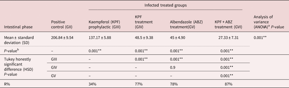
Prophylactic treatment with KPF (GIII) significantly decreased (P < 0.001) the mean count of adult worms (137.17 ± 5.88) with 34% efficacy compared to the untreated infected group (GII) (206.84 ± 9.54). A significant reduction in the mean adult worm count was identified in all treated groups (P < 0.001) when compared with the untreated infected group (GII). The lowest mean count of adult worms (27.33 ± 7.31) was found in GVI, which received both KPF and ABZ therapy, with a drug efficacy of 87%. In GV, which received ABZ, the mean adult worm count was (45 ± 4.90), with 78% drug efficacy, while the mean adult worm count was 48.5 ± 9.38 in GIV, which received KPF, with 77% reduction, with no statistically significant difference between the two groups (P = 0.9) (table 1).
Trichinella spiralis adult worm count in the small intestine.

The T. spiralis adult worm count is displayed as mean ± SD, while the percentage of reduction (R) is shown as a percentage depending on the following equation: R% = [(mean count in the infected, untreated control group − mean count in the experimental group)/mean count in the infected, untreated control group] × 100.
a P-value is significantly different when comparing groups depending on the one-way ANOVA.
b P-value significantly differs from the control group depending on Student's t-test.
cP-value significantly differs when comparing groups based on a post hoc test (Tukey HSD).
*Initial P-value < 0.05 is significant.
** Initial P-value < 0.01 is highly significant.
The impact of different treatment regimens on larvae counts in the muscles
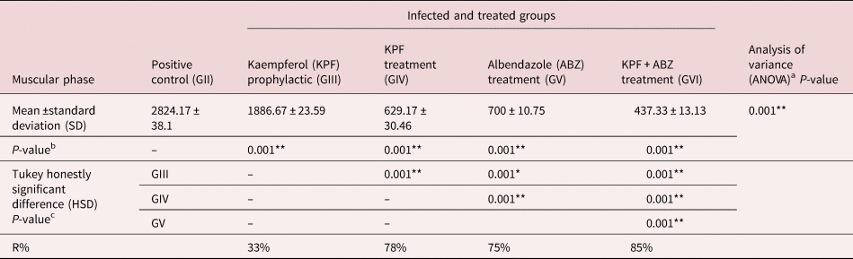
Regarding the medication's effects on the muscular phase, prophylactic administration of KPF to infected mice significantly decreased (P < 0.001) the mean larvae count per gram of muscle (1886.67 ± 23.59) with 33% efficacy compared to the positive control group (2824.17 ± 38.1). Compared to the positive control group, a significant decline in the mean larvae count per gram of muscle was found in all treatment groups (P < 0.001). The lowest mean count of larvae was detected in the mice group that received KPF and ABZ treatment (437.33 ± 13.13) with 85% efficacy, followed by the group that received KPF treatment (629.17 ± 30.46) with 78% efficacy. In the group that received ABZ treatment, the mean count of larvae was 700 ± 10.75, with a 75% reduction percentage (table 2).
Trichinella spiralis larvae count in muscles.

The T. spiralis larvae count per gram of muscles is displayed as the mean ± SD, while the reduction (R) is shown as a percentage depending on the following equation: R% = [(mean count in the infected, untreated control group − mean count in the experimental group)/mean count in the infected, untreated control group] × 100.
a P-value is significantly different when comparing groups depending on the one-way ANOVA.
b P-value significantly differs from the control group based on Student's t-test.
c P-value significantly differs when comparing groups depending on a post hoc test (Tukey HSD).
* Initial P-value < 0.05 is significant.
** Initial P-value < 0.01 is highly significant.
Histopathological intestinal changes
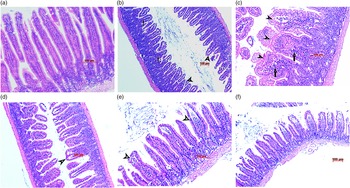
Small intestinal sections from the healthy control group (GI) were histopathologically examined after H&E staining, demonstrating typical architecture with an average crypt/villous ratio and healthy mucosa (fig. 1a). In contrast, the infected control group (GII) exhibited moderate to marked inflammatory infiltration with plasma cells, lymphocytes and macrophages, with scattered mast cells and eosinophils, as well as villous blunting, broadening and fusion (fig. 1b).
Histological photomicrographs of sections of the small intestine stained with haematoxylin and eosin. (a) Group I showed normal intestinal villous architecture (magnification: ×200); (b) Group II revealed intestinal villous shortening and broadening (short arrows) with a moderate inflammatory infiltrate (long arrow) (magnification: ×100); (c) Group III exhibited intestinal villous broadening and fusion (short arrows) with a moderate inflammatory infiltrate (long arrow) (magnification: ×200); (d) Group IV revealed focal mild shortening and broadening of intestinal villi (short arrows) with a mild inflammatory infiltrate (magnification: ×100); (e) Group V showed mild focal intestinal villous broadening (short arrows) with a mild inflammatory infiltrate (long arrow) (magnification: ×200); (f) Group VI exhibited almost normal intestinal villous architecture with minimal inflammatory infiltrate (magnification: ×100).

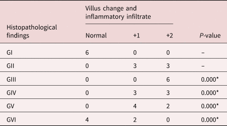
Examination of sections from various treated groups (GIII, GIV, GV and GVI) revealed a prominent decrease in intestinal inflammation and decreased affection for intestinal villi (fig. 1c–f). The most significant results were shown in group GVI (fig. 1f), demonstrating a significant statistical difference compared to the control group GII (P < 0.05) (table 3).
Histopathological grading in small intestinal haematoxylin and eosin-stained sections.

* Highly significant.
Normal: indicates normal villi architecture with villous width one-quarter of its length, lining revealing small basal regular nuclei and lamina displaying minimal lymphoplasmacytic infiltrate.
+1: indicates a mild increase in the width of villi with mild shortening, focal fusion, lining showing small regular basal nuclei and lamina revealing a mild lymphoplasmacytic infiltrate, mild oedema and few eosinophils.
+2: indicates a moderate increase in the villi width with moderate shortening, focal fusion, lining showing reactive nuclei and lamina, revealing a moderate lymphoplasmacytic infiltrate, a moderate eosinophils number and mild to moderate oedema.
The expression of NLRP3 in intestinal tissue of different groups
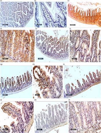
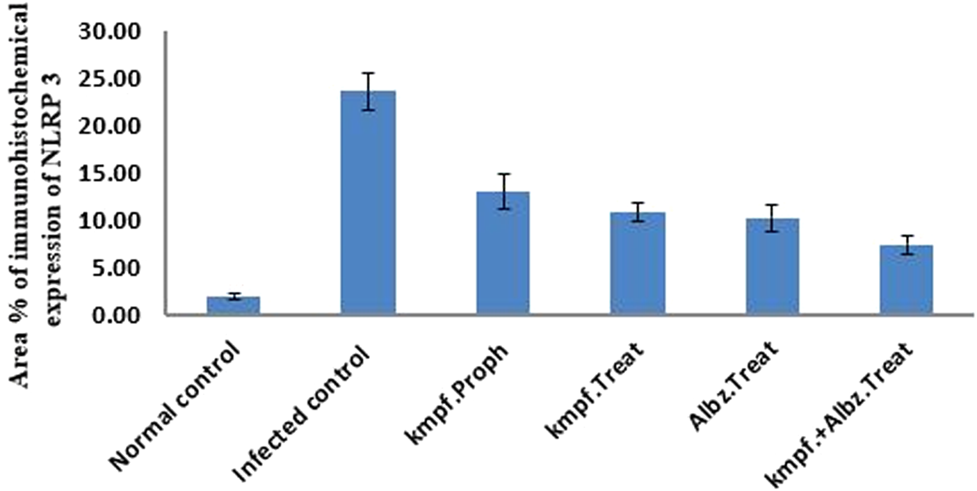
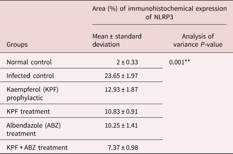
Quantitative analysis of sections from the small intestine demonstrated a considerable increase in NLRP3 levels in group II (infected, non-treated) (fig. 2c, d) relative to the group I (healthy control) samples (fig. 2a, b). In contrast, treated groups (GIII, GIV, GV and GVI) showed significantly lower levels of NLRP3 expression compared to group II (fig. 2e–l). The maximum decrease was found in group VI, which was administered KPF and ABZ (fig. 2k, l) (table 4 and fig. 3).
Immunohistochemical expression of NOD-like receptor-pyrin domain containing 3 (NLRP3) in the intestine of mice. (a, b) Group I (the healthy control) showed low expression of NLRP3; (c, d) Group II demonstrated an increase in NLRP3 levels; (e–l) Groups III, IV, V and VI showed reductions in NLRP3 expression.

The bar chart presents the mean ± standard deviation of the percentage area of immunohistochemical expression levels of NOD-like receptor-pyrin domain containing 3 in intestinal tissue.

The percentage area of NOD-like receptor-pyrin domain containing 3 (NLRP3) immunohistochemical expression in intestinal tissues in various groups.

** Initial P-value < 0.01 is highly significant.
Skeletal muscle histopathological changes examination
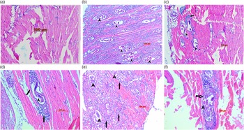
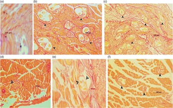
Sections of muscles from the healthy control group (GI) revealed a normal pattern of skeletal muscle upon histopathological investigation (figs 4a and 5a), while the infected control group (GII) showed several T. spiralis encysted larvae scattered throughout the sarcoplasm of muscle and cells of chronic inflammation, such as plasma cells, lymphocytes and histiocytes, encircling the encysted larvae and invading muscle bundles (fig. 4b), with continuous thick intact fibrotic capsules (fig. 5b).
Histological photomicrographs of muscle sections stained with haematoxylin and eosin. (a) Group I showed a normal pattern of skeletal muscle (magnification: ×200); (b) Group II had many viable larvae (short arrows) and mild-to-moderate inflammation (magnification: ×100); (c) Group III exhibited many viable larvae (short arrows) and mild inflammation (long arrow) (magnification: ×100); (d) Group IV demonstrated few larvae (short arrow) and mild inflammation (long arrows) (magnification: ×200); (e) Group V showed few degenerating larvae (short arrows) and moderate inflammation (long arrows) (magnification: ×200); (f) Group VI revealed scattered degenerated larvae (short arrow) and moderate inflammation (long arrow) (magnification: ×200).

Histological photomicrographs of muscle sections stained with picrosirius red. (a) Group I showed a normal pattern of skeletal muscle (magnification: ×200); (b) Group II demonstrated a continuous, thick, intact fibrotic capsule (arrows) (magnification: ×200); (c) Group III exhibited a continuous thick intact fibrotic capsule (arrows) (magnification: ×100); (d) Group IV had a thin capsule with an empty lumen replaced by fibrosis (arrows) (magnification: ×200); (e) Group V showed a thin, focally disrupted capsule (arrows) (magnification: ×200); (f) Group VI revealed marked capsular disruption with focal and near-total replacement by fibrous tissue (arrows) (magnification: ×200).

In the treated groups (GIII, GIV, GV and GVI), there were larval degeneration and death, thinned capsular tissue with focal disruption and varying intensities of pericapsular infiltrate consisting of eosinophils, histiocytes, mast cells, plasma cells and lymphocytes in different proportions (fig. 4c–f). Moreover, capsular disruption was observed in the picrosirius red stain (fig. 5c–f). Additionally, these changes were shown in GVI treated with the combination of KPF and ABZ (figs 4f and 5f), with a significant statistical difference compared to the positive control group (GII) (table 5).
Semiquantitative scoring of muscle tissue histopathological changes according to various treatment regimens.

* Highly significant.
.
Discussion
The treatment of various diseases, including parasitic infections, using medicinal plants has recently received considerable attention. Medicinal plants are well-tolerated with minimal adverse effects compared to synthetic drugs, making them a promising alternative (Elizondo-Luévano et al., Reference Elizondo-Luévano, Castro-Ríos, López-Abán, Gorgojo-Galindo, Fernández-Soto, Vicente, Muro and Chávez-Montes2021).
The preferred treatment for trichinellosis is ABZ, a benzimidazole. However, ABZ has been linked to several systemic adverse effects, including fatal encephalitis, epilepsy, severe drug eruptions and even death (Shalaby et al., Reference Shalaby, Moghazy, Shalaby and Nasr2010; Yadav, Reference Yadav2012).
Kaempferol, a dietary flavonoid, is an active compound with several activities, including antioxidant, anti-inflammatory, anti-microbial, anti-parasitic and anti-cancer properties (Devi et al., Reference Devi, Malar, Nabavi, Sureda, Xiao, Nabavi and Daglia2015). Moreover, KPF has a wide safety range. The chronic toxicity of KPF was assessed by administering 2000 mg/kg of KPF orally daily for 30 days. There were no visible signs of toxicity, nephrotoxicity, hepatotoxicity, haematotoxicity, or death in the mice (Somsak et al., Reference Somsak, Damkaew and Onrak2018).
Therefore, the present study aimed to evaluate the effect of KPF on different stages of T. spiralis and compare its efficacy against ABZ, the standard trichinellosis treatment. The number of adult worms recovered from the intestinal phase and the larvae recovered from the muscular phase were counted. Additionally, histopathological investigations were used to monitor treatment effectiveness.
Concerning the intestinal phase, the reduction percentage was 34% in the group prophylactically treated with KPF. Both ABZ and KPF monotherapy reduced the count of adult worms with 78% and 77% efficacy, respectively, while combined therapy with both ABZ and KPF induced the best response (87%).
Regarding the muscular phase, the best reduction in the mean count of larvae per gram of muscle was detected in the group that received a combination of KPF and ABZ treatment (85%), followed by the group treated with KPF (78%) and the group treated with ABZ (75%). In the KPF prophylactic group, the reduction percentage was 33%.
In accordance with the present study, several prior studies have investigated prophylactic treatments against T. spiralis (Abu El Ezz, Reference Abu El Ezz2005; Nada et al., Reference Nada, Mohammad, Moad, El-Shafey, Al-Ghandour and Ibrahim2018; El-Wakil et al., Reference El-Wakil, Abdelmaksoud, AbouShousha and Ghallab2021).
Numerous previous investigations have demonstrated the parasiticidal action of ABZ on T. spiralis intestinal worms, although the level of efficacy varied (Siriyasatien et al., Reference Siriyasatien, Yingyourd and Nuchprayoon2003; Huang et al., Reference Huang, Yao, Liu, Yang, Wang, Shi and Yang2020; El-Wakil et al., Reference El-Wakil, Abdelmaksoud, AbouShousha and Ghallab2021), which could be attributed to differences in dosage, duration and timing of treatment (Siriyasatien et al., Reference Siriyasatien, Yingyourd and Nuchprayoon2003). ABZ's parasiticidal effect is due to interference with the parasite's glucose metabolism and selective inhibition of microtubule assembly and polymerization (Vadlamudi et al., Reference Vadlamudi, Reddy and Raju2015).
The effect of KPF on the developmental stages of T. spiralis has not been previously investigated, although the parasiticidal effect of KPF has been demonstrated in other parasitic infections. KPF has been shown to possess a broad range of pharmacological and biological activities against Entamoeba histolytica, Giardia duodenalis, Leishmania and malaria parasites (Pérez-González et al., Reference Pérez-González, Gutiérrez-Rebolledo, Yépez-Mulia, Rojas Tom, Luna-Herrera and Jiménez-Arellanes2017; Somsak et al., Reference Somsak, Damkaew and Onrak2018; Argüello-García et al., Reference Argüello-García, Calzada, García-Hernández, Chávez-Munguía and Velázquez-Domínguez2020; Abdeyazdan et al., Reference Abdeyazdan, Mohajeri, Saberi, Mirzaei, Ayatollahi, Saghaei and Ghanadian2022). Moreover, KPF has demonstrated anthelmintic activity against schistosomiasis (Zhou et al., Reference Zhou, Zhang, Cheng, Li, Tang, Xue and Zhao2013).
It is postulated that KPF could inhibit oxidative stress, lipid peroxidation, reactive oxygen species formation, glycogen synthase kinase-3β and induce apoptosis (Somsak et al., Reference Somsak, Damkaew and Onrak2018).
In the present study, histopathological analysis of the small intestine of T. spiralis-infected, untreated mice revealed the presence of inflammatory infiltrates with villi broadening. At the same time, in all treated groups, these changes were improved, particularly in the group treated with combined therapy with both ABZ and KPF.
Regarding the histopathological results of muscle tissue, numerous larvae surrounded by thick capsules and inflammatory cells were observed in the infected, non-treated mice. Other researchers reported similar findings (El-Wakil et al., Reference El-Wakil, Abdelmaksoud, AbouShousha and Ghallab2021). In the treated groups, the capsule was disrupted and thinned with pericapsular infiltration of inflammatory cells and these effects were more noticeable in the group that received both medications. Histological changes in mice treated with ABZ in the intestinal and muscular phases were previously reported (Attia et al., Reference Attia, Mahmoud, Farrag, Makboul, Mohamed and Ibraheim2015; Nassef et al., Reference Nassef, Moharm, Atia, Brakat, Abou Hussien and Shamseldeen2019; El-Wakil et al., Reference El-Wakil, Shaker, Aboushousha, Abdel-Hameed and Osman2023).
The NOD-like receptor (NLR) is crucial for the host's defence against nematodes. One NLR that has received significant attention is NLRP3, essential for various parasitic diseases (Jin et al., Reference Jin, Bai, Yang, Ding, Shi, Fu, Boireau, Liu and Liu2020). Studies on Toxoplasma gondii and Neospora caninum have demonstrated that the absence of NLRP3 increases the parasite load (Moreira-Souza et al., Reference Moreira-Souza, Almeida-da-Silva, Rangel, Rocha, Bellio, Zamboni, Vommaro and Coutinho-Silva2017; Wang et al., Reference Wang, Gong and Zhang2017), and activation of NLRP3 is necessary for resistance to several Leishmania species (Lima-Junior et al., Reference Lima-Junior, Costa and Carregaro2013).
The NLRP3 was found to be upregulated by the parasite protein in T. spiralis (Jin et al., Reference Jin, Bai, Yang, Ding, Shi, Fu, Boireau, Liu and Liu2020). Similarly, in the present study, the level of NLRP3 in the intestine was high in the control and infected groups. However, in all treated groups, levels of NLRP3 were significantly decreased. The NLRP3 levels were correlated with the adult worm burden detected in the present study, indicating that the downregulation of NLRP3 is likely related to adult worm clearance.
The level of NLRP3 in the intestinal tissue of infected mice that were given either ABZ or KPF decreased. The inhibitory effect of ABZ on NLRP3 was documented previously, and the suggested mechanism is via nuclear factor kappa B inhibition, which regulates the expression of NLRP3, caspase 1 and pro-interleukin-1β (pro-IL-1β) (Ullah et al., Reference Ullah, Al Kury, Althobaiti, Ali and Shah2022). For KPF, the inhibitory effect on NLRP3 is through inhibition of IL-1β production and apoptosis-associated speck-like protein containing caspase recruitment domain oligomerization (Lim et al., Reference Lim, Min, Park and Kim2018).
Therefore, there was a marked decrease in the NLRP3 level in the group treated with combined therapy with both ABZ and KPF.
Conclusions
The results of the present study provide evidence that KPF may be a natural and safe alternative to ABZ in treating T. spiralis infections, with comparable efficacy in both the intestinal and muscular phases. Furthermore, when combined with ABZ, KPF demonstrated a superior response to ABZ alone. Further research is recommended to elucidate the mechanism of KPF's effect on trichinellosis and its potential synergistic action with ABZ.
Further studies are also recommended to clarify the mechanism underlying KPF's impact on trichinellosis and its synergistic effect when combined with the reference drug, ABZ.
Key findings
• The use of albendazole (ABZ) alone for treating trichinellosis is inadequate.
• Kaempferol has the potential to be effective against both the intestinal and muscular phases of Trichinella. spiralis and could act synergistically when combined with other anti-trichinellosis medications, especially ABZ.
• The group treated with combined drugs showed significant improvements in intestinal and muscular inflammation and a reduction in the thickness of the capsule.
Financial support
This research did not receive any specific grants from funding agencies in the public, commercial, or not-for-profit sectors.
Competing interests
None.
Ethical standards
The present study was approved by the ethical committee of the Faculty of Medicine for Girls, Al-Azhar University, Egypt, and the protocol's serial number is (FMG-IRB 2022081433).