INTRODUCTION
The larval stage of the pork tapeworm Taenia solium is responsible for cysticercosis. Humans are accidentally infected with T. solium by ingestion of eggs excreted with the faeces of individuals harbouring the adult tapeworm in the intestinal tract. The larvae migrate throughout the body, invade skeletal muscle, subcutaneous tissue or the central nervous system (the latter being neurocysticercosis), and encyst to form cysticerci. This disease is one of the re-emerging zoonoses worldwide, and it is the major aetiological agent of epileptic seizures in endemic areas, making it a major public health problem in most developing countries (Mahanty and Garcia, Reference Mahanty and Garcia2010).
Diagnosis of neurocysticercosis has been achieved by clinical criteria, computer tomography (CT) and nuclear magnetic resonance imaging (MRI) (Ito and Craig, Reference Ito and Craig2003). The imaging techniques are useful for diagnosis but may overlook the infection when the number of parasites is low and/or the figures are not clear or are not typical. Moreover, these techniques are really not suitable for the diagnosis of cysticercosis in endemic areas because of the high cost. Therefore, the development of an immunodiagnostic test that detects specific antibodies in either sera or cerebrospinal fluid (CSF) is necessary because of its simplicity and reliability especially when examining serum. Efforts have been directed towards identification and characterization of specific antigens, and glycoproteins in the cyst fluid of T. solium metacestodes have been widely considered for serodiagnostic purposes. Parkhouse and Harrison (Reference Parkhouse and Harrison1987) and Tsang et al. (Reference Tsang, Brand and Boyer1989) have demonstrated that lentil-lectin affinity chromatography-purified glycoproteins that showed seven bands around 15–30 kDa were highly specific to cysticercosis on immunoblot. These glycoproteins have been shown not to be suitable for ELISA antigens because of cross-reactivity. We have developed a method to purify diagnostic antigens also known as low-molecular-weight antigens (LMWAgs) by preparative isoelectric focusing electrophoresis (IEFE) from cyst fluid available for both immunoblot and ELISA (Ito et al. Reference Ito, Plancarte, Ma, Kong, Flisser, Cho, Liu, Kamhawi, Lightowlers and Schantz1998). However, as this method requires sophisticated and expensive equipment it is difficult to apply in endemic areas. We have also reported an affinity-purification method using polyclonal and monoclonal antibodies (Sato et al. Reference Sato, Sako, Nakao, Yamasaki, Nakaya and Ito2006), but this method was not in general use because it needs a specific antibody. Recombinant proteins or synthetic peptides of them are also available for immunodiagnostic purposes (Chung et al. Reference Chung, Bahk, Huh, Kang, Kong and Cho1999; Greene et al. Reference Greene, Hancock, Wilkins and Tsang2000; Sako et al. Reference Sako, Nakao, Ikejima, Piao, Nakaya and Ito2000; Hancock et al. Reference Hancock, Khan, Williams, Yushak, Pattabhi, Noh and Tsang2003); this is, however, confined to a small number of laboratories. Therefore a simple and low-cost method to prepare immunodiagnostic antigens, especially ELISA antigens, must be established urgently, because it is a key point for epidemiological studies and serodiagnosis in endemic areas. In this study, we report a novel and reliable preparation of immunodiagnostic antigens, LMWAgs, from cyst fluids using a cation-exchange chromatography (CEC), and the evaluation of LMWAgs as ELISA antigens.
MATERIALS AND METHODS
Parasite materials
All cysts were collected from naturally infected pigs in China and Ecuador. Fluid from T. solium cysts was centrifuged at 10 000 g for 30 min at 4 °C, the supernatant was recovered and kept at −80 °C until use.
Preparation of LMWAgs from cyst fluids
Cyst fluid from T. solium larva was dialysed against 10 mm HEPES (4-(2-hydroxyethyl)piperazine-1-ethanesulfonic acid) buffer (pH 8·0) containing 0·5 mm EDTA. After adding CHAPS (3-[(3-cholamidopropyl)dimethylammonio]propanesulfonate) to the dialysate to a final concentration of 2%, it was directly loaded on a HiTrap SP XL cation-exchange column (GE healthcare, Japan) pre-equilibrated with a start buffer (10 mm HEPES buffer, pH 8·0, containing 0·5 mm EDTA) and proteins were eluted with a gradient between 0 and 1·0 m NaCl in the start buffer or with 1·0 m NaCl in the start buffer by using Fast Protein Liquid Chromatography system (ÄKTA FPLC system, GE Healthcare, Japan). Alternatively, proteins bound to cation-exchange resins were recovered by a stepwise elution with the start buffer containing 1·0 m NaCl manually. The elutant was boiled for 20 min to precipitate the contaminants, then the supernatant was collected and kept at −20 °C.
Serum samples
A total of 60 serum samples from patients with cysticercosis (30 samples from China, 15 samples from Brazil and 15 samples from Ecuador), 30 serum samples from patients with alveolar echinococcosis (AE), 30 serum samples from patients with cystic echinococcosis (CE), and 41 serum samples from healthy persons were examined. Each diagnosis of cysticercosis, AE and CE had been carried out by imaging techniques, clinical findings, histological observations (if feasible) and/or serology. All cysticercosis patient sera were confirmed to be seropositive to LMWAgs purified by IEFE that has already been reported to be reliable (Ito et al. Reference Ito, Plancarte, Ma, Kong, Flisser, Cho, Liu, Kamhawi, Lightowlers and Schantz1998). Both AE and CE patient sera showed strong reaction to each immunodiagnostic antigens, recombinant Em18 (Sako et al. Reference Sako, Nakao, Nakaya, Yamasaki, Gottstein, Lightowers, Schantz and Ito2002) and recombinant Antigen B (Mamuti et al. Reference Mamuti, Yamasaki, Sako, Nakao, Xiao, Nakaya, Sato, Vuitton, Piarroux, Lightowlers, Craig and Ito2004), respectively.
SDS-polyacrylamide gel electrophoresis (SDS-PAGE) and immunoblot analysis
Proteins were treated with a SDS sample buffer (62·5 mm Tris-HCl, pH 6·8, 2·0% SDS, 50 mm dithiothreitol and 10·0% glycerol) at 100 °C for 5 min and separated in a 12·5 or 15·0% polyacrylamide gel. For immunoblot analysis, the separated proteins were transferred onto a polyvinylidene difluoride (PVDF) membrane sheet (Millipore). The sheet was blocked with blocking solution (20 mm Tris-HCl, pH 7·6, 150 mm NaCl, 1·0% casein, 0·1% Tween 20) and probed with LMWAgs-immunized rabbit sera or cysticercosis-patient sera followed by alkaline phosphatase-conjugated anti-rabbit IgG antibody (Novagen, USA) or peroxidase-conjugated recombinant protein G (Invitrogen, USA). Nitroblue tetrazolium/5-bromo-4-chloro-3-indoyl phosphate (KPL, USA) for alkaline phosphatase and 3,3′,5,5′-tetramethyl-benzidene (KPL, USA) for horseradish peroxidase were used for colour development.
Enzyme-linked immunosorbent assay (ELISA)
ELISA plates (Nunc-ImmunoTM plate MaxiSorpTM Surface, Nalge Nunc International, Japan) were coated with 100 μl of 1 μg/ml LMWAgs in PBS overnight at 4 °C. The wells were blocked with 300 μl of blocking solution (20 mm Tris-HCl, pH 7·6, 150 mm NaCl, 1·0% casein, 0·1% Tween 20) at 37 °C for 1–2 h. After the wells were rinsed twice with PBS containing 0·1% Tween 20 (PBST), 100 μl of serum samples diluted 1:100 in blocking solution were added and incubated at 37 °C for 1 h. The wells were washed five times with PBST, incubated with 100 μl of recombinant protein G conjugated with peroxidase (Invitrogen) at 37 °C for 1 h, washed five times with PBST and then rinsed PBS once. After incubation with 100 μl of substrate (0·4 μ m 2,2′-azino-di-[3-ethyl-benzthiazoline sulfonate] in 0·2 m citric acid buffer, pH 4·7) for 30 min at room temperature, the absorbance at 405 nm of each well was determined using ELISA plate reader (Immuno Mini NJ-2300, Biotec, Japan). ROC curve analyses with ELISA data of sera from cysticercosis patients and healthy persons were performed to determine optimal cut-off values.
Statistical analysis
The data of diagnostic performances obtained were tested by the Cochran's Q test and post hoc analyses were conducted with McNemar's test.
RESULTS AND DISCUSSION
Preparation of LMWAgs
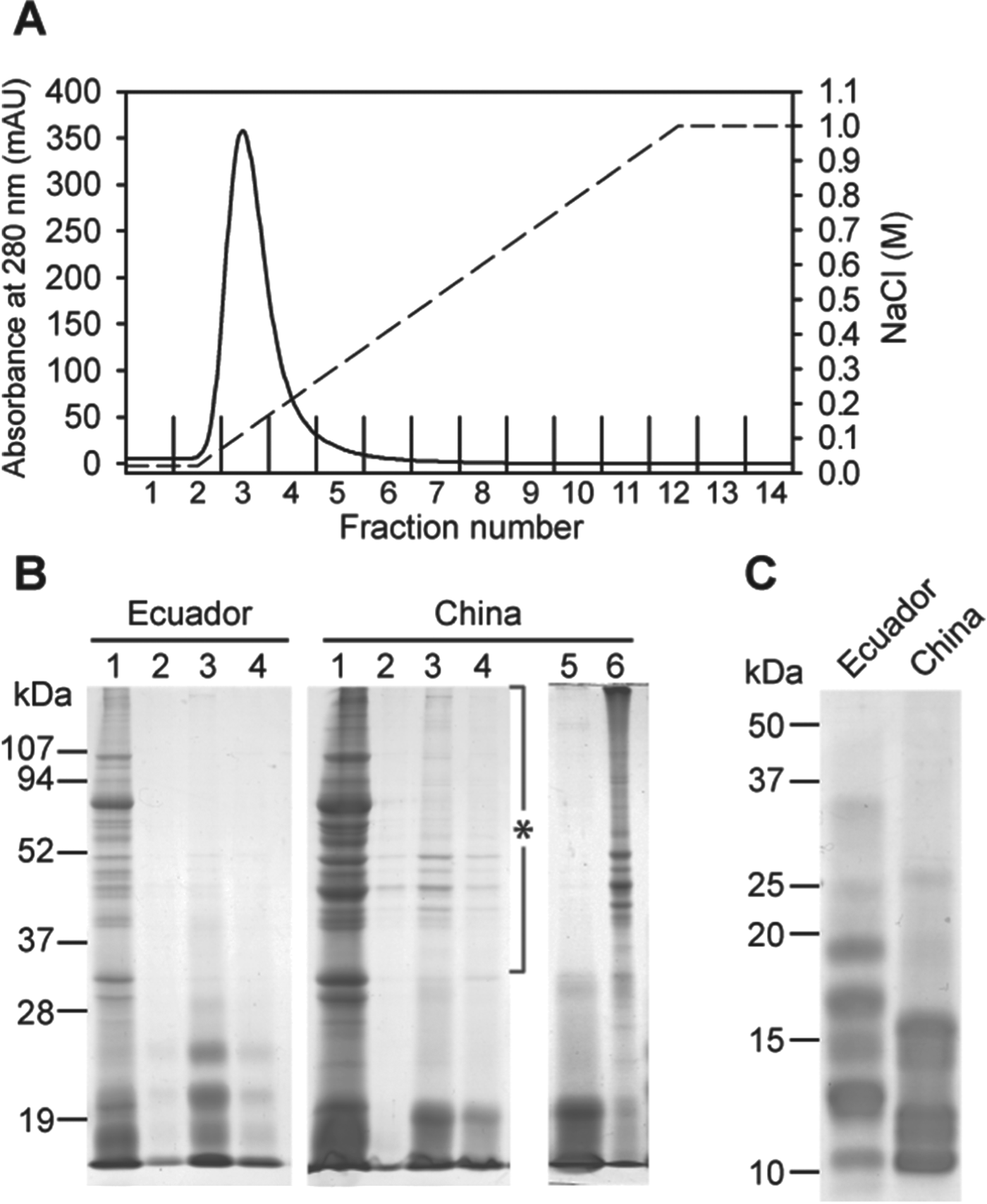
Because immunodiagnostic LMWAgs in the cyst fluid have a pI value of around 9·4 (Ito et al. Reference Ito, Nakao, Ito, Yuzawa, Morishima, Kawano and Fujii1999), we performed a CEC under pH 8·0 to purify them and a single sharp peak was resolved (Fig. 1A). Immunoblot analyses with sera from cysticercosis patients and from a rabbit immunized with LMWAgs revealed that resin-bound fractions (fractions 2–5) included LMWAgs (data not shown). There were no differences in between chromatographic profiles obtained from cyst fluids from Ecuador and China isolates (data not shown). By a single chromatographic procedure we could prepare LMWAgs sufficiently, but some contamination with high molecular size were sometimes observed although it depended on the batch of cyst fluid used for preparation (Fig. 1B). From the fact that Echinococcus Antigen B family to which LMWAgs belong is thermostable (Oriol et al. Reference Oriol, Williams, Perez Esandi and Oriol1971), we speculated that LMWAgs also had the thermostability and that, if the contaminated proteins were non-heat-resistant, the contaminants could be removed by heat-denatured precipitation and LMWAgs would be recovered in the supernatant. As expected, the heat-treatment precipitated contaminated proteins but not LMWAgs (Fig. 1B). The CEC-prepared LMWAgs migrated as a broad band between 10 to 25 kDa (Fig. 1C). There were critical differences in banding patterns between Ecuador and China isolates. Previously, we have demonstrated that: (1) T. solium could be divided into 2 genotypes, African/American and Asian, on the basis of mitochondrial DNA analyses (Nakao et al. Reference Nakao, Okamoto, Sako, Yamasaki, Nakaya and Ito2002); (2) each of the LMWAgs purified from cyst fluid of the two genotypes by antibody-affinity chromatography showed differences in banding patterns (Sato et al. Reference Sato, Sako, Nakao, Yamasaki, Nakaya and Ito2006); (3) such differences were lost on glycan removal with PNGase F (Sato et al. Reference Sato, Sako, Nakao, Yamasaki, Nakaya and Ito2006). In other words, glycoforms with different masses or a different number of N-linked oligosaccharides are the putative cause of the different banding patterns of glycoproteins from Asian, African or American geographical origin. Therefore, the differences in banding pattern of the CEC-prepared LMWAgs between the two genotypes might be caused by post-translational modification. Further analysis on post-translational modification of the CEC-prepared LMWAgs has to be performed, in addition to confirmation of reproducibility of preparation by using cyst fluids from several geographical different isolates.
Preparation of LMWAgs from T. solium cyst fluid. (A) Chromatographic profile of T. solium cyst fluid from the Ecuador isolate by FPLC system. Plain line, absorbance at 280 nm; dashed line, linear gradient of NaCl. (B) Each cation-exchange chromatography fraction, and the supernatant and the precipitate after heat-treatment were subjected to 12·5% SDS-PAGE and stained with Coomassie blue. Lane 1, crude CF; lane 2, fraction 2 from the CEC; lane 3, fraction 3 from the CEC; lane 4, fraction 4 from the CEC; lane 5, the supernatant after heat-denaturation treatment; lane 6, the precipitate after heat-denaturation treatment. The contaminants detected depended on the batch of cyst fluids and are indicated by an asterisk. (C) 15·0% SDS PAGE analysis of LMWAgs prepared from Ecuador and China isolates.

Evaluation of the CEC-prepared LMWAgs by ELISA
Evaluations of the CEC-prepared LMWAgs as ELISA antigens were carried out with serum samples from 60 cysticercosis patients, 30 AE patients, 30 CE patients and 41 healthy persons (Fig. 2 and Table 1). When crude CF antigens were used, the sera from more than half of echinococcosis patients and from one healthy person were positive and one cysticercosis case was negative. In contrast, all sera from cysticercosis cases showed positive reactions to both the CEC-prepared LMWAgs of Ecuador- and China-isolates, and some sera from alveolar and CE patients showed weak cross reactions, almost close to the cut-off borderline, to both antigens. The overall specificities of crude CF antigen and the CEC-prepared LMWAgs from Ecuador and China isolates were 66·3, 92·1 and 93·1%, respectively. There was no statistically significant difference in immunodiagnostic performance between the CEC-prepared LMWAgs from Ecuador and China isolates. In this study, we used only cysticercosis patient sera that were positive to IEFE-purified LMWAgs and echinococcosis patient sera with strong reactions to each homologous immunodiagnostic antigen for evaluation, which indicated that sera used were under unknown influences and sensitivities, and specificities based on the result of the ELISA did not necessarily reflect real cysticercosis and echinococcosis patient populations. Therefore, we need to evaluate this further with more sera from cysticercosis and echinococcosis patients, and also from patients with other parasitic infections to reveal immunodiagnostic performance quality of the CEC-prepared LMWAgs.
Evaluation by ELISA of three different antigens, crude CF from the Ecuador isolate, LMWAgs from the Ecuador isolate and LMWAgs from the China isolate. Sera from 60 patients with cysticercosis (red closed circle), 30 with alveolar echinococcosis (blue closed circle), 30 with cystic echinococcosis (green closed circle), and 41 healthy people (black closed circle) were used. The cut-off values for crude CF, LMWAgs from the Ecuador isolate, and LMWAgs from the China isolate are 0·310, 0·075 and 0·085, respectively, and are indicated by the dashed lines.
Results of ELISA using crude CF from Ecuador isolate, LMWAgs from Ecuador isolate and LMWAgs from China isolate
We have previously reported that the origin of the fluid from T. solium cysts may affect the antigenicity of LMWAgs (Sato et al. Reference Sato, Sako, Nakao, Yamasaki, Nakaya and Ito2006). As mentioned above, mitochondrial analyses revealed that T. solium can be divided into two genotypes, African/American and Asian (Nakao et al. Reference Nakao, Okamoto, Sako, Yamasaki, Nakaya and Ito2002). Furthermore, the purified LMWAgs could be differentiated into two banding patterns that correspond to the two genotypes. Although there were no differences in diagnostic sensitivities between LMWAgs from two genotypes, reactivities of sera from American patients to African/American LMWAgs were higher than those of Asian patients, and the inverse phenomenon was observed (Sato et al. Reference Sato, Yamasaki, Sako, Nakao, Nakaya, Plancarte, Kassuku, Dorny, Geerts, Benitez-Ortiz, Hashiguchi and Ito2003). This suggested that immunodiagnostic tests should be performed in each endemic area by using LMWAgs prepared from T. solium cysts collected in the appropriate endemic region. Therefore, establishment of a useful preparation of LMWAgs capable of easy application in endemic area is urgently required. Recently, a simple enrichment of 120- and 150-kDa immunodiagnostic antigen complexes of T. solium cyst fluids by trichroroacetic acid/acetone mixture precipitation has been reported (Lee et al. Reference Lee, Bae, Kim, Diaz-Camacho, Nawa and Kong2010). The antigens that were purified by this method showed immunodiagnostic sensitivity and specificity of 97·7 and 98·7% against active neurocysticercosis. However, this method requires use of a hazardous reagent, suggesting that it might be difficult to apply under general normal conditions. In contrast, the CEC method does not need such reagent and, moreover, can be performed manually with a syringe without expensive equipment (data not shown). This feature would make the CEC method easy to apply in endemic areas, especially in developing countries.
The CEC-prepared LMWAgs could be utilized to detect T. solium infections in pigs by immunoblot analysis and ELISA (data not shown). For this purpose, we need to consider the cross-reaction with sera from pigs infected with other infective agents, especially Taenia hydatigena. Previously, no cross-reaction to LMWAgs has been demonstrated in pigs infected with T. hydatigena by ELISA (Sato et al. Reference Sato, Yamasaki, Sako, Nakao, Nakaya, Plancarte, Kassuku, Dorny, Geerts, Benitez-Ortiz, Hashiguchi and Ito2003). However, the number of sera examined seems to be insufficient to conclude that there was no cross-reactivity. Therefore, a large-scale evaluation in pigs must also be performed.
Interestingly, the CEC method is applicable for the purification of LMWAgs family of proteins from Taenia saginata and Taenia asiatica cyst fluids (data not shown). Additionally, ELISA using sera from bovines experimentally infected with T. saginata and LMWAgs antigens prepared from T. saginata cyst fluids revealed that those antigens had high value to detect T. saginata infections with dramatic decreasing of background absorbance values obtained using crude cyst fluids (data not shown). More detailed characterization of the CEC-prepared LMWAgs from T. saginata and T. asiatica cyst fluids must be performed using more sera from animals with cysticercosis and other parasitic infections.
In conclusion, we have established a simple and reliable preparation of LMWAgs with high immunodiagnostic performance from T. solium cyst fluids by the combination of the CEC and heat-treatment. The CEC-prepared LMWAgs is highly specific to cysticercosis, but analyses on the B cell epitopes must be carried out to eliminate cross-reactivity with echinococcosis patient sera. The preparation method established would contribute to the successful control of cysticercosis in endemic areas.
ACKNOWLEDGEMENTS
We are grateful to W. Benitez-Ortiz (Ecuador) and T. Ikejima (China) for providing confirmed serum samples and T. solium cyst fluids.
FINANCIAL SUPPORT
This work was supported by the Grant-in-Aid for Scientific Research (A) (21256003, 24256002) to AI and (B) (21406009, 24406011) to MO, from the Japan Society for the Promotion of Science (JSPS); a grant from the Japan–China Medical Association (2012) to YS and by JSPS-Asia/Africa Scientific Platform Fund (2006–2011) and the Special Coordination Fund for Promoting Science and Technology from the Ministry of Education, Culture, Sports, Science & Technology in Japan (MEXT) (2003–2005, 2010–2012) to AI.