




No CrossRef data available.
Article contents
A cathepsin F of adult Clonorchis sinensis and its phylogenetic conservation in trematodes
Published online by Cambridge University Press: 01 March 2004
Abstract
A novel 28 kDa cysteine protease (Cs28CF) secreted by the hepatobiliary trematode, Clonorchis sinensis was identified. The protease was purified from the excretory-secretory products (ESP) of the adult worm using DEAE-ion exchange and Arginine-Sepharose 4B chromatography. It showed a high activity between pH 6·5 and 7·5 in a dithiothreitol (DTT)-dependent manner. Inhibitors specific to cysteine proteases down-regulated the activity. Addition of Cs28CF to monkey cholangiocyte cultures resulted in approximately 95% cell death after 7 days. The full-length cDNA (1078 bp) encoded a single peptide of 328 amino acids (aa) with an N-terminal hydrophobic sequence, an ERFNAQ motif in the propeptide and a mature domain. Expression of mRNA transcripts of Cs28CF was observed in both the metacercaria and adult stages. Bacterially expressed recombinant protein exhibited a specific antibody reaction with clonorchiasis sera. Deduced aa exhibited 52–76% sequence identity with the cathepsin F analogues from other organisms. A novel E/DXGTA motif was recognized in the propeptide region. Phylogenetic analysis of 63 papain family members revealed that the trematode cysteine proteases formed 2 major clades of cathepsins F and L. The trematode cysteine proteases classified as cathepsin F shared higher homology among themselves than those classified as cathepsin L. Cathepsin F is phylogenetically conserved in the trematode parasites as well as in mammals.
Keywords
Information
- Type
- Research Article
- Information
- Copyright
- 2004 Cambridge University Press
INTRODUCTION
Cysteine proteases are a large group of enzymes that catalyse the degradation of a variety of proteins and are significantly involved in the intracellular metabolic turnover (Chapman, Riese & Shi, 1999). In some cases, particularly in the helminth parasites, the cysteine proteases are not targeted to the intracellular compartments but are secreted outside through a mannose 6-phosphate independent pathway or by other mechanisms (Sajid & McKerrow, 2002). The parasitic cysteine proteases are believed to play pivotal roles not only in the stage transition (Chung et al. 1995; Lustigman et al. 1996; Hashmi et al. 2002) and invasion to the host tissue but also in nutrient uptake and immune evasion from the host (Carmona et al. 1993; Kong et al. 1994). The cysteine proteases are divided into distinct clans: clan CA (papain, calpain, viral cysteine proteases), CD (clostripain and caspase), CE (adenain), and CF (pyroglutamyl peptidase I), with the largest family being that of papain (family C1) (Barrett & Rawlings, 2001). The majority of parasite cysteine proteases belong to the papain family (family C1) within the clan CA. This family consists of 2 major subfamilies, cathepsins B and L. The former is characterized by the presence of an occluding peptide loop while the latter is characterized by the presence of an ERFNIN motif (Karrer, Peiffer & DiTomas, 1993; Illy et al. 1997; Sajid & McKerrow, 2002).
Cathepsin F is a new cysteine protease of the papain family found in mammals (Wang et al. 1998; Nagler, Sulea & Menard, 1999; Santamaria et al. 1999; Deussing et al. 2000). It possesses a long preproregion and a highly conserved ERFNAQ motif, which distinguish this enzyme from the other family members. The optimal pH for this enzyme is between 5·2 and 6·8. In mammals, cathepsin F targets the endosomal/lysosomal compartments via mannose-6-phosphate independent trafficking pathway (Wang et al. 1998; Deussing et al. 2000).
Clonorchis sinensis is a trematode parasite that causes hepatobiliary diseases in mammals including humans, and is widely distributed in Asian countries (Rim, 1987). It has been estimated that more than 7 million people are infected with the parasite worldwide (Crompton, 1999). The clinical symptoms of the patients with a light infection are often non-specific. However, a severe infection causes jaundice with an abnormal liver function, and even abdominal distension by the ascites. In patients with a chronic infection, the main pathological characteristics include periductal fibrosis combined with ductal dilatation and an adenomatous metaplasia of the ductal epithelium (Rim, 1987; Hong, Koh & Kim, 1993). It has also been suggested that there is an association between clonorchiasis and cholangiocarcinoma (Lee, Rim & Sell, 1997; Kim et al. 1999).
Several attempts to characterize C. sinensis cysteine proteases, which are thought to play important roles in the pathogenesis, have been carried out. An 18·5 kDa cathepsin B-like cysteine protease was purified from the adult worm (Song, Dresden & Rege, 1990). The cysteine protease activity in the acidic extracts of C. sinensis is differentially expressed during the developmental stage (Song & Rege, 1991). Other 17 and 24 kDa cysteine proteases have been shown to be involved in larval migration, parasite nutrition and lysis of the host tissue (Chung et al. 2000; Park et al. 2001). In addition, it has been reported that C. sinensis cysteine protease is highly antigenic and has potential for use as a serodiagnostic marker (Song et al. 1990; Na et al. 2002).
In this study, a new 28 kDa excretory cysteine protease of C. sinensis was purified. The enzyme was able to induce cytolysis in monkey cholangiocyte cultures in vitro. The full-length cDNA encoding the enzyme was subsequently cloned by screening a cDNA library. The recombinant form of this protein was highly reactive with the sera of patients with clonorchiasis. We also demonstrated that this protease is a member of the cathepsin F family, which is highly conserved in parasitic trematodes as well as in mammals.
MATERIALS AND METHODS
Preparation of adult C. sinensis and the excretory-secretory products (ESP)
C. sinensis metacercariae were collected from naturally infected freshwater fish, Pseudorasbora parva in an endemic area in Korea. The metacercariae were administered orally to white New Zealand rabbits. Two months later, the adult worms were harvested from the bile ducts of the infected rabbits and used to prepare the ESP as well as the RNA and DNA samples. The ESP was obtained by incubating 500 adult worms in physiological saline overnight in an air and 5% CO2 incubator at 37 °C. The medium was centrifuged at 20000 g for 1 h at 4 °C. The resulting supernatants were dialysed against distilled water for 2 h, lyophilized, and stored at −70 °C until use.
Assay of enzyme activity and determination of optimal pH and specific inhibitors
The enzyme activity was monitored using 5 mg Azocoll (Sigma, St Louis, MO, USA) in sodium phosphate buffer (100 mM, pH 7·0) in the presence of 5 mM DTT. One unit of the enzyme activity was defined as the amount of enzyme required to give a single unit (0·1) increase in the absorbance through a 1 cm light path at a wavelength of 540 nm, as described previously (Kong et al. 1997). The modulation of enzyme activity by pH was determined. A standard reaction mixture (500 μl) contained 2 μg of the purified enzyme and 5 mg of Azocoll supplemented with 5 mM DTT in sodium acetate buffer (100 mM) at pH 5·0, 5·5 and 6·0, in sodium phosphate buffer (100 mM) at pH 6·5 and 7·0 and in Tris–HCl buffer (100 mM) at pH 7·5 and 8·0. The effect of several inhibitors on the enzyme activity was observed essentially as described above except for the enzyme incubation with each inhibitor for 20 min at 37 °C prior to being subjected to the standard assay. The inhibitors used, which specifically acted on cysteine-, serine-, and metallo-protease, were transepoxy-succinyl-L-leucyl-amido(4-guanidino) butane (E-64) (10 μM), iodoacetamide (IAA) (1 mM), diisopropyl fluorophosphate (DFP) (1 mM), phenylmethylsulfonyl fluoride (PMSF) (1 mM), soybean trypsin inhibitor (SBTI) (1 mM) and ethylenediaminetetraacetic acid (EDTA) (2 mM). All the chemicals were purchased from Sigma. Each test was conducted in triplicate and was standardized by employing the appropriate buffer in each experiment. The values are expressed as a mean±S.D.
Purification of cysteine protease from adult ESP (Cs28CF)
The ESP, which was rehydrated in sodium acetate buffer (10 mM, pH 5·5), was applied to a 1·6×5 cm long DEAE-anion exchange column previously equilibrated with the same buffer. The bound proteins were eluted with a stepwise gradient salt concentration of 10, 20, 30, 40 and 50 mM. The fractions showing a high enzyme activity were dialysed against sodium phosphate buffer (10 mM, pH 7·0) and further subjected to 1·6×2 cm sized Arginine-Sepharose 4B affinity chromatography pre-equilibrated with the same buffer. The protein was eluted with the 10 mM salt concentration. The purified protein was monitored by sodium dodecyl sulfate (SDS)-PAGE stained with silver nitrate.
Gelatin-gel electrophoresis
The partially purified enzyme fraction (3 μg) was resolved by a 10–15% separating gel containing 0·2% (w/v) gelatin in non-reducing condition. After electrophoresis, the gel was washed with 2·5% (v/v) Triton X-100 (Sigma) for 1 h and incubated overnight at 37 °C in a sodium phosphate buffer (100 mM, pH 7·0) in the presence of 2 mM DTT to allow proteolysis. The proteolytic zone was visualized by Coomassie blue stain.
In vitro effect of Cs28CF on the growth of cultured monkey cholangiocytes
The monkey cholangiocytes were kindly provided by Dr K. Shogren, Mayo Foundation (Chicago, IL, USA). The cells (106) were cultured in 10 ml of α-minimum essential medium (MEM) supplemented with 3·6% fetal calf serum, 2 mML-glutamine, 0·1 μM insulin, 50 μg/ml gentamicin, and 100000 U/100 mg penicillin/streptomycin (GibcoBRL, Grand Island, NY, USA) in 6-well tissue culture plates (Nunc, Denmark). The plates were incubated at 37 °C in an air-5% CO2 equilibrated incubator. The cytopathic effect was examined in the presence of 2 μg/ml Cs28CF and 1 mM DTT. The control plates contained only 1 mM DTT. The medium was changed every 2–3 days. The growth of the cells was monitored by microscopy and trypan blue exclusion. The experiments were repeated 3 times and the results are expressed as a mean±S.D.
Construction and screening of an adult C. sinensis cDNA library
The total RNA was extracted from the adult C. sinensis using a TRIzol RNA isolation kit and the polyadenylated RNA was purified using an Oligotex mRNA purification kit following the manufacturer's protocol (Qiagen, Valencia, CA, USA). A cDNA library was constructed in a λZAP II expression vector using a λZAP II-cDNA synthesis kit according to the manufacturer's instruction (Stratagene, La Jolla, CA, USA). A cDNA fragment was amplified from the cDNA library by a polymerase chain reaction (PCR) using the sense primer, 5′-GTGTGGTTCATGTTGGGCGTTCTCG-3′, corresponding to a highly conserved region among the parasite cysteine proteases, and an anti-sense primer SKT7, 5′-TACCGGGCCCCCCCTCGAG-3′, which is complementary to the sequence between the T7 promoter and the Xho I site of the pBluescript SK-phagemid. The thermal cycler profile included 30 cycles at 94 °C (1 min), 59 °C (1 min) and 72 °C (1 min) followed by a final extension at 72 °C (7 min). The amplified products were purified using a QIAex II gel extraction kit (Qiagen) and were sequenced after cloning the products into the pT7Blue(R) TA vector (Novagen, Madison, WI, USA). The insert was labelled with an ECL direct nucleic acid labelling kit (Amersham-Pharmacia Biotech, Uppsala, Sweden) and was used to screen the cDNA library. The positive plaques were isolated and subjected to an in vivo excision using an ExAssist helper phage and a nonsuppressing Escherichia coli strain, SOLA (Stratagene). The DNA sequencing was conducted by a dideoxynucleotide chain termination method and an ABI-PRISM dye terminator cycle sequencing kit (Perkin-Elmer Applied Biosystems, Foster City, CA, USA) in an automated DNA sequencer (model 377; Perkin-Elmer).
Southern and Northern blot analysis
The high molecular weight genomic DNAs were isolated from the adult worms according to a standard phenol-chloroform extraction protocol (Sambrook, Fritsch & Maniatis, 1989) and were digested with the restriction enzymes including BamH I, EcoR I, Hinc II, Hind III, Msp I and Pst I (10 μg DNA for each enzyme). After separation on a 0·8% agarose gel, the DNA fragments were transferred to a Hybond N membrane (Amersham-Pharmacia Biotech). Southern blot analysis using a 32P-labelled full-length cDNA was done using the standard method (Sambrook et al. 1989).
The heat-denatured total RNAs (10 μg) resolved by 1·2% formaldehyde-agarose gel were blotted to a nylon membrane (Amersham-Pharmacia Biotech). The blot was hybridized with the full-length cDNA, which was radio-isotope labelled using a Rediprime DNA labelling system according to the manufacturers instructions (Amersham-Pharmacia Biotech). The filters were washed with high stringency and then exposed to Kodak X-AR film.
Reverse transcription (RT)-PCR
The total RNA was extracted from different developmental stages of the metacercaria and adult using the TRIzol reagent (Qiagen). The RT-PCR was done using the gene specific primers for Cs28CF (sense, 5′-GACTGCAGGAAATGGAACAG-3′; antisense, 5′-CGATTTCCTTCAACTTCTGTGC-3′). The tropomyosin gene (GenBank Accession no. L43918) was used as the positive control (sense, 5′-AGTCTCGTCTAGAAGCTGCTG-3′; antisense, 5′-GTAGCTGTGTCGGTTAATGAGG-3′). A total of 2 μg RNA, 5 mM MgCl2, 1 mM dNTP mixture, 20 U RNAase inhibitor, 1 μM each antisense primer, 2 μl 10×RNA PCR buffer and 5 U Avian murine leukaemia virus reverse transcriptase were used in a total reaction volume of 20 μl according to the manufacturers instructions (RNA PCR kit ver. 2.1. TaKaRa, Japan). The reaction mixture was incubated at 50 °C for 30 min. PCR was accomplished with the reverse transcribed products, as described above, after heating at 99 °C for 5 min. The PCR products were analysed on a 1·2% agarose gel.
Sequence alignment and phylogenetic analysis
A search of the non-redundant database at the National Center for Biotechnology Information (NCBI, http://www.ncbi.nlm.nih.gov) with the BLAST P algorithm identified more than 100 cysteine protease members, which showed a significant sequence homology to Cs28CF. They included the cathepsins F, K, L, S, and W from mammals, cathepsin Ls from arthropods, along with the trematode and filarial parasites, and cruzipains from protozoa. Sixty-three sequences contained the tertiary structures corresponding to the mature domain of the cysteine proteases and showed a >30% homology to Cs28CF. These sequences were aligned with CLUSTAL W (Thompson, Higgins & Gibson, 1994) and were optimized by manual trimming using the GenDoc program (Nicholas & Nicholas, 1997). A phylogenetic analysis was done using the PROTDIST and NEIGHBOR of PHYLIP and the phylogram was constructed with TreeView (Page, 1996). Bootstrapping analysis with 1000 replicates was conducted to evaluate the statistical significance of each branching point.
Expression of recombinant protein (rCs28CF)
A cDNA (648 bp) encoding the putative mature protein was amplified by PCR using the full-length cDNA clone as a template. The forward primer used was 5′-AGATCTGACGATGGATAACGAG-3′, which matched the first 12 nucleotides following the initiation codon. The reverse primer was SKT7 as described above. The thermal cycler profile included 30 cycles of 94 °C (1 min), 62 °C (1 min), 72 °C (1 min), and a final extension at 72 °C (7 min). The PCR product was gel purified, subcloned into the pT7Blue(R) TA vector (Novagen) and sequenced. An insert harboring the correct sequence was isolated by digestion with EcoR I and Xho I and placed into the corresponding sites of the pGEX4T-2 expression vector (Amersham-Pharmacia Biotech), which expresses the recombinant protein as a fusion with glutathione S-transferase (GST). The expression fidelity was confirmed by direct sequencing. The recombinant plasmid was transformed into E. coli BL21 and induced with 0·5 mM isopropyl β thiogalactoside (IPTG; Calbiochem, La Jolla, CA, USA). The bacteria were lysed by sonication and the supernatant was collected after centrifugation. The protein was purified by a passage over a column of glutathione agarose beads (Amersham-Pharmacia Biotech) with 20 mM reduced glutathione in Tris–HCl (100 mM, pH 9·0). The GST carrier in the fusion protein was further removed by the thrombin treatment.
Purification of anti-rCs28CF antibody
The polyclonal antiserum against C. sinensis adult worm extracts was generated by immunizing the New Zealand White rabbit 3 times with Freund adjuvants (Sambrook et al. 1989). A monospecific antibody against the recombinant protein was purified using a SulfoLink Kit according to the manufacturers protocol (Pierce, Rockford, IL, USA). Briefly, 10 mg of the recombinant protein was dialysed against sodium acetate buffer (100 mM, pH 6·0), followed by mixing with 6 mg of 2-mercaptoethanol. The mixture was then packed into a column pre-equilibrated with Tris–HCl (50 mM, pH 8·5). After loading the polyclonal rabbit antiserum (5 ml), the column was extensively washed to remove the unbound antibodies. The monospecific antibodies bound to the recombinant protein were eluted by glycine buffer (100 mM, pH 2·5). The antibodies were dialysed against PBS (100 mM, pH 7·2) overnight and stored at −70 °C until use.
Western blotting
The proteins resolved by 10–15% SDS–PAGE under reducing conditions were transblotted to an Immobilon-P transfer membrane (Millipore, Bedford, MA, USA). The blot was incubated with the purified monospecific antibodies for 1 h at a dilution of 1[ratio ]400. The membrane was further incubated with peroxidase-conjugated anti-rabbit IgG (Cappel, West Chester, PA, USA). The reaction was developed with an enhanced chemiluminescence reagent (Pierce).
Western blotting was also done using the serum samples from clonorchiasis (n=35), paragonimiasis (n=20), fascioliasis (n=10), schistosomiasis japonicum (n=10) and the normal controls (n=25) to assess the diagnostic applicability of the rCs28CF. The patients were diagnosed either by a stool examination (clonorchiasis), computed tomography of the chest and ELISA (paragonimiasis), or ELISA (fascioliasis and schistosomiasis). The recombinant protein was resolved by 12% SDS–PAGE and was transferred to an Immobilon-P membrane. It was cut into strips and incubated with the respective serum overnight in a dilution of 1[ratio ]100. The strips were further incubated with 1[ratio ]1000 diluted peroxidase conjugated anti-human IgG (Cappel) for 3–4 h. The colour reactions were developed with 0·03% (w/v) 4-chloro-1-naphthol (4C1N) chromogen in phosphate buffer (100 mM, pH 7·2).
RESULTS
Purification and biochemical properties of a 28 kDa cysteine protease of C. sinensis ESP
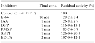
DEAE-anion exchange and Arginine-Sepharose 4B affinity columns were used to purify the cysteine protease of adult C. sinensis ESP. DTT was required to maintain the catalytic activity, as in the other cysteine proteases. The purified enzyme migrated to 28 kDa by SDS-PAGE analysis (Fig. 1A). The gelatin substrate gel electrophoresis using a partially purified enzyme (bound fraction of 20 mM salt in DEAE column) also showed a distinct proteolytic zone approximately 28 kDa at pH 7·0 as shown in Fig. 1B. The enzymatic assay exhibited significant proteolytic activity against Azocoll. A total of 0·8 mg of the protease was purified from 17·8 mg of the crude protein with a recovery rate of 14·9%. Specific activity increased 3·4-fold (data not shown). The purified enzyme was incubated with an Azocoll substrate supplemented with DTT at different pH to observe an optimal pH for the enzyme activity. The peak activity was observed at pH 7·0. However, the protease retained approximately 80% activity between pH 5·5 and 8·0 (Fig. 1C). Table 1 summarizes a modulation of the enzyme activity by different types of protease inhibitors. E-64 and IAA, both of which specifically act on the cysteine proteases, were found to significantly inhibit the enzyme activity, while no inhibitory modulation was observed with the inhibitors specific to the serine proteases (DFP, PMSF, SBTI) or metalloproteases (EDTA). This result suggests that the protease purified was a member of the cysteine protease superfamily.

Fig. 1. (A) Determination of the homogeneity of a 28 kDa cysteine protease purified from the excretory-secretory products (ESP) of the adult Clonorchis sinensis by 10–15% SDS-PAGE under reducing conditions. The bound fraction of the 20 mM salt concentration (lane DEAE), where a high enzyme activity was shown, was dialysed against sodium phosphate buffer (100 mM, pH 7·0), lyophilized, and subjected to Arginine-Sepharose 4B affinity chromatography. The bound protein, eluted with a 10 mM salt concentration, exhibited a single band at 28 kDa with high enzyme activity (lane Arg, 1 μg loaded). Mr, molecular weight in kDa; Lanes ESP, crude ESP; Crude, crude extracts of the adult C. sinensis (30 μg loaded). (B) The proteolytic activity of the partially purified enzyme fraction shown by gelatin substrate gel electrophoresis. After the electrophoresis of 3 μg protein by 10–15% SDS-PAGE in non-reducing conditions, the gel was incubated in 100 mM sodium phosphate buffer at pH 7·0 overnight at 37 °C. A distinct zone of proteolysis appeared at approximately 28 kDa. (C) Modulation of the enzyme activity by pH. A total of 2 μg purified enzyme was incubated with 5 mg Azocoll supplemented with 5 mM DTT in 100 mM sodium acetate buffer at pH 5, 5·5 and 6, in 100 mM sodium phosphate buffer at pH 6·6 and 7 and in 100 mM Tris–HCl buffer at pH 7·5 and 8. Mean±S.D., n=3.
Table 1. Modulation of the enzyme activity by various inhibitors (See Materials and Methods for detailed description. Values are expressed in mean±S.D. of 3 independent experiments and were standardized using a blank for each experiment. Abbreviations used are: DTT, dithiothreitol; E-64, transepoxy-succinyl-L-leucyl-amido(4-guanidino) butane; IAA, iodoacetamide; DFP, diisopropyl fluorophosphate; PMSF, phenylmethylsulfonyl fluoride; SBTI, soybean trypsin inhibitor; EDTA, ethylenediaminetetraacetic acid.)
We examined whether this protease affects the growth of the cultured monkey cholangiocyte. As shown in Fig. 2A, the cholangiocytes incubated with the purified enzyme revealed noticeable morphological changes, such as a detachment of the cell layers and a loss of the cellular polarity, in 3–7 days. In contrast, the cells cultured in the control media that only contained 1 mM DTT exhibited normal growth. The quantification of the cell proliferation by trypan blue exclusion revealed that less than 5% of the cells had survived 7 days after beginning the culture, whereas the numbers in the control group increased more than 2-fold (Fig. 2B).

Fig. 2. In vitro cytolytic effect of Cs28CF on monkey cholangiocytes. A total of 106 cells were cultured in the presence of 2 μg/ml Cs28CF supplemented with 1 mM DTT. (A) Microscopic illustration of the morphological changes in the cells cultured with Cs28CF. (B) Trypan blue exclusion analysis of the cell proliferation rate at different times. [bull ], cholangiocyte cultured without Cs28CF; ○, cells cultured with Cs28CF.
Sequence analysis of the gene encoding C. sinensis cysteine protease
We obtained 650 bp cDNA fragments by PCR amplification of adult C. sinensis cDNA library using the primers derived from the conserved sequences among the cysteine proteases. Sequence analysis revealed that this fragment was closely related to but clearly distinct from the cysteine proteases of other organisms including helminth parasites. This fragment was used to screen the cDNA library. Approximately 5×10⁴ plaques were hybridized and 42 plaques showing a positive reaction were obtained. After a second and third screening, 36 clones possessing an identical sequence in the overlapping region were isolated. One clone possibly harbouring a full-length cDNA was identified by sequence analysis. The clone was composed of 1078 bp, which contained a 987 bp with a complete open reading frame (ORF; 328 aa). A short poly(A) stretch (AATAAA) at 3′ end was 13 bp upstream from the poly(A) addition site.
A comparison of the primary structures of Cs28CF with the other related members showed that the protease might be translated as an inactive preproenzyme and be processed into a mature active protease making the Trp resulted in the seventh position of the putative mature enzyme (Karrer et al. 1993). The protein had a preponderance of hydrophobic residues in the first 19 aa of the protein, which was expected for the signal peptide commonly found at the N-terminus of the cysteine proteases (Dufour, 1988). The predicted cleavage sites between the signal sequence and the pro-domain, and between the pro- and mature-domain were recognized between Ala (at position −97) and Arg (−96), and between Met (−1) and Asp (+1), respectively (the first aa of the putative mature domain, Asp, was numbered +1). Four small domains possibly constituting α-helical structures were present in Asn (−88) through to Try (−76), Asn (−70) through to Leu (−65), Lys (−60) through to Glu (−47) and Ser (−31) through to Lys (−23) (Fig. 3).

Fig. 3. Alignment of the predicted amino acid sequence of a Cs28CF with other related members of cathepsin F. The conserved amino acids are shaded and gaps (-) are introduced to optimize the alignment. Prepro- and pro-region are indicated by the arrows. Four α-helical domains in the pro-region are underlined. The NH2-terminal sequence is marked by a double underline. The cysteine protease catalytic triad (C, H, and N) is shown by bold-underline. Putative sites for mannose 6-phosphate N-glycosylation are indicated by the boxes. Substrate-binding amino acid residue (G) is shown with the arrowheads. The asterisks are showing the 7 Cys, which are essential for forming tertiary structures by a disulfide linkage. The ERFNAQ motif is also shown. In addition, a small motif of E/DXGTA, indicated by the double-lined box is recognized in cathepsin Fs except for that from Schistosoma mansoni, which is followed by the ERFNAQ motif. The numbers in parentheses start from the N terminus of Cs28CF. Cs, Cs28CF (AF093243); Pw, Paragonimus westermani cruzipain (U70537); Sm, Schistosoma mansoni cathepsin L1 (U07345); Ce, Caenorhabditis elegans cathepsin L (NM_072814); hum, human cathepsin F (NM_003793); mou, mouse cathepsin F (MMU131851); Nt, Nicotina tabacum pre-pro-cysteine proteinase (AJ242994); Ib, Ipomoea batatas papain-like cysteine protease isoform (AF138264).
Further analysis of the 93 aa of the putative propeptide region, which contained a block of conserved aa in cathepsins L, F, and W, revealed that cathepsin F molecules from both trematodes and mammals preserved the ERFNAQ motif between Asp (−68) and Gln (−49) except for 1 sequence of Glu, which was replaced by Asp (−68) (Wang et al. 1998; Nagler et al. 1999; Deussing et al. 2000; Yun et al. 2000). In contrast, members that could be classified into cruzipain and cathepsin L subfamilies from trematodes, filaria, arthropod and other vertebrates conserved the ERFNIN motif (Karrer et al. 1993). Importantly, a small domain of the E/DXGTA motif between Glu (−45) and Ala (−42) (between the 3rd and 4th α-helical domain) was identified only in cathepsins F and W except for Gln (−46), which was replaced by Val in the cathepsin F of S. mansoni (double-lined box in Fig. 3).
The mature domain (216 aa) possessed 3 essential residues of Cys (+25), His (+161) and Asn (+181), which are critical for composing a catalytic triad of the cysteine proteases; His is required for ion-pair formation with Cys at an active site (Lewis, Johnson & Shafer, 1981; Groves et al. 1996). The 2 putative N-linked glycosylation sites of Asn-Arg-Ser and Asn-Glu-Ser present in the middle region (at positions +99 to +101, and +108 to +110) suggests a possibility that these glycosylation sites might have some effect on the proteolytic activity of the native enzyme (Wang et al. 1998; Deussing et al. 2000). The mature domain also contained 7 Cys at positions 22, 25, 56, 63, 96, 154 and 204, which are crucial for cathepsins L and F to form tertiary structures by a disulfide linkage (Wang et al. 1998; Santamaria et al. 1999). The potential S1 subsites were recognized at Try (+67), Pro (+68), Ala (+162) and Ala (+211) and those of S2 at Pro (+69), Ser (+211) and Ile (+214), respectively. The putative S1′ sites were found at Gln (+19) and Trp (+177). The Gly at position +65 might serve as a single substrate-binding site (Heussler & Dobbelaere, 1994). The mature domain encoded a predicted molecular weight of 23760 Da, which is similar to the size of the native protease that was purified from the ESP after making an allowance for its post-translational modification. All these structural features suggested that the protease cloned in this study is a novel cysteine protease of the clan CA, possibly belonging to the cathepsin F subfamily. We designated this enzyme as C. sinensis 28 kDa cathepsin F (Cs28CF). The gene was registered in the GenBank database with the accession number AF093243.
Expression pattern of the Cs28CF gene
Southern blot analysis was carried out using a full-length cDNA as a probe in order to determine the genomic distribution of the Cs28CF gene. As shown in Fig. 4A, the 2 or 3 bands recognized in the genomic DNA digested with various enzymes suggested that this gene might be as a single copy of the genome. The total RNA extracted from the adult parasite was examined for Cs28CF expression. A single transcript of 1·1 kb in size was hybridized with the cloned cDNA by Northern blot analysis, which was consistent with the size of the Cs28CF cDNA (data not shown).

Fig. 4. Genomic distribution and expression of Cs28CF by Southern (A), RT-PCR (B) and Western blot analysis (C). (A) The genomic DNA was digested with the restriction enzymes of BamH I, EcoR I, Hinc II, Hind III, Msp I or Pst I and hybridized with the full-length cDNA. BamH I cuts at position +312 yields fragments of 7 kb and a large fragment of >15 kb. EcoR I at position +860 shows fragments of 3·1 kb and 3·5 kb. Hinc II and Hind III (no cut at cDNA) give rise to fragments of 4·2 kb and >15 kb, respectively. (B) RT-PCR with the gene specific primers for Cs28CF shows that the mRNA transcript of Cs28CF was expressed in both the metacercaria and adult stages. The tropomyosin gene, which was used as a control, also appears to be constantly expressed in both stages. M, 100 bp ladder. (C) Western blots of the recombinant and native proteins. The proteins were resolved by 10–15% SDS-PAGE, transblotted to an Immobilon-P transfer membrane and reacted with the monospecific antibodies against the recombinant protein. Mr, molecular weight in kDa; mat, thrombin cleaved mature domain; fusion, mature domain linked with GST carrier; crude ESP, crude ESP of C. sinensis.
Comparative RT-PCR with the gene specific primers for Cs28CF was done to observe the expression pattern of the mRNA transcripts according to the developmental stages. As shown in Fig. 4B, the mRNA of Cs28CF was expressed in both the metacercaria and the adult stages, which suggested that the metacercariae preserve Cs28CF in the intestine or excretory granules and secrete the enzyme when they excyst.
Expression of rCs28CF and Western blot analysis
A 648 bp fragment encoding the putative mature domain was cloned into the pGEX 4T-2 vector and expressed in E. coli as a fusion with the GST. The fusion protein was purified from the soluble bacterial extracts by affinity chromatography using a glutathione-Sepharose 4B column. The purified GST-fusion protein migrated to approximately 48 kDa. After removing the GST carrier by thrombin digestion, the purified protein present in SDS–PAGE was approximately 23 kDa (data not shown), which is consistent with the molecular mass predicted from the cDNA sequence of the mature domain. However, neither the 48 kDa nor the 23 kDa protein exhibited any proteolytic activity in the enzyme assays (data not shown). Western blot analysis using monospecific antibodies against the recombinant protein showed that both the 48 kDa and the 23 kDa molecules exhibited strong reactivity (Fig. 4C). The monospecific antibodies also recognized a single approximately 28 kDa band in the crude ESP of the adult worm (lane crude ESP).
Reactivity of rCs28CF against serum samples from trematode infections
Fig. 5 shows representative results of the immunoblot evaluation of the rCs28CF with the sera from the patients with clonorchiasis, paragonimiasis, fascioliasis, schistosomiasis japonicum and those from the normal controls. Strong antibody reactions against rCs28CF were observed in 30 of 35 clonorchiasis sera (panel Cs). Meanwhile, 2 of 20 and 1 of 10 samples from paragonimiasis and fascioliasis showed positive reactions (panels Pw and Fh). None of the serum samples from the schistosomiasis japonicum (10 samples) and normal controls (25 cases) exhibited a positive reaction. The overall sensitivity and specificity were determined to be 85·7% (30 of 35 samples) and 95·4% (62 of 65 cases), respectively.

Fig. 5. Immunoblot analysis of rCs28CF against serum samples of trematode infections. After transfer-blot of rCs28CF separated by 12% SDS-PAGE, each strip was incubated with the respective serum in a dilution of 1[ratio ]100, subsequently with 1[ratio ]1000 diluted peroxidase conjugated anti-human IgG. The colour reactions were developed with 4C1N chromogen. Cs, clonorchiasis; Pw, paragonimiasis; Fh, fascioliasis; Sj, schistosomiasis japonicum; Normal, normal controls.
Phylogenetic analysis of Cs28CF
The aa sequences were aligned to maintain the blocks of homology and to align the cysteines that form disulfide bridges in the cysteine proteases from other organisms. The alignment of the Cs28CF aa sequence with the other related members showed that it shared a significant homology with those of C. sinensis, Paragonimus westermani, Schistosoma mansoni, Ipomoea batatas (potato), Nicotina tabacum (tabaco) and cathepsin F molecules from human and mice by 52·0–76·0%. The members that could be classified to trematode cathepsin L, such as Fasciola hepatica and S. japonicum cysteine proteases, exhibited 36·0 and 37·1% sequence identity (Table 2).
Table 2. Comparison of the identies between Clonorchis sinensis cathepsin F and the other related members (The sequence used for comparison included the whole region of the preproenzyme except for the C. sinensis cysteine proteinase (U85984) which contained the mature domain only. The sequences were retrieved using the BLAST P program.)

The phylogenetic tree was constructed using the aa sequences of the mature domain of 63 members to examine the evolutionary relationship of Cs28CF with the other members belonging to cathepsins L and F. Fig. 6 shows that several tightly conserved clades of the parasite cysteine proteases, such as the trematode cathepsin F, protozoan cruzipain, trematode cathepsin L, and filarial cathepsin L, were monophyletically formed along with the evolutionary positions of their host organisms. The statistical significance of each branching points clustering the genetic elements into these conserved clades was well supported by a bootstrap analysis. The cysteine proteases of the trematodes formed 2 major clades of cathepsins F and L. The cysteine proteases from the parasitic trematodes including C. sinensis and P. westermani formed a tightly clustered clade, which was most closely related to those of the mammalian cathepsins F and W. These findings confirm that the cloned gene encoded the cathepsin F of C. sinensis (Cs28CF). In addition, this study also demonstrated that cathepsin F is highly conserved not only in higher mammals but also in trematode parasites.

Fig. 6. Phylogenetic relationship of Cs28CF to the other cathepsin L complexes based on the deduced mature domain. The tree was constructed using the neighbour-joining algorithm in PHYLIP package and was unrooted. The numbers at branching node represent the bootstrap value of 1000 replicates. The numbers of each element correspond to the following (GenBank Accession numbers are given in parentheses): 1, Toxocara canis cathepsin L-like cysteine proteinase (U53172); 2, C. sinensis cysteine proteinase (U85984); 3, C. sinensis cysteine proteinase 1 precursor (AF093243, in this study); 4, C. sinensis cysteine proteinase (U85983); 5, C. sinensis cysteine proteinase (AB020036); 6, P. westermani neutral thiol protease (D21124); 7, P. westermani cysteine proteinase (AF071801); 8, P. westermani pre-procathepsin L (AF362769); 9, P. westermani cruzipain (U70537); 10, human cathepsin F (NM_003793); 11, P. westermani cysteine proteinase (U69120); 12, Leishmania major cathepsin L-like protease (U43706); 13, Schistosoma mansoni Puerto Rican preprocathepsin L (U07345); 14, mouse cathepsin W (NM_009985); 15, human cathepsin W (NM_001335); 16, Leishmania donovani cathepsin L-like protease (AF004593); 17, Trypanosoma cruzi cruzain (M84342); 18, T. brucei cathepsin L-like protease (X54353); 19, T. congolense cysteine protease (L25130); 20, L. chagasi cathepsin L (AF217087); 21, L. mexicana cysteine protease (AJ319727); 22, L. mexicana lmcpb2.8 (Z49962); 23, Hypera postica cysteine proteinase (AF157961); 24, Tetrahymena pyriformis (D86408); 25, Cryptosporodium parvum cryptopain (AF091366); 26, Schistosoma mansoni cathepsin L (Z32529); 27, S. japonicum preprocathepsin L (U38476); 28, Spirometra mansoni cathepsin L (D63670); 29, Fasciola hepatica secreted cathepsin L 1 (U62288); 30, F. gigantica cathepsin L1 (AF112566); 31, F. hepatica cathepsin L (AB009306); 32, Fasciola sp. cysteine protease (S70380); 33, F. hepatica cathepsin L-like protease (L33772); 34, F. gigantica cathepsin L (AF239264); 35, F. hepatica cathepsin L (AF271385); 36, F. gigantica cathepsin L1C (AF239265); 37, F. gigantica cathepsin L1D (AF239266); 38, F. hepatica secreted cathepsin L2 (U62289); 39, F. hepatica cathepsin L-like protease (Z22765); 40, F. hepatica procathepsin L3 (AJ279093); 41, F. gigantica cathepsin L (AF419329); 42, mouse preprocathepsin P (AF158182); 43, zebrafish cathepsin L (Y08321); 44, mouse cathepsin L (M20495); 45, rat cathepsin L (NM_013156); 46, pig cathepsin L (AJ315771); 47, African green monkey cysteine protease (AF201700); 48, human cathepsin L (NM_001912); 49, human cathepsin L (M20496); 50, human pro-cathepsin L (X12451); 51, human cathepsin S (M90696); 52, Brugia pahangi cathepsin L (AF031819); 53, Onchocerca volvulus cathepsin L (AF331036); 54, B. malayi cathepsin L (AF331035); 55, Litomosoides sigmodontis (AF331037) cathepsin L; 56, human cathepsin K (NM_000396); 57, human cathepsin O (X82153); 58, Artemia franciscana (shrimp) cathepsin L (AF147207); 59, Heterodera glycines cathepsin L (Y09498); 60, Drosophila melanogaster cysteine proteinase 1 (U75652); 61, Sarcophaga peregrina (insect) cathepsin L (D16533); 62, Frankliniella occidentalis (western flower thrips) cathepsin L (AF410883); 63, Acanthamoeba healyi cysteine proteinase (AF462309).
DISCUSSION
The cysteine proteases of the helminth parasites are believed to play important roles in parasitic infections. These molecules have been implicated in various pathological processes, particularly in a host tissue invasion and penetration by hydrolysing the extracellular matrix proteins and the basement membrane components (Berasain et al. 1997). It has also been hypothesized that the cysteine proteases act as key factors responsible for degrading the host immunoglobulins, thus helping the parasites to evade antibody-dependent cell cytotoxicity provoked by the host (Carmona et al. 1993; Kong et al. 1994; Wilson et al. 1998).
Despite substantial advances in pathobiological studies of clonorchiasis, molecular interactions between the parasite and the host are not well understood. This study characterized a novel 28 kDa cysteine protease from the ESP of adult C. sinensis that is expressed in both the metacercaria and adult stages. An in vitro experiment showed that the enzyme was able to inhibit the growth of monkey cholangiocytes, which suggests the involvement of this protease in the pathophysiological changes in the bile duct epithelial cells in clonorchiasis. A cDNA clone encoding the protease was isolated and its molecular structure was determined. A bacterially expressed recombinant protein was highly reactive to the sera of patients with clonorchiasis. Phylogenetic analysis of the deduced aa sequence and the domain organization with other cysteine proteases strongly suggests that this enzyme is a member of cathepsin F and is highly conserved among the parasitic trematodes as well as in mammals.
Characterization of the interaction between the cysteine proteases and cholangiocytes would be an important step toward understanding the basic pathogenic mechanisms of clonorchiasis because the periductal fibrosis and ductal hyperplasia are the most prominent morphological alterations in clonorchiasis (Rim et al. 1987; Hong et al. 1993; Kim et al. 1999). Therefore, the in vitro effect of Cs28CF on the growth of cultured monkey cholangiocytes was examined to understand the potential role of Cs28CF in inducing the pathological changes of the biliary epithelium. The gradual degradation of the cholangiocytes cultured with the purified protease suggests that the protease excreted/secreted by the living parasite might directly damage the biliary epithelium, and cause pathological changes. This means that under in vivo conditions, low levels of continuously released protease might continually irritate the ductal epithelium, which finally leads to alterations in the bile duct epithelium that is characteristic of clonorchiasis. This hypothesis will require further observation.
Comparative analysis of the primary structures of Cs28CF with other related cysteine proteases revealed a typical structural feature common to all the known members of the papain family including a N-terminal hydrophobic signal sequence, a propeptide domain and a mature enzyme. A search for sequence similarity using the deduced aa exhibited a significant identity (36·0–76·0%) with cathepsins L and F from other organisms. Further observation of the prodomain suggested that it belongs to the cathepsin F family bearing the ERFNAQ motif instead of the ERFNIN motif (Karrer et al. 1993; Deussing et al. 2000). Consistent with these structural characteristics, a phylogenetic analysis based on the mature domain established that Cs28CF is confined to the cathepsin F clade. In addition, we identified a small domain of E/DXGTA, which was located downstream of ERFNAQ, was tightly conserved in cathepsins F and W except for Glu, which was replaced by Val in S. mansoni cathepsin F. Together with the ERFNAQ motif, this domain might act as a scaffold of the proregion and would be important for maintainance of the inhibitory specificity of α-helical structures of the cathepsins F and W (Guo et al. 2000). This presumption awaits further elucidation.
In general, prepropeptide of cathepsin F is composed of 3 different domains; a C-terminal peptide similar to cathepsin L followed by a linker peptide and an N-terminal segment that shares significant homology with cystatin, which suggests that the protease might be considered to be a hybrid of cathepsin L and cystatin (Nagler et al. 1999; Wex, Wex & Bromme, 1999). However, Cs28CF did not contain the cystatin motif (Fig. 3). It is likely that the 4 small α-helical domains in the propeptide region are responsible for the stability of the enzyme from autoactivation as the cystatin domain did in the other cathepsin F molecules (Groves et al. 1996; Nagler et al. 1999; Ho et al. 2002). Recently, Park et al. (2001) and Na et al. (2002) cloned and expressed cysteine proteases of 24 and 28·5 kDa from C. sinensis, respectively. While they did not purify the native proteases from the parasite, they expressed the active enzymes and showed that the cloned enzymes were the cysteine proteases belonging to the cathepsin L clade. Sequence analysis revealed a high degree of similarity between these enzymes and our Cs28CF (76·0 and 63·0%). These 3 enzymes also formed a tightly conserved clade of cathepsin F, as shown by the phylogenetic analysis. This result strongly suggests that C. sinensis expresses several species of cathepsin F-like cysteine proteases.
Phylogenetic analysis employing the aa sequences of the mature protein revealed that the trematode cysteine proteases formed 2 major clades of cathepsin F and L. The cysteine proteases of the trematodes including C. sinensis and P. westermani, which can be classified into the cathepsin F family, formed a tightly clustered clade, which was most closely related to those of mammalian cathepsins F and W. A pairwise sequence comparison demonstrated that the members of the trematode cathepsin F clade are more closely related to each other (60·4–76·0%) than those in the trematode cathepsin L family (36·0–37·1%). This suggests that cathepsin F is phylogenetically conserved not only in a high animal taxa including human and mice but also in lower invertebrate organisms including trematode parasites.
In this study, we could not determine the N-terminal aa sequence of the purified enzyme despite attempting to analyse the partial aa sequence of the purified enzyme several times. Therefore, we expressed a recombinant form of the putative mature domain. Using antigen-matrix affinity chromatography, we purified monospecific antibodies from the rabbit antiserum generated against the crude extracts. Immunoblot analysis employing this antibody recognized a single band at approximately 28 kDa of the crude ESP, which suggests that the recombinant protein is identical to that from the ESP. In addition, Southern hybridization showed that the gene encoding the protease might exist as a single copy, which also suggests that the cDNA characterized in this study represented the gene encoding the 28 kDa enzyme of the ESP.
Previous studies have suggested that the detection of specific antibodies against the cysteine proteases of C. sinensis could be available to diagnose the infected patients (Song & Rege, 1990; Na et al. 2002). The C. sinensis-specific protease, such as recombinant Cs28CF, could be a target for making a diagnosis. In this study, the recombinant Cs28CF reacted with the specific antibodies circulating in sera of patients with clonorchiasis. The purified antibodies against the recombinant protein might be used to identify the locality of this protease in the worm tissue. These results would be useful to exploit a vulnerable target to which a vaccine or an anthelminthic drug could be directed (Engel et al. 1998; McKerrow, Engel & Caffrey, 1999).
In conclusion, this study identified a new cysteine protease of 28 kDa from the oriental liverfluke, C. sinensis (Cs28CF). An analysis of the structural features of the full-length cDNA and the deduced aa sequence showed that this enzyme is a cathepsin F, which is phylogenetically conserved in various organisms including invertebrates. The ability of Cs28CF to degrade cultured monkey cholangiocytes suggests a plausible role in the pathological changes of the host biliary epithelium during the course of the infection. In addition, the recombinant protein specifically reacted with the sera from the clonorchiasis patients, which further suggests the possibility of the enzyme being secreted from the parasite in vivo. Characterization of the Cs28CF should aid in understanding the roles of the cysteine proteases in the pathogenesis as well as in developing the serodiagnostics and anti-parasitic agents through a structural analysis in the future.
T.H.K. and D.H.Y. contributed equally to the work. Dr L. Ma, Louisiana State University Health Science Center (New Orleans, LA, USA) is greatly acknowledged for his critical reading of the manuscript. We are grateful to Dr K. Shogren, Mayo Foundation (Chicago, IL, USA) for the kind donation of the monkey cholangiocytes. Thanks are also due to Dr W. M. Sohn (Gyeongsang National University, Korea) who sent us the living metacercariae of C. sinensis. This work was supported by the Health Technology Planning and Evaluation (HMP-96-M-2-1059) and Anti-Communicable Diseases Program of the National Institute of Health (NIH 348-6111-215), Ministry of Health and Welfare, Korea.
References
REFERENCES
BARRETT, A. J. &
RAWLINGS, N. D.
(2001).
Evolutionary lines of cysteine peptidases.
Biological Chemistry 382, 727–733.CrossRefGoogle Scholar
BERASAIN, P.,
GONI, F.,
McGONIGLE, S.,
DOWD, A.,
DALTON, J. P.,
FRANGIONE, B. &
CARMONA, C.
(1997).
Proteinases secreted by Fasciola hepatica degrade extracellular matrix and basement membrane components.
Journal of Parasitology 83, 1–5.Google Scholar
CARMONA, C.,
DOWD, A. J.,
SMITH, A. M. &
DALTON, J. P.
(1993).
Cathepsin L proteinase secreted by Fasciola hepatica in vitro prevents antibody-mediated eosinophil attachment to newly excysted juveniles.
Molecular and Biochemical Parasitology 62, 9–17.CrossRefGoogle Scholar
CHAPMAN, H. A.,
RIESE, R. J. &
SHI, G. P.
(1999).
Emerging roles for cysteine proteases in human biology.
Annual Review of Physiology 59, 63–88.Google Scholar
CHUNG, Y. B.,
CHUNG, B. S.,
CHOI, M. H.,
CHAI, J. Y. &
HONG, S. T.
(2000).
Partial characterization of a 17 kDa protein of Clonorchis sinensis.
Korean Journal of Parasitology 38, 95–97.CrossRefGoogle Scholar
CHUNG, Y. B.,
KONG, Y.,
JOO, I. J.,
CHO, S. Y. &
KANG, S. Y.
(1995).
Excystment of Paragonimus westermani metacercariae by endogenous cysteine protease.
Journal of Parasitology 81, 137–142.CrossRefGoogle Scholar
CROMPTON, D. W.
(1999).
How much human helminthiasis is there in the world?
Journal of Parasitology 85, 397–403.Google Scholar
DEUSSING, J.,
TISLJAR, K.,
PAPAZOGLOU, A. &
PETERS, C.
(2000).
Mouse cathepsin F: cDNA cloning, genomic organization and chromosomal assignment of the gene.
Gene 251, 165–173.CrossRefGoogle Scholar
DUFOUR, E.
(1988).
Sequence homologies, hydrophobic profiles and secondary structures of cathepsins B, H and L: comparison with papain and actinidin.
Biochimie 70, 1335–1342.CrossRefGoogle Scholar
ENGEL, J. C.,
DOYLE, P. S.,
HSIEH, I. &
McKERROW, J. H.
(1998).
Cysteine protease inhibitors cure an experimental Trypanosoma cruzi infection.
Journal of Experimental Medicine 188, 725–734.CrossRefGoogle Scholar
GROVES, M. R.,
TAYLOR, M. A. J.,
SCOTT, M.,
CUMMINGS, N.,
PICKERSGILL, R. W. &
JENKINS, J. A.
(1996).
The prosequence of procaricain forms an α-helical domain that prevents access to the substrate-binding cleft.
Structure 4, 1193–1203.CrossRefGoogle Scholar
GUO, Y. L.,
KURZ, U.,
SCHULTZ, J. E.,
LIM, C. C.,
WIEDERANDERS, B. &
SCHILLING, K.
(2000).
The 1/2 helical backbone of the prodomains defines the intrinsic inhibitory specificity in the cathepsin L-like cysteine protease subfamily.
FEBS Letter 469, 203–207.CrossRefGoogle Scholar
HASHMI, S.,
BRITTON, C.,
LIU, J.,
GUILIANO, D. B.,
OKSOV, Y. &
LUSTIGMAN, S.
(2002).
Cathepsin L is essential for embryogenesis and development of Caenorhabditis elegans.
Journal of Biological Chemistry 277, 3477–3486.CrossRefGoogle Scholar
HEUSSLER, V. T. &
DOBBELAERE, D. A.
(1994).
Cloning of a protease gene family of Fasciola hepatica by the polymerase chain reaction.
Molecular and Biochemical Parasitology 64, 11–23.CrossRefGoogle Scholar
HO, J. D.,
MELTSER, Y.,
BUGGY, J. J.,
PALMER, J. P.,
ELROD, K. C.,
CHAN, H.,
MORTARA, K. D. &
SOMOZA, J. R.
(2002).
Expression, purification, crystallization and preliminary X-ray diffraction studies of human cathepsin F complexed with an irreversible vinyl sulfone inhibitor.
Acta Crystallographica D58,
2187–2190.CrossRefGoogle Scholar
HONG, S. T.,
KHO, W. G. &
KIM, W. H.
(1993).
Turnover of biliary epithelial cells in Clonorchis sinensis infected rats.
Korean Journal of Parasitology 31, 83–89.CrossRefGoogle Scholar
ILLY, C.,
QURAISHI, O.,
WANG, J.,
PURISIMA, E.,
VERNET, T. &
MORT, J. S.
(1997).
Role of the occluding loop in cathepsin B activity.
Journal of Biological Chemistry 272, 1197–1202.CrossRefGoogle Scholar
KARRER, T. M.,
PEIFFER, S. L. &
DITOMAS, M. E.
(1993).
Two distinct gene subfamilies within the family of cysteine protease genes.
Proceedings of National Academy of Sciences, USA 90,
3063–3067.CrossRefGoogle Scholar
KIM, K. H.,
KIM, C. D.,
LEE, H. S.,
LEE, S. J.,
JEEN, Y. T.,
CHUN, H. J.,
SONG, C. W.,
LEE, S. W.,
UM, S. H.,
CHOI, J. H.,
RYU, H. S. &
HYUN, J. H.
(1999).
Biliary papillary hyperplasia with clonorchiasis resembling cholangiocarcinoma.
American Journal of Gastroenterology 94, 514–517.CrossRefGoogle Scholar
KONG, Y.,
CHUNG, Y. B.,
CHO, S. Y. &
KANG, S. Y.
(1994).
Cleavage of immunoglobulin G by excretory-secretory cathepsin S-like protease of Spirometra mansoni plerocercoid.
Parasitology 109, 611–621.CrossRefGoogle Scholar
KONG, Y.,
KANG, S. Y.,
KIM, S. H.,
CHUNG, Y. B. &
CHO, S. Y.
(1997).
A neutral cysteine protease of Spirometra mansoni plerocercoid invoking an IgE response.
Parasitology 114, 263–271.CrossRefGoogle Scholar
LEE, J. H.,
RIM, H. J. &
SELL, S.
(1997).
Heterogeneity of the “oval-cell” response in the hamster liver during cholangiocarcinogenesis following Clonorchis sinensis infection and dimethylnitrosamine treatment.
Journal of Hepatology 26, 1313–1323.CrossRefGoogle Scholar
LEWIS, S. D.,
JOHNSON, F. A. &
SHAFER, J. A.
(1981).
Effect of cysteine-25 on the ionization of histidine-159 in papain as determined by proton nuclear magnetic resonance spectroscopy. Evidence for a His-159-Cys-25 ion pair and its possible role in catalysis.
Biochemistry 6, 48–51.Google Scholar
LUSTIGMAN, S.,
McKERROW, J. H.,
SHAH, K.,
LUI, J.,
HUIMA, T.,
HOUGH, M. &
BROTMAN, B.
(1996).
Cloning of a cysteine protease required for the molting of Onchocerca volvulus third stage larvae.
Journal of Biological Chemistry 271, 30181–30189.CrossRefGoogle Scholar
McKERROW, J. H.,
ENGEL, J. C. &
CAFFREY, C. R.
(1999).
Cysteine protease inhibitors as chemotherapy for parasitic infections.
Bioorganic and Medicinal Chemistry 7, 639–644.CrossRefGoogle Scholar
NA, B. K.,
LEE, H. J.,
CHO, S. H.,
LEE, H. W.,
CHO, J. H.,
KHO, W. G.,
LEE, J. S.,
LEE, J. S.,
SONG, K. J.,
PARK, P. H.,
SONG, C. Y. &
KIM, T. S.
(2002).
Expression of cysteine proteinase of Clonorchis sinensis and its use in serodiagnosis of clonorchiasis.
Journal of Parasitology 88, 1000–1006.CrossRefGoogle Scholar
NAGLER, D. K.,
SULEA, T. &
MENARD, R.
(1999).
Full-length cDNA of human cathepsin F predicts the presence of a cystatin domain at the N-terminus of the cysteine protease zymogen.
Biochemical and Biophysical Research Communication 257,
313–318.CrossRefGoogle Scholar
NICHOLAS, K. B. &
NICHOLAS, H. B. jr.
(1997).
GenDoc: a tool for editing and annotation of multiple sequence alignment. Distributed by authors (www.cris.com/~ketchup/genedoc.shtmi).
PAGE, R. D.
(1996).
Tree view: an application to display phylogenetic trees on personal computers.
Computerized Applied Biosciences 12,
357–358.CrossRefGoogle Scholar
PARK, S. Y.,
LEE, K. H.,
HWANG, Y. B.,
KIM, K. Y.,
PARK, S. K.,
HWANG, H. A.,
SAKANARI, J. A.,
HONG, K. M.,
KIM, S. I. &
PARK, H.
(2001).
Characterization and large-scale expression of the recombinant cysteine proteinase from adult Clonorchis sinensis.
Journal of Parasitology 87, 1454–1458.CrossRefGoogle Scholar
RIM, H. J.
(1987).
The current pathobiology and chemotherapy of clonorchiasis.
Korean Journal of Parasitology 24 (Suppl.),
1–141.Google Scholar
SAJID, M. &
McKERROW, J. H.
(2002).
Cysteine proteases of parasitic organisms.
Molecular and Biochemical Parasitology 120,
1–21.CrossRefGoogle Scholar
SAMBROOK, J.,
FRITSCH, E. F. &
MANIATIS, T.
(1989).
Molecular Cloning: A Laboratory Manual,
2nd Edn. Cold Spring Harbor Laboratory Press, Cold Spring Harbor, NY.
SANTAMARIA, I.,
VELASCO, G.,
PENDAS, A. M.,
PAZ, A. &
LOPEZ-OTIN, C.
(1999).
Molecular cloning and structural and functional characterization of human cathepsin F, a new cysteine proteinase of the papain family with a long propeptide domain.
Journal of Biological Chemistry 274,
13800–13809.CrossRefGoogle Scholar
SONG, C. Y.,
DRESDEN, M. H. &
REGE, A. A.
(1990).
Clonorchis sinensis: Purification and characterization of a cysteine proteinase from adult worms.
Comparative Biochemistry and Physiology (B) 97,
825–829.CrossRefGoogle Scholar
SONG, C. Y. &
REGE, A. A.
(1991).
Cysteine proteinase activity in various developmental stages of Clonorchis sinensis: a comparative analysis.
Comparative Biochemistry and Physiology (B) 99,
137–140.CrossRefGoogle Scholar
THOMPSON, J. D.,
HIGGINS, D. G. &
GIBSON, T. J.
(1994).
CULSTAL W: improving the sensitivity of progressive multiple sequence alignment through sequence weighting, position-specific gap penalties and weight matrix choice.
Nucleic Acid Research 22, 4873–4880.Google Scholar
YUN, D. H.,
CHUNG, J. Y.,
CHUNG, Y. B.,
BAHK, Y. Y.,
KANG, S. Y.,
KONG, Y. &
CHO, S. Y.
(2000).
Structural and immunological characteristics of a 28-kilodalton cruzipain-like cysteine protease of Paragonimus westermani expressed in the definitive host stage.
Clinical and Diagnostic Laboratory Immunology 7, 932–939.CrossRefGoogle Scholar
WANG, B.,
SHI, G. P.,
YAO, P. M.,
LI, Z.,
CHAPMAN, H. A. &
BROMME, D.
(1998).
Human cathepsin F. Molecular cloning, functional expression, tissue localization, and enzymatic characterization.
Journal of Biological Chemistry 273, 32000–32008.CrossRefGoogle Scholar
WEX, T.,
WEX, H. &
BROMME, D.
(1999).
The human cathepsin F gene-a fusion product between an ancestral cathepsin and cystatin gene.
Biological Chemistry 380, 1439–1442.CrossRefGoogle Scholar
WILSON, L. R.,
GOOD, R. T.,
PANACCIO, M.,
WIJFFELS, G. L.,
SANDEMAN, R. M. &
SPITHLL, T. W.
(1998).
Fasciola hepatica: Characterization and cloning of the major cathepsin B protease secreted by newly excysted juvenile liver fluke.
Experimental Parasitology 88, 85–94.CrossRefGoogle Scholar
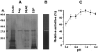
Fig. 1. (A) Determination of the homogeneity of a 28 kDa cysteine protease purified from the excretory-secretory products (ESP) of the adult Clonorchis sinensis by 10–15% SDS-PAGE under reducing conditions. The bound fraction of the 20 mM salt concentration (lane DEAE), where a high enzyme activity was shown, was dialysed against sodium phosphate buffer (100 mM, pH 7·0), lyophilized, and subjected to Arginine-Sepharose 4B affinity chromatography. The bound protein, eluted with a 10 mM salt concentration, exhibited a single band at 28 kDa with high enzyme activity (lane Arg, 1 μg loaded). Mr, molecular weight in kDa; Lanes ESP, crude ESP; Crude, crude extracts of the adult C. sinensis (30 μg loaded). (B) The proteolytic activity of the partially purified enzyme fraction shown by gelatin substrate gel electrophoresis. After the electrophoresis of 3 μg protein by 10–15% SDS-PAGE in non-reducing conditions, the gel was incubated in 100 mM sodium phosphate buffer at pH 7·0 overnight at 37 °C. A distinct zone of proteolysis appeared at approximately 28 kDa. (C) Modulation of the enzyme activity by pH. A total of 2 μg purified enzyme was incubated with 5 mg Azocoll supplemented with 5 mM DTT in 100 mM sodium acetate buffer at pH 5, 5·5 and 6, in 100 mM sodium phosphate buffer at pH 6·6 and 7 and in 100 mM Tris–HCl buffer at pH 7·5 and 8. Mean±S.D., n=3.
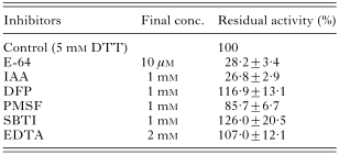
Table 1. Modulation of the enzyme activity by various inhibitors
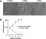
Fig. 2. In vitro cytolytic effect of Cs28CF on monkey cholangiocytes. A total of 106 cells were cultured in the presence of 2 μg/ml Cs28CF supplemented with 1 mM DTT. (A) Microscopic illustration of the morphological changes in the cells cultured with Cs28CF. (B) Trypan blue exclusion analysis of the cell proliferation rate at different times. [bull ], cholangiocyte cultured without Cs28CF; ○, cells cultured with Cs28CF.
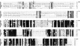
Fig. 3. Alignment of the predicted amino acid sequence of a Cs28CF with other related members of cathepsin F. The conserved amino acids are shaded and gaps (-) are introduced to optimize the alignment. Prepro- and pro-region are indicated by the arrows. Four α-helical domains in the pro-region are underlined. The NH2-terminal sequence is marked by a double underline. The cysteine protease catalytic triad (C, H, and N) is shown by bold-underline. Putative sites for mannose 6-phosphate N-glycosylation are indicated by the boxes. Substrate-binding amino acid residue (G) is shown with the arrowheads. The asterisks are showing the 7 Cys, which are essential for forming tertiary structures by a disulfide linkage. The ERFNAQ motif is also shown. In addition, a small motif of E/DXGTA, indicated by the double-lined box is recognized in cathepsin Fs except for that from Schistosoma mansoni, which is followed by the ERFNAQ motif. The numbers in parentheses start from the N terminus of Cs28CF. Cs, Cs28CF (AF093243); Pw, Paragonimus westermani cruzipain (U70537); Sm, Schistosoma mansoni cathepsin L1 (U07345); Ce, Caenorhabditis elegans cathepsin L (NM_072814); hum, human cathepsin F (NM_003793); mou, mouse cathepsin F (MMU131851); Nt, Nicotina tabacum pre-pro-cysteine proteinase (AJ242994); Ib, Ipomoea batatas papain-like cysteine protease isoform (AF138264).
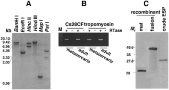
Fig. 4. Genomic distribution and expression of Cs28CF by Southern (A), RT-PCR (B) and Western blot analysis (C). (A) The genomic DNA was digested with the restriction enzymes of BamH I, EcoR I, Hinc II, Hind III, Msp I or Pst I and hybridized with the full-length cDNA. BamH I cuts at position +312 yields fragments of 7 kb and a large fragment of >15 kb. EcoR I at position +860 shows fragments of 3·1 kb and 3·5 kb. Hinc II and Hind III (no cut at cDNA) give rise to fragments of 4·2 kb and >15 kb, respectively. (B) RT-PCR with the gene specific primers for Cs28CF shows that the mRNA transcript of Cs28CF was expressed in both the metacercaria and adult stages. The tropomyosin gene, which was used as a control, also appears to be constantly expressed in both stages. M, 100 bp ladder. (C) Western blots of the recombinant and native proteins. The proteins were resolved by 10–15% SDS-PAGE, transblotted to an Immobilon-P transfer membrane and reacted with the monospecific antibodies against the recombinant protein. Mr, molecular weight in kDa; mat, thrombin cleaved mature domain; fusion, mature domain linked with GST carrier; crude ESP, crude ESP of C. sinensis.
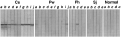
Fig. 5. Immunoblot analysis of rCs28CF against serum samples of trematode infections. After transfer-blot of rCs28CF separated by 12% SDS-PAGE, each strip was incubated with the respective serum in a dilution of 1[ratio ]100, subsequently with 1[ratio ]1000 diluted peroxidase conjugated anti-human IgG. The colour reactions were developed with 4C1N chromogen. Cs, clonorchiasis; Pw, paragonimiasis; Fh, fascioliasis; Sj, schistosomiasis japonicum; Normal, normal controls.
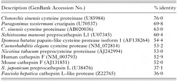
Table 2. Comparison of the identies between Clonorchis sinensis cathepsin F and the other related members
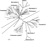
Fig. 6. Phylogenetic relationship of Cs28CF to the other cathepsin L complexes based on the deduced mature domain. The tree was constructed using the neighbour-joining algorithm in PHYLIP package and was unrooted. The numbers at branching node represent the bootstrap value of 1000 replicates. The numbers of each element correspond to the following (GenBank Accession numbers are given in parentheses): 1, Toxocara canis cathepsin L-like cysteine proteinase (U53172); 2, C. sinensis cysteine proteinase (U85984); 3, C. sinensis cysteine proteinase 1 precursor (AF093243, in this study); 4, C. sinensis cysteine proteinase (U85983); 5, C. sinensis cysteine proteinase (AB020036); 6, P. westermani neutral thiol protease (D21124); 7, P. westermani cysteine proteinase (AF071801); 8, P. westermani pre-procathepsin L (AF362769); 9, P. westermani cruzipain (U70537); 10, human cathepsin F (NM_003793); 11, P. westermani cysteine proteinase (U69120); 12, Leishmania major cathepsin L-like protease (U43706); 13, Schistosoma mansoni Puerto Rican preprocathepsin L (U07345); 14, mouse cathepsin W (NM_009985); 15, human cathepsin W (NM_001335); 16, Leishmania donovani cathepsin L-like protease (AF004593); 17, Trypanosoma cruzi cruzain (M84342); 18, T. brucei cathepsin L-like protease (X54353); 19, T. congolense cysteine protease (L25130); 20, L. chagasi cathepsin L (AF217087); 21, L. mexicana cysteine protease (AJ319727); 22, L. mexicana lmcpb2.8 (Z49962); 23, Hypera postica cysteine proteinase (AF157961); 24, Tetrahymena pyriformis (D86408); 25, Cryptosporodium parvum cryptopain (AF091366); 26, Schistosoma mansoni cathepsin L (Z32529); 27, S. japonicum preprocathepsin L (U38476); 28, Spirometra mansoni cathepsin L (D63670); 29, Fasciola hepatica secreted cathepsin L 1 (U62288); 30, F. gigantica cathepsin L1 (AF112566); 31, F. hepatica cathepsin L (AB009306); 32, Fasciola sp. cysteine protease (S70380); 33, F. hepatica cathepsin L-like protease (L33772); 34, F. gigantica cathepsin L (AF239264); 35, F. hepatica cathepsin L (AF271385); 36, F. gigantica cathepsin L1C (AF239265); 37, F. gigantica cathepsin L1D (AF239266); 38, F. hepatica secreted cathepsin L2 (U62289); 39, F. hepatica cathepsin L-like protease (Z22765); 40, F. hepatica procathepsin L3 (AJ279093); 41, F. gigantica cathepsin L (AF419329); 42, mouse preprocathepsin P (AF158182); 43, zebrafish cathepsin L (Y08321); 44, mouse cathepsin L (M20495); 45, rat cathepsin L (NM_013156); 46, pig cathepsin L (AJ315771); 47, African green monkey cysteine protease (AF201700); 48, human cathepsin L (NM_001912); 49, human cathepsin L (M20496); 50, human pro-cathepsin L (X12451); 51, human cathepsin S (M90696); 52, Brugia pahangi cathepsin L (AF031819); 53, Onchocerca volvulus cathepsin L (AF331036); 54, B. malayi cathepsin L (AF331035); 55, Litomosoides sigmodontis (AF331037) cathepsin L; 56, human cathepsin K (NM_000396); 57, human cathepsin O (X82153); 58, Artemia franciscana (shrimp) cathepsin L (AF147207); 59, Heterodera glycines cathepsin L (Y09498); 60, Drosophila melanogaster cysteine proteinase 1 (U75652); 61, Sarcophaga peregrina (insect) cathepsin L (D16533); 62, Frankliniella occidentalis (western flower thrips) cathepsin L (AF410883); 63, Acanthamoeba healyi cysteine proteinase (AF462309).
 You have
Access
- 34
- Cited by