Introduction
The commercial success of in vitro-derived embryos is compromised by inadequate culture conditions that produce morphologically impaired embryos (Hansen, Reference Hansen2006). This fact is reflected by poor embryo viability and subsequent low post-thaw embryo survival and low pregnancies rates (Polard & Leibo, 1994; Lonergan et al., Reference Lonergan, Rizos, Gutierrez-Adan, Moreira, Pintado, de la Fuente and Boland2003). This quality seems to be determining factor in the limited use of in vitro-produced bovine embryos (Enright et al., Reference Enright, Lonergan, Dinnyes, Fair, Ward, Yang and Boland2000; Rizos et al., Reference Rizos, Lonergan, Boland, Arroyo-Garcia, Pintado, de la Fuente and Gutierrez-Adan2002). Culture media used for mammalian embryos vary greatly in its composition (Whitten, Reference Whitten1957; Xu et al., Reference Xu, Yadav, Rorie, Plante, Betteridge and King1992; Krisher et al., Reference Krisher, Gibbons and Gwazdauskas1998), its ability to support development (Bavister et al., Reference Bavister, Rose-Hellekant and Pinyopummintr1992; Keskintepe et al., Reference Keskintepe, Burnely and Brackett1995; Thompson et al., Reference Thompson, Partridge, Houghton, Cox and Leese1996) and its resulting embryo quality, assessed by the expression level of developmentally important gene transcripts (Wrenzycki et al., Reference Wrenzycki, Herrmann, Keskintepe, Martins, Sirisathien, Brackett and Niemann2001). Addition of serum or the use of somatic cell co-culture in mammalian embryo culture further complicates a proper assessment and evaluation of the role of individual culture media components (Bavister, Reference Bavister1992). The use of defined or semi-defined media seems to alleviate the problem, however (Lane et al., Reference Lane, Gardner, Hasler and Hasler2003a). Lane and colleagues reported that bovine embryos that were produced in the G1.2/G2.2 culture system resulted in pregnancies, following both fresh transfer and cryopreservation at equivalent rates to co-culture. Attempts at delipidation of bovine embryos, in order to improve embryo quality, showed beneficial effects on their tolerance to freezing and thawing. However, as a result, this altered the developmental potential of delipidated blastocysts (Diez et al., Reference Diez, Heyman, Bourhis, Guyader-Joly, Degrouard and Renard2001). It seems obvious that parameters for optimal in vitro embryo development have been vague and parameters have been not identified as the first reported successful culture of rabbit embryos (Brachet, Reference Brachet1913).
In an attempt to improve the quality of in vitro-produced embryos we used the biological polymer hyaluronan, which is a main component of the extracellular matrix in which most tissues differentiate and develop (Toole, 1997). Hyaluronan is present transiently in both the cell nucleus and cytoplasm (Entwistle et al., Reference Entwistle, Hall and Turley1996) and is a glycosaminoglycan (GAG), which is composed of uronic acid and an amino sugar, and unlike other GAGs is not sulphoester. Until very recently HA was only modestly investigated in bovine oocyte/embryo culture. The role of HA in embryonic development starts very early (Eppig, Reference Eppig1981; Salustri et al., 1998), and its levels are associated with normal oocyte development and intracellular events (Tirone et al., Reference Tirone, D'Alessandris, Hascall, Siracusa and Salustri1997; Bergqvist et al., Reference Bergqvist, Yokoo, Heldin, Frendin, Sato and Rodríguez-Martínez2005). Many of those processes are mediated through cell-surface receptors for hyaluronan, such as CD44, RHAMM and ICAM-1 (Turley et al., Reference Turley, Austen, Vandelight and Clary1991; Entwistle et al., Reference Entwistle, Hall and Turley1996; Knudson et al., Reference Knudson, Aguiar, Hua and Knudson1996). Recently, CD44 and RHAMM HA receptors have been identified in bovine preimplantation embryos (Furnus et al., Reference Furnus, Valcarcel, Dulout and Errecalde2003; Stojkovic et al., Reference Stojkovic, Krebs, Kolle, Prelle, Assmann, Zakhartchenko, Sinowatz and Wolf2003). It is now apparent that hyaluronan plays a central role in the physical and chemical properties of intracellular and extracellular environments of the developing embryo.
Extracellularly, hyaluronan in aqueous solutions has a tendency to a form three-dimensional network (Scott, Reference Scott and Presti1992; Kobayashi et al., Reference Kobayashi, Okarnoto and Nishinari1994). It can be assumed that hyaluronan provides a sensitive means of retaining critical concentrations of necessary metabolites in the diminutive volume of fluid surrounding the oocyte or embryo. In addition, the role of hyaluronan always seems to be associated with membranes. Contrary to other glycosaminoglycans, which are synthesized in the Golgi apparatus, hyaluronan is synthesized in the plasma membrane (Prehm, Reference Prehm1984). The present studies were designed to investigate the effect of high molecular weight hyaluronan on the developmental competence, viability and gene expression patterns of in vitro-produced bovine embryos cultured in TCM-199 medium.
Materials and Methods
Oocyte maturation (IVM) and fertilization (IVF) and embryo culture (IVC)
Cumulus–oocyte complexes (COC) were aspirated from follicles 2 to 7 mM in diameter from slaughterhouse-derived ovaries and assigned randomly to maturation media. After selection based on morphological evaluation (Lonergan et al., Reference Lonergan, Crolan, Van Langendonck, Donnay, Kathir and Mermillod1996), COC were washed three times in a modified Tyrode's-lactate solution, and once in TCM-199 medium (Sigma; that contains the surfactant Tween-80) supplemented with 10% fetal calf serum (FCS) and 10 ng/ml epidermal growth factor (EGF), which was the final maturation medium. Oocytes were matured in 500 μl medium (approximately 50 per drop) without paraffin oil in Nunclon dishes (Nunc). All media were supplemented with 50 IU penicillin-G and 50 μg streptomycin sulphate per ml. After 24 h in the maturation medium, oocytes were inseminated in 20 μl Fert-Talp with 30 μl of 1 × 10⁶ spermatozoa per ml selected by swim-up through hyaluronan (Palasz et al., Reference Palasz, Alkemade and Mapletoft1996) into Fert-Talp medium supplemented with 6 mg/ml fatty acid-free BSA, 5.0 μM/ml heparin, 1.0 μM hypotaurine, 1.0 um epinephrine and 1.0 mM sodium pyruvate (Parrish et al., Reference Parrish, Susko-Parrish, Winer and First1988). Swim-up medium was prepared in Fert-Talp with 6 mg/ml BSA, 1.0 mM of sodium pyruvate and 1 mg/ml high molecular weight (600 ± 100 kDa) HA (MAP-5; Bioniche Inc.). Presumptive zygotes were cultured in 40 μl drops (approximately 25 zygotes per drop) of TCM-199 medium supplemented with 0.25 mM sodium pyruvate and 15 μg/ml insulin, transferrin, selenium (ITS) and 4 mg/ml BSA, in Falcon dishes (Becton Dickinson & Co.) under paraffin oil. All oocytes and embryos were incubated at 39 °C in humidified air with 5% CO2, 5% O2 and 90% N2. Cleavage rates were evaluated 46 h post-insemination and the proportion of embryos developing in culture was recorded on days 4, 6, 7, 8 and 9 (insemination = day 0). Embryo developmental stage and morphology was assessed under a phase-contrast microscopy (200×) and quality was determined by embryo morphology, total cell count, by the post-thaw viability and mRNA gene expression patterns.
Assessment of embryo development and morphology
Embryo quality was evaluated by quantitative and qualitative methods described separately in each experiment. Blastocyst developmental stages were determined by the size of blastocoel cavity: (i) early blastocyst – clearly visible; (ii) blastocyst up to two-thirds volume of the embryo; (iii) full blastocyst – 100% volume of the embryo without visible thinning of the zona pellucida; (iv) expanded blastocyst – occupied 100% volume of the embryo with visible thinning of the zona pellucida; (v) hatching blastocyst – zona pellucida open and part of embryo proper out of the zona; (vi) hatched blastocyst – entire embryo is out the zona pellucida.
Determination cell numbers
The total number of cells in each embryo was determined by nuclei counts after day 8 of culture. A total of 8–10 embryos at the same stage of development were taken from each treatment group in each replicate. Embryos were washed twice in a 100 μl drop of PBS containing 0.5 mg/ml surfactant pluronic F-68 and then fixed in a 100 μl drop of 4% paraformaldehyde solution (w/v) in PBS, (pH 7.4) for 1 h at room temperature. Embryos were then washed three times in 100 μl drops of PBS with pluronic F-68 and transferred to a 50 μl microdrop of Hoechst 33342 dye. After 10 min of staining, embryos were transferred in minimum volume to a glass slide coated previously with Sigma coat, allowed to settle and then a cover slip was placed over the drop. Slides were stored in the dark at 4 °C until examined microscopically. The total number of cells was counted under fluorescence microscope (λ 320 nm). All chemicals were purchased from Sigma Chemical Co. unless otherwise stated.
Embryo freezing and thawing
As a measure of viability day 7 and 8 blastocysts and expanded blastocysts were cryopreserved in TCM-199 medium with HEPES supplemented with 10% propylene glycol and 0.2 M sucrose. Embryos were equilibrated in a single step at room temperature for 10 min during which three to five blastocysts were loaded into 0.25 ml straws (IMV, L'Aigle, France. Straws were placed directly into the freezing chamber of an alcohol freezer (Bio-Cool 3; FTS-Systems, Inc.) at −5 °C, and after 1 min were seeded with cotton Q-tip, held for 10 min, cooled at 0.5 °C/min to −35 °C, and then plunged into liquid nitrogen. Embryos were thawed for 10 s in the air and then in a 35 °C water bath without stirring until the ice was melted (approximately 10 s). Embryos were expelled into TCM-199 medium with 0.5 M sucrose and held for 3 min at 25 °C. Then embryos were washed twice in HEPES and once in TCM-199 medium containing 0.3% BSA. The embryos were placed in TCM-199 medium containing 0.3% BSA, embryo morphology and development were evaluated under the stereoscope after 2 and 24 h. Embryos that re-expanded within 2 h after thawing and progressed to the next developmental stage in culture were considered as viable.
RNA extraction and reverse transcription (RT)
Poly(A) RNA was extracted from four to five pools of 10 embryos, following the manufacturer's instructions using the Dynabeads mRNA Direct KIT (DYNAL Biotech, Oslo, Norway). Briefly, samples were lysed in 50 μl lysis/binding buffer (DYNAL). After vortexing and a brief centrifugation, the samples were incubated at room temperature for 10 min. Ten microlitres of prewashed Dynabeads oligo(dT)25 were added to each sample. After 5 min of hybridization, the beads were separated from the binding buffer using the Dynal magnetic separator and beads were washed in buffers A and B (DYNAL) and the poly(A) RNA was eluted from the beads by adding 11 μl of DEPC-treated water. The resultant poly(A) mRNA was used in the RT-PCR in a total volume of 20 μl using 2.5 μM random hexamer primers, 1× RT buffer, 20 IU RNase inhibitor, 50 IU mMLV reverse transcriptase enzyme (Promega, Madrid, Spain), 5 mM MgCl2 and 1 mM of each dNTP. Tubes were heated to 70 °C for 5 min to denature the secondary RNA structure before the addition of RT enzyme. The reverse transcription reactions were incubated at room temperature for 10 min and then at 42 °C for 60 min to allow the reverse transcription of RNA, followed by 93 °C for 1 min to denature the RT enzyme.
Quantitative RT-PCR
The quantification of all gene transcripts was carried out by real time quantitative RT-PCR in three replicate PCR experiments. PCR was performed using a Rotorgene 2000 Real Time CyclerTM (Corbett Research, Sydney, Australia) and SYBR Green (Molecular Probes, Eugene, OR) as a double-stranded DNA-specific fluorescent dye. The PCR reaction mixture (25 μl) contained 2.5 μl 10× buffer, 3 mM MgCl2, 2 U Taq Express (MWGAG Biotech, Ebersberg, Germany), 100 μM of each dNTPs, and 0.2 μM of each primer.
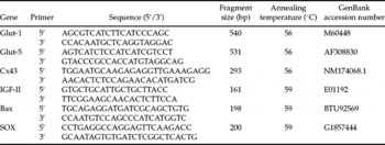
In addition, the double-stranded DNA dye, SYBR Green I, (1:3000 of 10 000× stock solution) was included in each reaction. The PCR protocol included an initial step of 94 °C (2 min), followed by 40 cycles of 94 °C (15 s), 56 to 59 °C (30 s) and 72 °C (30 s). Fluorescence data were acquired at 85 °C. The melting protocol consisted of holding at 40 °C for 60 s and then heating from 50 to 94 °C holding at each temperature for 5 s while monitoring fluorescence. Product identity was confirmed by ethidium-bromide-stained 2% agarose gel electrophoresis. As negative controls, tubes were prepared in which RNA or reverse transcriptase was omitted during the RT-reaction. The comparative CT method was used for quantification of expression levels. The quantification was normalized to the endogenous control histone H2a. Fluorescence was acquired in each cycle in order to determine the threshold cycle or the cycle during the log–linear phase of the reaction at which fluorescence increased above background for each sample. Within this region of the amplification curve, each difference of one cycle is equivalent to a doubling of the amplified product of the PCR. According to the comparative Ct method, the ΔCt value was determined by subtracting the H2a Ct value for each sample from each gene Ct value of the sample. Calculation of ΔΔCt involved using the highest sample ΔCt values (i.e., the sample with the lower target expression) as an arbitrary constant to subtract from all other ΔCt sample values. Fold changes in the relative gene expression of target was determined by using the formula, 2-ΔΔCt. The details of primers used for RT-PCR are shown in Table 1.
The effect of TCM-199 medium with or without BSA and/or HA added to the culture drops on day 4 of culture on embryo development

a −cPercentages with different superscripts differ significantly (p < 0.05).
Experimental design
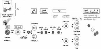
A total of 676 presumptive zygotes in five replicates were cultured in TCM-199 medium + BSA or TCM-199 medium alone for 96 h. On day 4 of culture, half of the embryos in each group were placed in fresh culture drops (no change) and the other half were placed in fresh culture drops supplemented with 1 mg/ml HA (MAP-5) in a 2 × 2 factorial design (Fig. 1).
Experimental design.

At least seven blastocysts from each treatment group were fixed for nuclei counts. Five day 7 blastocysts from each replicate from each treatment were frozen at −79 °C (Wrenzycki et al. Reference Wrenzycki, Herrmann, Carnwath and Niemann1999) for the evaluation of molecular composition expressed by mRNA transcription of six developmentally important genes: apoptosis (Bax), oxidative stress (SOX), growth factor (IGF-II), cell connection (Cx43) and transport and metabolism, glucose (Glut-1) and fructose (Glut-5) gene expression patterns. The remaining embryos in each group that developed to the blastocyst stage were cryopreserved to evaluate cryotolerance and viability.
Statistical analysis
Differences between percentages of embryos developing in culture were compared by chi-square analysis with the correction by Fisher's Exact test. Differences in blastocyst cell numbers were compared by ANOVA. Data on mRNA expression were analysed using the SigmaStat (Jandel Scientific, San Rafael, CA) software package. One-way repeated-measures ANOVA (followed by multiple pair-wise comparisons using Student–Newman–Kleus method) were used for the analysis of differences in mRNA expression assayed by quantitative RT-PCR.
Results
There were no differences in cleavage rates on day 2 (p = 0.12) and embryo development to the 16-cell stage on day 4 (p = 0.45) between zygotes cultured in TCM-199 medium or TCM-199 medium + BSA. The addition of HA on day 4 to TCM-199 medium containing BSA resulted in the highest proportion of embryos developing to the blastocyst stages on days 7, 8 and 9 (p < 0.05; Table 2). Whilst TCM-199 medium alone resulted in the lowest blastocyst production rate; the other two groups were intermediate and not different (Table 1).
Primers used for RT-PCR

The mean number of cells in day 7 blastocysts cultured in TCM-199 medium only (101 ± 18; n = 15) was significantly lower (p < 0.05) than in TCM-199 medium + HA (129 ± 17; n = 12), TCM-199 medium + BSA (136 ± 11; n = 12); and in TCM-199 medium + BSA + HA (151 ± 15; n = 18), which were not different.
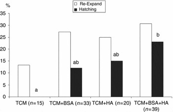
The addition of HA on day 4 of culture to TCM-199 medium containing BSA resulted in a significant increase (p < 0.05) in embryo development to the hatching stage after freezing and thawing, as compared with the group without both BSA and HA (Figure 2).
The effect of embryo culture in TCM-199 medium with or without BSA and on day 4 of culture with HA on embryo survival following freeze–thawing procedures. Bars with different superscripts differ significantly (p < 0.05).

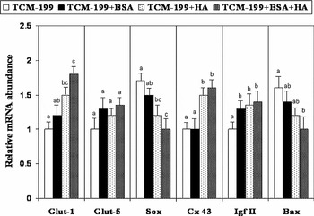
Differences in mRNA expression were found for genes related to glucose transport and metabolism (Glut-1, peroxidation of lipid membranes (SOX), cells apoptosis (Bax) and cell-to-cell adhesion related to communication trough gap junction (Cx-43). No differences were found in the level of expression genes for transport and metabolism of fructose (Glut-5) and growth factor (IGF-II). Expression of SOX was reduced significantly (p < 0.05) in TCM-199 medium supplemented with HA, whether or not BSA was present, and Bax expression was significantly lower in TCM-199 medium plus BSA than in other groups (Figure 3).
Relative abundance of six developmentally important gene transcripts in bovine blastocysts developed in TCM-199 medium with or without BSA and with or without HA added to culture drops on day 4 of culture. Bars with different superscripts differ significantly (p < 0.05).

The level of expression of glucose transport and metabolism (Glut-1) gene was significantly higher in blastocysts produced in TCM-199 medium plus HA, regardless of whether BSA was present in the medium. The level of expression of cell-to-cell adhesion gene Cx-43 was significant higher in both HA groups than in TCM-199 medium alone or plus BSA groups (p < 0.05). However, the level of expression of transport and metabolism of fructose (Glut-5) gene was not affected, while the expression of IGF II growth factor gene was significantly lower in TCM-199 medium, alone as compared with the other three groups (p < 0.05).
Discussion
Culture of embryos with HA resulted in an improved blastocyst development, higher post-thaw survival and improved gene expression patterns compared with those embryos cultured without HA. The use of HA in TCM-199 medium showed a profound effect on the expression of a set of developmentally important genes indicative of embryo metabolism, apoptosis, cell compaction and oxidative stress. Previously, Wrenzycki et al. (Reference Wrenzycki, Herrmann, Keskintepe, Martins, Sirisathien, Brackett and Niemann2001) had shown that the expression of developmentally important genes of day 7 blastocysts cultured in TCM-199 medium (without HA) was inferior to embryos cultured in SOF medium. In the present studies, addition of HA to TCM-199 medium showed a visible improvement in embryo molecular make-up as compared with TCM-199 medium supplemented with BSA only. In our study, we used a concentration of 1 mg/ml of HA, which was based on our previous dose titration trial (Palasz et al., Reference Palasz, Alkemade and Mapletoft2000). This concentration may appear high, when compared with the physiological dose of approximately 0.5 mg/ml of HA in the cumulus cell matrix surrounding the oocytes during ovulation (Salustri et al., Reference Salustri, Yanagishita, Underhill, Laurent and Hascall1992) or for epidermal HA (0.1–0.2 μg/ml). However, HA concentration may be higher (∼2.5 mg/ml) in the area surrounding the cells, because of restriction of HA to the small extracellular spaces (Tammi et al., Reference Tammi, Ripellino, Margolis, Maibach and Tammi1989). Previously, Stojkovic et al. (Reference Stojkovic, Krebs, Kolle, Prelle, Assmann, Zakhartchenko, Sinowatz and Wolf2003) used a concentration as high as 6 mg/ml for bovine embryo culture and in other studies a dose as low as 0.125 mg/ml of HA was used (Lane et al., Reference Lane, Maybach, Hooper, Hasler and Gardner2003b). It is relatively well established that cell movement and proliferation requires sequential cell attachment and detachment at localized areas, processes in which HA seems to play a critical role. The function of HA is hydration and separation. Hydration because it holds on to water and separation because it acts as a structural component throughout the body. Such hydration is largely dependent on molecular weight and at a specific concentration HA can restricts water movement, which gives rise to the formation of large cell-free spaces at the time of cell movement. It is proposed that these spaces are avenues for cell migration and are closely correlated with hyaluronan configuration and concentration (Shu et al., Reference Shu, Liu, Luo, Roberts and Prestwich2002). Therefore, further studies are required to investigate the effects of HA concentration on embryo culture.
Addition of HA to TCM-199 medium supplemented with BSA 4 days after fertilization produced higher quality embryos, as reflected by improved molecular characteristics and improved embryo development after freezing. Improvement in post-thaw embryo survival and developmental competence may be attributed to the improved molecular composition of embryos cultured with HA and enhancement of embryo fine structure, as reported previously (Palasz et al., Reference Palasz, Rodriguez-Martinez, Beltran-Breña, Perez-Garnelo, Martinez, Gutierrez-Adan and De la Fuente2006). In that study, embryos cultured in SOF medium and HA accumulated less lipids and the distribution of lipid deposits appeared more normal, with no residual bodies/phagosomes. Hyaluronan C44 transmembrane and RHAMM intracellular receptors may also play a role in the enhancement of embryo fine structure. Specifically, it has been shown that RHAMM interacts with microtubules and actin filaments (Assmann et al., Reference Assmann, Jenkinson, Marshall and Hart1999), which may have far reaching consequences regarding organization and cryotolerance of the cytoskeleton. Increased re-expansion and hatching rates after cryopreservation of bovine embryo previously cultured in HA with different macromolecular supplements has been reported (Furnus et al., Reference Furnus, de Matos and Martínez1998; Lane et al., Reference Lane, Gardner, Hasler and Hasler2003a). In the present study, only embryo hatching rates were affected by the presence of HA in culture, but the number of embryos re-expanding after freeze/thawing was also higher than in the other groups. The relatively low number of embryos frozen in this study resulted from uneven embryo development among the groups, which made it difficult to make any further comparisons.
The positive effect of HA on embryo development in culture may also be related to any changes that HA may create in the microenvironment immediately surrounding zygotes. In aqueous solutions, cross-linked HAAHA forms hydrogel in three-dimensional structures (Scott et al., Reference Scott, Cummings, Brass and Chen1991; Tian et al., Reference Tian, Hou, Ma, Zhang, Xu, Lee, Li, Spector and Cui2005), similar to the in vivo conditions in which the embryos develop. The movement of the cells, determination of cell fate, signal transduction and numerous other biological functions can not be replicated in existing two-dimensional in vitro culture systems. Cell surface receptors that are expressed on the culture medium side of the embryo cells have better access to active components of the medium than cell receptors expressed on cells that face the surface of the Petri dish (Zhang, Reference Zhang2004). The uneven distribution of receptors in two-dimensional in vitro culture systems may affect communication between cells. It is believed that HA may change the local concentration of intracellular signals and influence cell growth and differentiation by binding polypeptide growth factors (Ruoslahti & Yamaguchi, Reference Ruoslahti and Yamaguchi1991). The poly-aminoglycans are good candidates for use in the development of three-dimensional cultures (Cukierman et al., Reference Cukierman, Pankov and Yamada2002) and hyaluronan could be the best candidate. It is well known that HA is the main component of extracellular matrix (ECM) and its role and concentration increases in proportion to fetus development (Fenderson et al., Reference Fenderson, Stamenkovic and Aruffo1993). However, regulation of ever-changing hyaluronan concentrations needed for cell movement within the zona and differentiation in vitro may present a formidable challenge. In addition, if we accept the theory that the cytoplasm of living cells is a gel, rather than an aqueous solution (Pollack, Reference Pollack2001), HA may possess all the necessary properties to create a gel-like intracellular environment and the three-dimensional architecture extracellularly.
We were able to demonstrate that HA in the culture medium had a profound effect on the expression of selected developmentally important genes: glucose (Glut-1) and fructose (Glut-5), transport and metabolism, cell-to-cell adhesion (Cx-43), growth factor (IGF-II), peroxidation of lipid membranes (SOX) and apoptosis (Bax). The expression levels of the oxidative stress gene (SOX) and cell apoptosis gene (Bax), situated on autosomal chromosomes, were considerably lower in blastocysts cultured with HA. In the case of SOX, this effect was more profound when BSA was present. Oxidative modification of cell components, due to the action of reactive oxygen species, is one of the most potentially damaging processes, including increase in DNA fragmentation, in in vitro embryo culture. The expression of many genes may be affected by reactive oxygen species (Guerin et al., Reference Guerin, Mouatassim and Menozo2001). Interestingly, the glycol group in the glucuronate residue of the HA structure has been shown to be resistant to periodate oxidation (Scott et al., Reference Scott, Cummings, Brass and Chen1991). Studies using in vivo and in vitro culture of cells have confirmed the scavenging properties of HA, which inhibits the release of free oxygen radicals (Presti & Scott, Reference Presti and Scott1994). The expression of Bax was reduced significantly in blastocysts cultured in TCM-199 medium supplemented with HA and BSA than in TCM-199 medium with or without BSA. The frequency of apoptosis has been reported to be higher in in vitro- than in vivo-derived blastocysts (Gjorret et al., Reference Gjorret, Avery, Larsson, Schellander and Hyttel2001). Therefore, the reduction in Bax expression in blastocysts cultured in HA may prevent early embryonic death, deformation of fetus or early abortions associated with in vitro-derived embryos (Brill et al., Reference Brill, Torchinsky, Carp and Toder1999). In bovine ovaries, Cx43, a protein related to gap junctions, is essential for normal follicular growth (Nuttinck et al., Reference Nuttinck, Peynot, Humblot, Massip, Dessy and Flechon2000) and in preimplantation embryos it is important for active transport of fluids through embryonic cell membranes (Rizos et al., Reference Rizos, Lonergan, Boland, Arroyo-Garcia, Pintado, de la Fuente and Gutierrez-Adan2002). In the present study Cx43 mRNA was strongly expressed in both HA groups (with and without BSA) and was considerably weaker in groups without HA. The high level of expression of Cx43 mRNA in blastocysts derived in vivo has been reported previously (Rizos et al., Reference Rizos, Lonergan, Boland, Arroyo-Garcia, Pintado, de la Fuente and Gutierrez-Adan2002), and this is further supported by embryo fine structure following cryopreservation (Fair et al., Reference Fair, Lonergan, Dinnyes, Cottell, Hyttel, Ward and Boland2001). The level of expression of Glut-5 mRNA, an important fructose transporter and responsible for nucleotide synthesis after zygotic transcription, did not differ among the groups and there was a moderate effect of HA on the expression of IGF-II mRNA. Conversely, expression of Glut-1 mRNA was significantly higher in HA culture groups than in those groups that did not include HA. Increased Glut-1 mRNA expression has also been demonstrated with low (2%) oxygen culture (Harvey et al., Reference Harvey, Kind, Pantaleon, Armstrong and Thompson2004). Glucose is readily taken up and utilized by preimplantation embryos of different species, and its uptake is controlled by the expression of Glut-1, the mRNA transcripts of which have been shown to be 10-fold higher in in vivo mouse blastocysts than those produced in vitro (Leppens-Luisier et al., Reference Leppens-Luisier, Urner and Sakkas2001).
It can be concluded that the diverse properties of hyaluronan resulted in the production of high quality embryos, both in the presence and absence of BSA. The relative abundance and pattern of expression of selected, developmentally important marker genes used in this study strongly indicates a positive effect of HA in the in vitro culture of bovine embryos in TCM-199 medium. Further exploration of the unique and highly versatile properties of hyaluronan may contribute to the development of in vitro culture systems that better mimic in vivo conditions.
Acknowledgment
This study was supported by grant # OT2006-002 and in part by grant AGL2003-05783 from the Ministerio de Educación y Ciencia, Madrid, Spain. We would like to acknowledge and thank Bioniche Animal Health, Belleville, ON, Canada for providing MAP-5 used in this study.