I. INTRODUCTION
Quasi-one-dimensional (1D) nanostructures of metals have drawn considerable attention because of their unique physical properties and potential applications in fabricating nanoscale electronic devices as both interconnectors and active components.Reference Xia, Yang, Sun, Wu, Mayers, Gates, Yin, Kim and Yan1, Reference Ma, Zhang, Xi, Zhang and Qian2 Among the 1D nanostructure candidates, metal nanowires have attracted particular interest in that they are expected to prospectively play an imperative role in fabricating nanoscale electronic,Reference Sun3 optoelectronic,Reference Guo, Qiu, Bao, Wiley, Yang, Zhang, Ma, Yu and Tong4 or magnetic devices.Reference Sun, Searson and Chien5 They offer an ideal model system to experimentally investigate physical phenomena such as quantized conductance and localization effects.Reference Grado-Caffaro and Grado-Caffaro6 The function of silver in particular has been extensively exploited in photonics,Reference Sun3, Reference Dickson and Lyon7 electronics,Reference De, Higgins, Lyons, Doherty, Nirmalraj, Blau, Boland and Coleman8 photography,Reference Pestryakov, Bogdanchikova and Knop-Gericke9 catalysis,Reference Lu, Xue and Li10 biological labeling,Reference Zong, Cao, Jia, Bao and Lu11 and surface-enhanced Raman scattering.Reference Zhao, Fu, Tian, Ren and Tian12 When silver takes the form of 1D nanostructures, its performance in many aspects could be potentially enhanced.Reference Sun3 On the nanometer scale, however, metallic materials are normally sensitive to air and moisture, so mounting an inertial sheath can protect the liable material, avoiding the degradation of performance of nanodevices.
Carbon, from diamond, graphite (graphene), fullerenes, nanotubes to diamond-like ultrathin films, is a versatile, fascinating material of fundamental value and technological importance.Reference Hoheisel, Schrettl, Szilluweit and Frauenrath13–Reference Hu, Wang, Wu, Yu, Antonietti and Titirici16 For its inertness, the material as coating offers proper protection of active substances. Nanoengineering the two materials, silver and carbon, in the configuration of a Ag-core and a carbon-shell offers a new kind of nanostructures or coaxial nanocables as a promising candidate for nanodevices, which have resultantly received noteworthy awareness.Reference Hu, Wang, Wu, Yu, Antonietti and Titirici16–Reference Jin, Kuang, Jiang, Xu, Xie and Zheng19 Such heterostructural nanoassembly can have more advantages in protecting the core material from oxidation and corrosion, dodging direct contact between biological tissues and the core material, affording an easy, effective chemical process, and offering enhanced functions and properties.Reference Zheng, Meng and Tang20–Reference Wang, Xiong, Chen, Xi, Zhou and Zhang23 Several methods have been applied, with the assistance of catalysts or surfactants, to synthesize silver–carbon (Ag–C) nanocables. For example, a cetyltrimethylammonium bromide-assisted hydrothermal approach was adopted to fabricate core–shell nanoparticles and nanocables with a silver core coated by a carbonaceous shell.Reference Fang, Tang, Lei and Li24 In the meantime, the surfactant of sodium dodecyl sulfate was used in the hydrothermal system to control the reaction process and serve the oriented growth of the silver nanowire core.Reference Song, Zhao, Zheng, Yang, Fu and He25
In this work, we attempt a surfactant-free hydrothermal process to grow ultralong Ag–C nanocables with Ag nanowires as the cores and carbon as the sheaths through the mediation of H2SO4. Silver nitrate is picked to act as the silver source, whereas glycerol is selected both as the reducing agent and as the carbon source to form a carbonaceous shell. The formation of the nanocables is mediated by H2SO4 to control the oriented growth of the silver nanowire core and to promote the carbon deposition. In the investigation, relevant Ag–C nanostructures were systematically and extensively studied as a function of both temperature and H2SO4 concentration to determine the optimal conditions for preparing high-quality Ag–C nanocables. The characterization exclusively proves the efficient synthesis of the Ag–C nanocables. The most favorable reaction conditions for the generation of ultralong, robust and uniform Ag–C coaxial nanocables were found to be at T = 180 °C and 0.5 M H2SO4. Of interest, the nanocables manifest unusual ferromagnetism at room temperature, which is analyzed in terms of the modified Langevin equation. In addition, the chain-like hydrogen sulfate compounds owing to the H2SO4 mediation is mechanistically reckoned for the formation of Ag–C nanocables.
II. EXPERIMENTAL
A. Materials
The precursors used for the one-pot hydrothermal growth of Ag–C nanostructures include glycerol [CH2(OH)CH(OH)CH2OH, analytic grade >99.0%, Sinopharm Chemical Reagent Co., Shanghai, China], silver nitrate (AgNO3, >99.95%, The First Chemical Reagents, Shanghai, China), sulfuric acid (H2SO4, analytic grade 95–98%, Shanghai Zhenxin No. 2 Chemical Plant Co., Shanghai, China) and absolute ethanol (CH3CH2OH). All reagents were used as received without further purification.
B. Preparation of Ag–C nanostructures mediated by H2SO4
In a typical experiment, 0.015 mol glycerol dissolved in 30 ml H2SO4 solution with different acid concentrations and 0.0015 mol AgNO3 were added to a beaker under vigorous stirring. After becoming transparent, the solution was poured into a stainless steel autoclave with a Teflon liner of 45 ml capability and maintained at 150, 160, 170, and 180 °C for 10 h. Several additional temperatures, 140, 190, and 200 °C, were selected for studying the formation mechanism of Ag–C nanocables. After the reaction was completed, the autoclave was allowed to cool down to room temperature naturally. A gray jelly-like solid product was obtained, which was centrifuged and washed with distilled water and absolute ethanol three times in sequence. Subsequently, the products were dried at 60 °C for 8 h for characterization.
C. Characterization of the Ag–C nanostructures mediated by H2SO4
The crystal structures of as-synthesized products were studied by x-ray diffractometry (XRD; MAP 18XAHF or X’pert PRO SUPER, Cu Kα radiation λ = 1.54056 Å). The morphology of the Ag–C nanostructures was examined by field emission scanning electron microscopy (FEI Sirion 200 and Hitachi S4800, Tokyo, Japan), and the Ag–C nanocables were analyzed in detail by transmission electron microscopy (TEM; JEM-100SX and Hitachi H-800, Tokyo, Japan) and high-resolution TEM (HRTEM; JEM-100SX and Hitachi H-800, Tokyo, Japan). Moreover, the TEM–EDX (energy-dispersive x-ray analysis) characterization was conducted to identify the composition of the nanostructure. The Raman spectra of the as-prepared samples were acquired at ambient temperature on a Renishaw RM1000 Raman microspectrometer (Renishaw, Gloucestershire, UK) with an Ar laser at an excitation wave length of 514.5 nm, whereas thermogravimetric analyses (TGA) were performed on a Perkin-Elmer TGA Q2000 thermogravimetric analyzer (TA Instruments, New Castle, DE) in the temperature range from ambient temperature to 800 °C at a heating rate of 20 °C min−1 under an atmosphere of nitrogen. The Fourier transform infrared (FTIR) spectra of the nanostructures were recorded with a spectrophotometer Nicolet 870 between 4000 and 400 cm−1 after the individual material crushed with a pestle in an agate mortar and compressed into a 2 mm semitransparent disk with KBr (IR spectroscopy grade, Merck, Germany). Subsequently, pertinent ultraviolet–visible (UV–vis) spectra were measured with a TU-1901 model UV–vis double beam spectrophotometer. Additionally, magnetic characterization was performed on the dried samples to evaluate the magnetic properties of the nanostructures at room temperature by a vibrating sample magnetometer (VSM; Model-155, Digital Measurement System, Inc., Beijing, China).
D. Magnetic analysis on the hysteresis curves of the Ag–C nanocables
The modified Langevin equation was used to analyze the ferromagnetic hysteresis curves of the Ag–C nanocables.
E. Formation mechanism of the Ag–C nanocables
Based on the evidences supplied by the thorough experiments, a plausible mechanistic rationale was proposed for the formation of the Ag–C nanocables.
III. RESULTS AND DISCUSSION
The Ag–C coaxial nanocables and other types of nanostructures have been hydrothermally grown in the AgNO3–glycerol–H2SO4–water reaction system and systematically investigated as a function of both temperature and H2SO4 concentration. In the hydrothermal synthesis, glycerol acts as both reducing agent and carbon source for carbonization in the form of the carbon nanoshell, and H2SO4 is used to mediate the directional growth of the silver nanowire core and to drive the carbonization of the glycerol molecules in constructing the Ag–C coaxial nanocables. After the reaction, the products were purified for further analysis. As the characterization shown in the next sections, the silver ions can be easily reduced to the elemental Ag atoms in the hydrothermal process, consistent with the findings in the AgNO3–glycerol reaction system.Reference Sinha and Sharma26
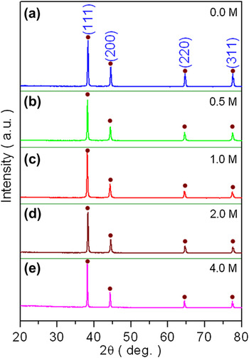
Figure 1 shows the phase and purity of representative products from the hydrothermal processing at the temperature of 180 °C with a wide range of H2SO4 concentrations from 0.0 to 4.0 M. All diffraction patterns have almost the same peak positions at 38.2°, 44.4°, 64.6°, and 77.4°, which can be assigned to the cubic silver (JCPDS no. 04-0783) and/or the hexagonal silver (JCPDS no. 41-1402).Reference Song, Zheng, Liu and Zhang17, Reference Sinha and Sharma26 The rather sharp diffractions suggest the high crystallinity of the silver crystals. In addition, there is no obvious diffraction peak corresponding to silver oxides, despite the easiness of oxidation of the element. The outcome hints the possible protecting effect of a carbon nanoshell formed on the surface of the silver nanostructures as a result of the carbonization of the glycerol molecules in the assistance of H2SO4. The absence of the peaks relevant to graphite and diamond might be due to the fact that the carbon as deposited is low in crystallinity, most likely in an amorphous state. Two other possibilities could make additional contributions to the outcome. One comes from the strong scattering of the Ag crystalline composition which overshadows the relatively weak carbon, whereas the other is attributable to the overlapping in certain diffractions shared by them. Nonetheless, the carbon nanoshell is going to be directly proved in the microstructural and spectroscopic analysis below. We note that the x-ray diffraction examination of the other Ag–C nanostructures prepared in this work shows similar patterns as reported in Fig. 1.
X-ray diffractometry analysis of Ag–C nanostructures hydrothermally fabricated at the reaction temperature of 180 °C as a function of H2SO4 concentration. Diffraction patterns obtained at (a) 0.0, (b) 0.5, (c) 1.0, (d) 2.0, and (e) 4.0 M.

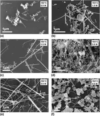
Figure 2 shows the SEM morphological evolution of the Ag–C nanostructures grown hydrothermally at the temperature of 150 °C as a function of the H2SO4 concentration. As revealed in Fig. 2(a), the product prepared at the low concentration of 0.5 M H2SO4 is dominated by irregular nanoparticles with a small number of Ag nanowires (nanocables or nanorods). The number of the Ag nanowires is increased when the acid concentration is raised to 1.0 M, but the specimen is still composed in a large quantity of nanoparticles of different sizes and shapes [Fig. 2(b)]. As shown in Fig. 2(c), nevertheless, the nanowires or nanocables turn to be the major product of the hydrothermal synthesis at the moderate concentration of 1.5 M H2SO4 though the diameter of the nanowires varies significantly. Rather uniform nanowire-like result could be clearly observed in Fig. 2(d) at the concentration of 2.0 M H2SO4, indicating the prevailing formation of the silver nanowire cores. Yet, there remain many nanoparticles in the product. After further increasing the concentration of H2SO4 to 3.0 M, Fig. 2(e) shows that the sample is mostly compiled of nanocable-like products as the carbon shell becomes thick enough to be directly visualized. In contrast, the nanocables rapidly disappeared and Ag nanospheres were generated when the H2SO4 concentration reached 4.0 M [Fig. 2(f)]. It is likely that the tendency of the hydrothermal reduction of silver ions and glycerol carbonization into the nanospheres becomes overwhelmed when the concentration of H2SO4 is continued to increase. As observed above, it is apparent that the H2SO4 concentration has demonstrated the substantial effects on the shape and percentage of the Ag–C nanostructures from nanoparticles to nanocables in the reaction system at the given temperature of 150 °C.
Scanning electron microscopy (SEM) morphological analysis of the Ag–C nanostructures grown hydrothermally at the temperature of 150 °C as a function of the H2SO4 concentration. (a) 0.5, (b) 1.0, (c) 1.5, (d) 2.0, (e) 3.0, and (f) 4.0 M.

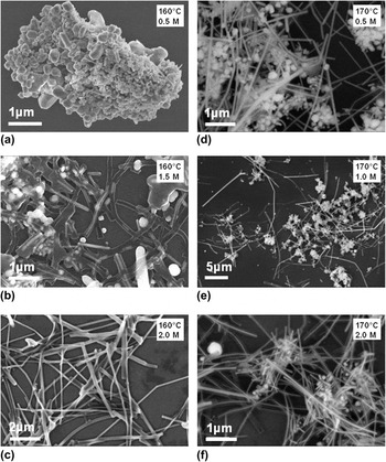
In Fig. 3, the factors of temperature and acid concentration were probed more on the shape tailoring of the Ag–C nanostructures in the hydrothermal treatments. Figures 3(a)–3(c) show the morphologic changes of the products harvested at 160 °C with the H2SO4 concentration adjusted from 0.5 to 2.0 M. Clearly, the predominant outcome under the condition of 0.5 M H2SO4 is asymmetrical nanoparticles of different sizes [Fig. 3(a)], in striking contrast to the robust nanowires acquired from the preparation process at 2.0 M H2SO4 [Fig. 3(c)]. In Fig. 3(b), the transition in the morphologies is evident as the H2SO4 concentration is switched to 1.5 M. In the cases of the reaction system proceeded at the temperature of 170 °C, the effect of the reaction temperature becomes more noticeable. As shown in Fig. 3(d), the process under the concentration of 0.5 M H2SO4 produces a significant amount of nanowires with quite regular dimension, in addition to nanoparticles that are uniform and finer when compared to both 150 and 160 °C counterparts. The status extends to the acid concentration of 1.0 M [Fig. 3(e)]. Under the condition of 2.0 M H2SO4, the product becomes Ag–C nanocables for the most part, with short nanocables visible in the image [Fig. 3(f)].
Comparison SEM study of the morphological evolution of the Ag–C nanostructures prepared at the temperatures of 160 and 170 °C as a function of the H2SO4 concentration. Images obtained at 160 °C for (a) 0.5, (b) 1.5, and (c) 2.0 M; images garnered at 170 °C for (d) 0.5, (e) 1.0, and (f) 2.0 M.

The reaction temperature was further raised to 180 °C to search for optimized conditions in preparation of high-quality Ag–C coaxial nanocables and the results are given in Fig. 4. As the H2SO4 concentration was set to 0.5 M, highly uniform and ultralong Ag–C nanocables were adequately obtained [Fig. 4(b)]. At a lower concentration of 0.25 M, however, the morphology of the product is radically different, consisting of both nanocables and nanoparticles of huge difference in size [Fig. 4(a)]. In the cases of higher H2SO4 concentrations, 1.0 and 1.5 M, both nanocables and nanoparticles exist in favor of the former, but the quality is far from satisfaction [Figs. 4(c) and 4(d)]. In Fig. 4(e) for the 2.0 M case, smaller nanoparticles are found to decorate on larger nanospheres, in addition to the occurrence of nanocables. Further increase in the acid concentration to 3.0 M produces more nanospheres than nanocables [Fig. 4(f)].
SEM morphological analysis of the Ag–C nanostructures at the temperature of 180 °C as a function of the H2SO4 concentration. (a) 0.25, (b) 0.5, (c) 1.0, (d) 1.5, (e) 2.0, and (f) 3.0 M.

The results and analysis above have lucidly expressed the trend in the evolution of the Ag–C nanostructures from nanoparticles to nanocables as a consequence of the changes in the reaction temperature and the concentration of sulfuric acid. The characterization appropriately allows us to draw conclusions that a higher concentration of H2SO4 can promote the formation of high-purity nanocables at lower temperature, whereas a lower H2SO4 concentration is more constructive at higher temperature.
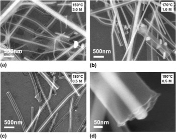
As shown in Fig. 5, the core–shell or coaxial nanostructuring of the Ag–C nanocables are clearly demonstrated in the enlarged SEM images. The formation of the carbon nanoshell takes places at all temperatures investigated in the presence of H2SO4 and is owing to the H2SO4-assisted hydrothermal dehydration of glycerol, in opposite to the case in which the minimal carbonization of glycerol was observed in the hydrothermal reaction without using H2SO4.Reference Sinha and Sharma26 Prepared at 150 °C and 3.0 M H2SO4, Fig. 5(a) provides a plain contour of the silver nanowire cores in the bright contrast covered by the carbonaceous nanosheaths in the relatively light color. Evidently, the core diameter varies to a great extent and the same happens to the shell thickness. Adjusting the acid concentration to 1.0 M and raising the temperature to 170 °C, the nanostructure of the Ag–C nanocables appear similarly, but with more nanoparticles present [cf. Figs. 5(b) and Figs. 3(e)]. As already displayed in Fig. 4(b) in the low magnification, excellent Ag–C nanocables could be made when the experimental conditions tuned to 180 °C and 0.5 M H2SO4. Figure 5(c) illustrates correspondingly the uniform core–shell nanoconfiguration of the Ag–C nanocables at an expanded scale, whereas Fig. 5(d) highlights the cross-sectional outline of one single fractured nanocable acquired from the process of the sample preparation, with the robust Ag nanorod inside the semitransparent carbonaceous nanoshell.
Magnified SEM observation of representative Ag–C coaxial nanocables as a function of temperature (T) and H2SO4 concentration (C A): (a) T = 150 °C and C A = 3.0 M, (b) T = 170 °C and C A = 1.0 M, and (c) T = 180 °C and C A = 0.5 M. (d) A fractured nanocable obtained at T = 180 °C and C A = 0.5 M, showing the core–shell nanostructuring.

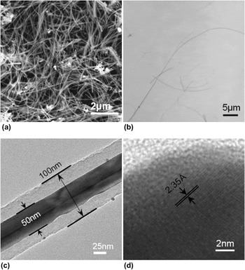
As the optimal Ag–C nanocables were generated under the conditions of 180 °C and 0.5 M H2SO4, more detailed analysis is conducted on the product. Figure 6(a) is the low-magnification SEM image of the as-prepared product in a large quantity, showing the general morphology of the nanocables packing like supple thin threads. It is found that the nanocables have a high purity (>90%) and superior uniformity, indicative of the efficiency of preparing the Ag–C coaxial nanocables through the mild hydrothermal process mediated by H2SO4 but without utilizing a surfactant as commonly done. The TEM examination affords more insights into the morphology and microstructures of the products. As shown in Fig. 6(b), the strong, ultralong Ag–C nanocables in a great aspect ratio are corroborated by TEM observation. The nanocables unveil comparatively smooth contouring and can be as long as 100 μm or more. Of relevance, they show outstanding flexibility attributing to the high aspect ratio of the length to the diameter of a nanocable. The assessment implies prospective good machinability of such Ag–C nanocables for future nanodevice fabrication.
Morphological mapping and microstructural analysis of the Ag–C nanocables from the hydrothermal growth under the conditions of 180 °C and 0.5 M H2SO4. (a) Low-magnification SEM image showing the uniform, flexible Ag–C nanocable network. (b) Low-magnification bright-field transmission electron microscopy (TEM) imaging, revealing ultralong Ag–C nanocables. (c) High-magnification bright-field TEM imaging of a single Ag–C nanocable, showing the core–shell nanoarchitecture. (d) High-resolution TEM image of the core material in the Ag–C nanocable.

A section of a representative nanocable is exhibited in Fig. 6(c), in which light and dark contrast is unmistakably observed along the radial direction. The different contrast suggests a different phase composition, indicating the core–shell cable structure. The dark contrast suggests nanocrystalline Ag as a result of the larger mass in the core region, in contrast to the light contrast portion outside the core region which indicates the carbon sheath layer. The diameter of the silver nanowire core is ∼50 nm, while the carbon nanoshell is measured to be ∼25 nm thick. The high crystallinity of the core material is unambiguously substantiated by the high-resolution imaging, as verified by the lattice spacing of 2.35 Å in Fig. 6(d), which corresponds to cubic Ag (111). Nonetheless, the lattices showing the carbonaceous material of the nanoshell are not apparent in the high-resolution imaging, which is consistent with the XRD outcome discussed above and attributed to the low scattering power of the shell material and likely amorphization of carbon under destructive irradiation from the electron beam.
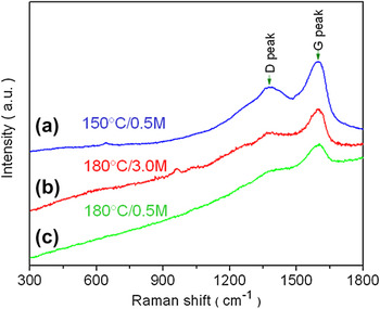
The technique of Raman spectroscopy as an effective tool to probe the surface properties was applied to explore surface information of the Ag–C core–shell nanostructures prepared in this work. Figure 7 compares the Raman spectra acquired for the specimens synthesized under the conditions of (a) 150 °C and 0.5 M H2SO4, (b) 180 °C and 3.0 M H2SO4, and (c) 180 °C and 0.5 M H2SO4. All specimens encompass, more or less in the intensities, two strong peaks positioning at ∼1590 and 1380 cm−1, respectively. The former can be attributed to the graphite-like carbon (G-peaks), while the latter is assigned to diamond-like carbon (D-peaks).Reference Fang, Tang, Lei and Li24 In specific, the two peaks in the case of 150 °C and 0.5 M H2SO4 (curve a) are the strongest and turn to be the weakest in the case of 180 °C and 0.5M H2SO4 (curve c), with the intermediate strengths for the case of 180 °C and 3.0 M H2SO4 (curve b). The phenomena could be caused by the different deposited carbon quantities in the products and stronger scattering power in a larger granular Ag nanostructure.
Raman spectra of Ag–C nanostructures prepared at different hydrothermal conditions: (a) 150 °C and 0.5 M H2SO4, (b) 180 °C and 3.0 M H2SO4, and (c) 180 °C and 0.5 M H2SO4. The positions of diamond-like and graphite-like peaks are indicated by the arrows, respectively.

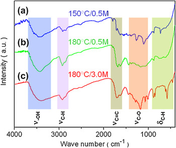
The Ag–C nanostructures were further examined by FTIR, as shown in Fig. 8 for the cases of (a) 150 °C and 0.5 M H2SO4, (b) 180 °C and 0.5 M H2SO4, and (c) 180 °C and 3.0 M H2SO4. The nanomaterials produced via the facile hydrothermal process are rich in the infrared features.Reference Kubo, Tan, White, Antonietti and Titirici27 Independent of the specimens from the different synthetic conditions, the spectra share the similar IR bands, indicating that the nanomaterials have a similar chemical nature. The bands at 600–875 cm−1 are assigned to the C–H bond out-of-plane bending vibrations (δ C–H), while the bands in the 1000–1450 cm−1 correspond to the C–O stretching vibrations (ν C–O). Moreover, the bands between 1630 and 1700 cm−1 can be attributed to the C=C stretching vibrations (ν C=C), whereas the bands around 2800 and 2900 cm−1 match to the C–H stretching vibrations (ν C-H). The -OH stretching vibrations (ν –OH) account for the strong IR signatures at ∼3400 cm−1. We note that possible band shifting and overlapping exists in the spectra, which could partly be attributed to changes in the elastic constants of the relevant bonds sitting on the nanostructure surface of high curvature owing to the small nanostructure dimension. As the redundant glycerol molecules and other residues were removed by the purification procedure, the phenomena indicate the covering of the carbonaceous materials on the surface of the Ag–C nanostructures. As a result, the vibration fingerprints render it obvious the carbon covering on the silver surface from the hydrothermal carbonaceous method. The functional groups such as hydroxyl and carbon–carbon double bonds on the surface of the Ag–C nanostructures grant a potential avenue to load other functional groups, molecules, ions, nanoparticles, or even to formulate other core–shell or hollow nanostructures.Reference Zong, Cao, Jia, Bao and Lu11
FTIR spectra of Ag–C nanostructures prepared at different hydrothermal conditions: (a) 150 °C and 0.5 M H2SO4, (b) 180 °C and 0.5 M H2SO4, and (c) 180 °C and 3.0 M H2SO4. The different characteristic vibrations are assigned as indicated.

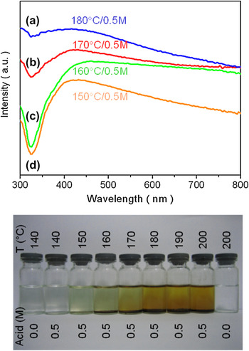
We now handle the optical properties of representative Ag–C nanostructures as a consequence of surface plasmon (SP) ensuing from nanostructured silver.Reference Murray and Barnes28, Reference Jung, Seo, Park, Ryu and Song29 Figure 9 shows the UV–vis spectra of various specimens in water, including the products obtained under the conditions of (a) 180 °C and 0.5 M H2SO4, (b) 170 °C and 0.5 M H2SO4, (c) 160 °C and 0.5 M H2SO4, and (d) 180 °C and 0.5 M H2SO4. All spectra show broad absorption peaking around 420 nm, which is attributed to the SP occurring transversely across the diameter of the nanocables and the existence of the silver nanoparticles in the specimens.Reference Jung, Seo, Park, Ryu and Song29 In the case of the 180 °C sample (curve a), the solution has strong absorption in a wide range from 200 to 600 nm, probably boosted by the surrounding carbonaceous shell. The absorption is reduced upon lowering the reaction temperature to 170 °C (curve b). Comparative to the previous cases, the 160 °C sample sees a flat-out absorption with the shallow resonance red-shifted to 450 nm (curve c). In contrast, the band shape of the 150 °C sample is more distinct with a sharp absorption at the position of ∼420 nm, which could arise from the dominated Ag nanoparticles, as evidenced in the relevant studies of Ag nanoparticles and nanowires.Reference Bhui, Bar, Sarkar, Sahoo, De and Misra30, Reference Chen, Wang, Jiang, Zhou, Chen, Yu and Yang31 In principle, the peak position and band shape of the plasmon resonance observed in the aqueous solution of the Ag–C nanostructures are tuned by the nanostructure shape and dimension as well as the surrounding carbonaceous dielectric medium.
UV–vis absorption spectra of Ag–C nanostructures prepared at various hydrothermal conditions: (a) 180 °C and 0.5 M H2SO4, (b) 170 °C and 0.5 M H2SO4, (c) 160 °C and 0.5 M H2SO4, and (d) 150 °C and 0.5 M H2SO4. Photoimages show the color changes of the aqueous solutions of the Ag–C nanostructures prepared in a wide range of hydrothermal conditions.

The photoimages in the figure show the color changes of the aqueous solutions of the Ag–C nanostructures prepared over a broad range of hydrothermal conditions. Located at the beginning and end in the image rows, the solutions appear colorless when no acid was added and the reaction temperatures were set to 140 and 200 °C separately. But after the addition of 0.5 M sulfuric acid, the color of the solutions turn gradually from near colorless to brown with the increasing temperature from 140 to 200 °C. Simultaneously, the solutions steadily became cloudier as the temperature. The outcomes imply different Ag–C nanostructures and bear out the role of H2SO4 in catalyzing the carbonization process of glycerol and signify that the glycerol molecules were carbonized much more easily at a higher temperature in the catalytic action of H2SO4. Interestingly, the color of the aqueous solutions imply the hydrophilicity of the nanostructure surface, which permits further processing of such nanostructures in an aqueous medium without additional surface decoration.
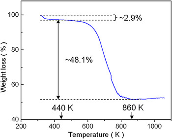
The thermal stability and the overall mass ratio of the Ag–C nanocables were evaluated for the case of 180 °C and 0.5 M H2SO4 by TGA. As shown in Fig. 10, the TGA curve can be divided into three phases. In the first phase between room temperature and 440 K, there is a weight loss of ∼2.9%, attributable to the evaporation of physically adsorbed water and other volatile compositions. In the second phase from 440 to 860 K, the depletion of the carbonaceous composition accounts for the weight loss of ∼48.1% of the specimen under heating. From 860 K upward, the specimen has an almost constant mass, with a slight mass increase probably as a consequence of nitrogenation of silver. So the nanocables possess an adequate thermal stability prior to ∼600 °C.Reference Fu, Huang, Huang, Pan, Zhu and Tang32
Thermogravimetric analyses curve of the Ag–C nanocables fabricated under the conditions of 180 °C and 0.5 M H2SO4.

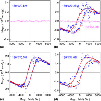
Different from the corresponding bulk diamagnetic characteristics of the materials as silver and carbon (both diamond and graphite) are diamagnetic at room temperature, nanostructuring can lead to the introduction of ferromagnetism, as observed in other Ag–C composite nanomaterials.Reference Caudillo, Gao, Escudero, Jose-Yacaman and Goodenough33 As shown in Fig. 11, the magnetic properties of the specimens prepared under different conditions, one at 150 °C and 0.5 M H2SO4 and the others at 180 °C and a series of H2SO4 concentrations, were comparatively recorded in the individual hysteresis curves obtained by VSM at room temperature. It is evident from the curves that the two samples made at 150 °C/0.5 M H2SO4 and 180 °C/0.0 M H2SO4 show nonmagnetic behavior at room temperature, whereas the samples obtained at 180 °C under the action of H2SO4 give in opposite a definite ferromagnetic response at room temperature in the companion of a diamagnetic signature. Since the samples were exactly prepared except for the temperatures and different H2SO4 concentrations used for the hydrothermal reactions, the likelihood of impurities responsible for the ferromagnetism in the samples obtained, for instance, at 180 °C and 0.5 M H2SO4 could be securely dismissed, in reference to the nonmagnetic state of the samples fabricated under the conditions of 150 °C/0.5 M H2SO4 or 180 °C/0.0 M H2SO4. For concise discussion and analysis as proceeded below, we concentrate on the case of the sample fabricated at 180 °C and 0.5 M H2SO4 (other cases can be similarly tackled). As addressed above, the silver nanocore in the product is ∼50 nm in diameter and the carbon nanoshell is ∼25 nm in thickness, which results in a much larger interface area between the silver and carbon than that in the cases of nanoparticles. The nanosizing renders the interfacial effects to become important and is conceived to underpin the ferromagnetism observed, which is sustained by the magnetic analysis below. In light of the observations in Fig. 4(b), Fig. 5(c), and Fig. 6, the Ag–C nanocables obtained at 180 °C and 0.5 M H2SO4 are nearly uniform, and thus the individual nanocables may be magnetically treated as single giant spins. Consequently, the corresponding magnetization is expressed by a modified Brillouin function or Langevin function on the nanocable ensemble in consideration of the setting-in coercivity,Reference Liu, Hou, Zhang, Kim and Wu34

Magnetic analysis of the hysteresis curves for the Ag–C nanostructures prepared under the different hydrothermal conditions: (a) 150 °C and 0.5 M H2SO4; (b) 180 °C and 0.25 M H2SO4, in comparison to the case of 180 °C without using H2SO4; (c) 180 °C and 0.5 M H2SO4; and (d) 180 °C and 1.0 M H2SO4. Experimental data in dots and the fitting results in curves.

where

In the equations, M/M s is the magnetization (M) normalized to the saturation magnetization (M s), H the applied magnetic field, H c the coercivity, T the temperature and kB the Boltzmann constant. The magnetic moment μ of the individual nanocables concerned can be obtained from fitting of the Langevin function to the experimental data. Since there are diamagnetic and paramagnetic contributions in the measurements which can be represented by a common term, magnetic susceptibility coefficient χ, the overall magnetization as a function of the applied magnetic field is

where

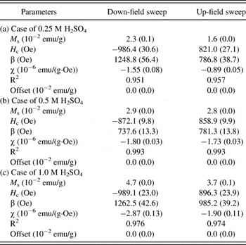
We apply Eq. (3) to the hysteresis curves obtained for the specimen prepared at 180 °C and 0.5 M H2SO4. The fitting results are plotted in Fig. 11(c), which have the correlation coefficients better than 0.99. In the figure, the fittings are represented by the curves and the experimental data are given in dots, separately. In the single-phase fitting, the magnetic susceptibility coefficient χ, the saturation magnetization M s, the coercivity H c, and the switching parameter β show almost no significant difference in both modes of down-field and up-field sweeping within the errors of the measurements. For the down-field sweeping, the parameters are −(1.80 ± 0.03)×10−6 emu·g−1Oe−1, (2.90 ± 0.03)×10−2 emu·g−1, −872.1 ± 9.8 Oe, and 737.6 ± 13.3 Oe for χ, M s, H c, and β, and for the up-field sweeping they become −(1.73 ± 0.03)×10−6 emu·g−1Oe−1, (2.83 ± 0.03)×10−2 emu·g−1, 858.9 ± 9.9 Oe, and 781.3 ± 13.8 Oe, correspondingly. The negative values of magnetic susceptibility coefficient χ clearly reflect the intrinsic contributions from the volume diamagnetic properties of the silver and carbon compositions. It is remarkable that the nanocables show sizeable ferromagnetism at room temperature, with the coercivity as high as ∼870 Oe. Table I sums up the results analyzed in Fig. 11. Obviously, the three cases tabulated for the samples prepared at 180 °C and different H2SO4 concentrations show similar tendencies of the magnetic responses.
Parameters derived from the fitting of the experimental hysteresis curves to the modified Langevin equation for the samples prepared at 180 °C and different H2SO4 concentrations.

Note:
Fitting errors in the brackets.
As elucidated above, the relatively weak saturation magnetization shores up the proposition on the origin of the ferromagnetism from the interfacial interactions. Since silver and carbon commonly show weak diamagnetic behavior, the observation of ferromagnetism in the Ag–C nanocables is interesting. Unusual magnetic phenomena have been discovered in nanoscaled materials and discussed in alloy nanoparticles, organic monolayers, and giant anisotropy at the nanoscale.Reference Wu, Min, Liu, Cho and Kim35–Reference Hernando, Crespo and Garcia37 A similar analysis could be applicable in this work. The anomalous magnetism is conceived energetically as a consequence of spin–orbital interactions, which are in turn due to the ensuing effects of nanostructuring. The interaction energy may be expressed as  , where J, L, and S are the respective magnitudes of the total, orbital, and spin angular moments, J, L, and S. The interaction parameter, given as
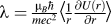 , depends on the potential energy U(r), which also could be a function of an applied magnetic field (here m and e are the electron mass and charge, μ B is the Bohr magneton, c the speed of light,
 the Planck constant divided by 2π and r the orbital radius). Dependent on λ, the interaction can be either ferromagnetic or diamagnetic.Reference Wu, Min, Liu, Cho and Kim35–Reference Hernando, Crespo and Garcia37 In this work, we are interested in the case in which the spin–orbital interaction is favored when L is parallel to S for λ > 0, leading to a ferromagnetic state. Indeed, the unique nanostructure in the Ag–C nanocables renders more surface and nanograin boundaries available for pinning and spin–orbital interactions, and therefore, the interfacial interactions are in favor of ferromagnetism as observed. Moreover, the nearly perfect fit proves the well-defined ferromagnetic and diamagnetic behavior of the Ag–C nanocables made, for example, at 180 °C and 0.5 M H2SO4 and strongly suggests the probable existence of a single ferromagnetic phase. We emphasize that magnetic phenomena reported here are worthy more efforts to explore.
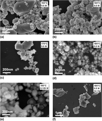
As pointed out in the analysis in the precedent sections, both temperature and the acid concentration have considerable effects on the formation of the Ag–C nanostructures. To evaluate the critical function of H2SO4 in the fabrication of the nanocables, we performed more discriminative experiments to provide additional evidences for the mechanistic study. First, the specimens were hydrothermally prepared at four different temperatures from 150, 160, 170 to 180 °C without addition of H2SO4 and the results were presented in Figs. 12(a)–12(d). It is straightforward from the images that all hydrothermal products are in the form of powders and there is no discernible indication for the formation of coaxial nanostructures. The outcome is similar to a previous study which produced purely Ag nanoparticles from glycerol and AgNO3 without an acid and other additives.Reference Sinha and Sharma26 Though no fundamental diversification was observed when the system did not contain sulfuric acid, the powder size reduces and becomes more homogeneous when the reaction temperature was raised from 150 to 180 °C. In conclusion, no nanocables were obtained if H2SO4 were not used while the amount of AgNO3 and glycerol were kept the same, indicating that H2SO4 has played a crucial role in the formation of the Ag–C nanocables.
SEM morphological analysis of Ag–C nanostructures prepared under special hydrothermal conditions: (a) 150 °C and no H2SO4, (b) 160 °C and no H2SO4, (c) 170 °C and no H2SO4, (d) 180 °C and no H2SO4, (e) 180 °C and 0.125 M H2SO4, and (f) 180 °C and 0.5 M H2SO4 without AgNO3.

In the meantime, one extra experiment was conducted at 180 °C with the addition of H2SO4 at an acid concentration as low as 0.125 M with the objective to well resolve the essential functioning of the acid. As shown in Fig. 12(e), actually, there are short carbon-coated silver nanorods as well as carbon-shelled nanoparticles, definitely showing the incipient stage of the Ag–C nanocables in the present facile hydrothermal synthesis. The carbon coating on the silver nanoparticles at such a low acid concentration shows the crystal clear catalytic effect of the acid on the carbonization of the glycerol molecules. On another aspect, the likelihood of the nanocable shape resulting from a possible nanotubular structure of glycerol during the carbonization of the hydrothermal process is assessed on the basis of another experiment executed under the same conditions as in the case of fabricating the optimal Ag–C nanocables but without adding AgNO3. Figure 12(f) represents the SEM inspection, showing large and irregular carbon flakes and offering no evidence for a tubular assembly. So, the prospect of the glycerol molecules in directly shaping the form of the nanocables could almost certainly be ruled out.
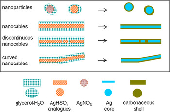
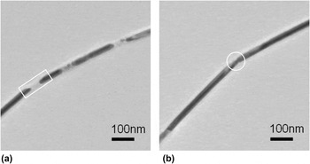
On account of the experimental findings and analysis above, H2SO4 plays a decisive role in formation of the Ag–C nanocables and is benign to the glycerol carbonation. As a matter of fact, the AgNO3 and H2SO4 molecules in the reaction system can form a series of hydrogen sulfate compounds such as AgHSO4, Ag(H3O)(HSO4)2, and Ag2(HSO4)2(H2SO4).Reference Stiewe, Kemnitz and Troyanov38 In addition to the dimers, trimers, and tetramers formed through the hydrogen bonding and the coordination of the silver and HSO4− ions, the compounds can result in chain-like structures,Reference Stiewe, Kemnitz and Troyanov38 which in turn develop into nanobundles under the action of the glycerol and water molecules and surrounded by them. In our reaction system, the silver ions can be reduced by glycerol and simultaneous carbonization reaction of the glycerol molecules takes place through the promotion of H2SO4. Since the change in the H2SO4 concentration has resulted in the morphological change of the Ag–C nanostructures, pH might play an important role, either in a direct or indirect way, in the formation of the Ag–C nanomaterials. As illustrated in Fig. 13, schematic routes are proposed for the growth of the Ag–C nanostructures (nanocables including defect ones and nanoparticles), in which the construction of the nanocables is H2SO4-mediated via the formation of AgHSO4 and analog compounds. The mechanism as proposed favors Ag–C nanostructures of high crystallinity, as evidenced by the strong, sharp diffractions observed in the XRD patterns (Fig. 1) and the TEM analysis (Fig. 6). Though the silver particles could be obtained with and without H2SO4, only a very thin carbonaceous shell was possible to form when without adding H2SO4 (Fig. 12).Reference Sinha and Sharma26 In the presence of H2SO4, Ag–C core–shell nanostructures in form of particles, short and long nanocables are produced dependent on the acid concentration [Figs. 2–4 and 12(e)]. Two kinds of defect nanocables covered with discernibly thin carbon, discontinuation and bending, are shown in Fig. 14. We remark that the carbon covering on the silver is owing to the fact that materials with low surface energy generally have a tendency to spread over substances with high surface energy and the tests showed the much lower graphene layers surface energy than that of silver nanowires.Reference Fu, Huang, Huang, Pan, Zhu and Tang32, Reference Deng, Xu, Chen, Song and Chen39
Schematic routes for the growth of the Ag–C nanostructures (nanocables including the defect cases and nanoparticles). The construction of the nanocables is H2SO4-mediated via the formation of AgHSO4 and analog compounds.

TEM images of defect nanocables obtained under the condition of 180 °C and 0.5 M H2SO4. (a) Discontinuation as indicated by the frame. (b) Bending in the position as circled.

IV. CONCLUSIONS
In summary, we have conducted systematic and exensive studies on the development of the Ag–C nanostructures over a broad range of temperature and as a function of H2SO4 concentration, to locate the optimal conditions for preparing high-quality Ag–C nanocables. The characterization clearly demonstrated the simple and efficient synthesis of nanocables with silver nanowires as the cores and carbon as the sheaths, without introduction of a surfactant. In the hydrothermal process, glycerol acts as both reducing agent and carbon source, while H2SO4 is used in the system to mediate the directional growth of the silver nanowire core and to catalyze the deposition of carbon on the silver surface in constructing the Ag–C coaxial nanocables. The formation and nanostructuring of the Ag–C nanocables was evidently supported by the XRD patterns, SEM and TEM images, HRTEM lattice imaging, TGA, Raman, FTIR UV–vis, and spectral analysis. The most favorable reaction conditions for the generation of ultralong, robust and uniform Ag–C coaxial nanocables were found to be at T = 180 °C and 0.5M H2SO4. Of significance, the nanocables manifest extraordinary ferromagnetism at room temperature. Furthermore, the plausible mechanism of forming Ag–C nanocables was proposed, as a result of the chain-like hydrogen sulfate compounds owing to the H2SO4 mediation. This green, surfactant-free facile hydrothermal process could be valuable for future bulk synthesis and functionalizing various carbon-related nanomaterials of technical importance. Such coaxial nanocables comprising highly conducting metallic cores and relatively insulating sheathes could be used as components and interconnects in nanoelectronic devices. Further extension of this approach and investigation of the electrically conducting properties of the quasi-one-dimensional nanocables obtained are of great interest in the ongoing research.
ACKNOWLEDGMENTS
This work was supported in part by the National Science Foundation of China (Nos. 20875001, 20905001, and 50901074), the Natural Science Foundation of Anhui Province (Nos. 090414200 and KJ2010A014), the 211 Project of Anhaui University, the Scientific and Technological Development Projects, Science and Technology Department of Henan Province, China (Nos. 092102210004, 092300410031, and 11230040011), the Natural Science Research Foundation Grants, Education Department of Henan Province, China (Nos. 2009A150004 and 2009A150005), the Pioneer Research Center Program through the National Research Foundation of Korea funded by the Ministry of Education, Science and Technology, South Korea (No. 2010-0002190), and the Industrial Core Technology Development Program funded by the Ministry of Knowledge Economy, South Korea (No. 10033183).