Introduction
The family Bucephalidae is a diverse and cosmopolitan group of parasites, with hundreds of species infecting fish hosts. The family is characterized by the configuration of the genitalia, a ventrally located mouth and the presence of an anterior attachment rhynchus (Overstreet & Curran, Reference Overstreet, Curran, Gibson, Jones and Bray2002). This family is also characterized by its life cycle, which includes three hosts: a bivalve as the first intermediate host and fish as secondary intermediate and definitive hosts. Eggs are passed in the faeces, and a miracidium larva hatches from the egg. The miracidial surface has a locomotor apparatus consisting of plates and jointed appendages (Woodhead, Reference Woodhead1929) that allows it to move through the water to infect a first intermediate host; the motile cercaria larvae then emerge from the bivalve to find a fish. The larvae penetrate the skin of the fish, where they encyst. They then become metacercariae and can be found in muscles, fins, gills or any other part of the fish host. When it preys on the infected fish, the definitive host becomes parasitized by the bucephalid digenean.
Because digeneans in different developmental stages can be morphologically distinct from one another, experimental infections and molecular analyses are useful for confirming their life cycles (e.g. Nolan & Cribb, Reference Nolan and Cribb2004; Pina et al., Reference Pina, Barandela, Santos, Russell-Pinto and Rodrigues2009). The life cycles of a few species of Bucephalidae have been elucidated (Stunkard, Reference Stunkard1976; Gargouri-Ben Abdallah & Maamouri, Reference Gargouri-Ben Abdallah and Maamouri2002; Pina et al., Reference Pina, Barandela, Santos, Russell-Pinto and Rodrigues2009; da Costa-Marchiori et al., Reference da Costa Marchiori, Magenta-Magalhães and Pereira Junior2010), although the specific host species for many digenean species of this family are completely unknown. Thus, the objective of this study was to describe the life cycle of Prosorhynchoides carvajali Muñoz & Bott, Reference Muñoz and Bott2011 through natural and experimental infections and molecular analyses, to determine all possible hosts involved in the development of this parasite.
The definitive hosts of P. carvajali are labrisomid fish that live in the intertidal rocky zone (Muñoz & Bott, Reference Muñoz and Bott2011). Therefore, the rocky intertidal bivalves Perumytilus purpuratus and Semimytilus algosus (Mytilidae) are candidates for harbouring Prosorhynchoides species. Previously, these two bivalves have been shown to be infected with sporocysts of Bucephalidae (Szidat, Reference Szidat1963; Lasiak, Reference Lasiak1991; Muñoz et al., Reference Muñoz, López and Cárdenas2013). Labrisomid fish are piscivorous and prey on intertidal fish such as blenniids and other labrisomids (unpublished data) that are also potential secondary hosts for P. carvajali. However, several other fish species of the intertidal rocky zone of central Chile should also be evaluated as potential host species.
Materials and methods
Natural infections in bivalves and fish
A total of 3482 mytilids were collected in Montemar (32°57′S, 71°29′W) and Caleta Abarca (33°02′S, 71°31′W), central Chile. Each bivalve was dissected to determine the presence of bucephalid sporocysts according to the features described by Muñoz et al. (Reference Muñoz, López and Cárdenas2013).
Ten intertidal fish species from the families Blenniidae (Scartichthys viridis, Hypsoblennius sordidus), Tripterygiidae (Helcogrammoides chilensis, H. cuninghami), Labrisomidae (Auchenionchus variolosus, A. microcirrhis), Kyphosidae (Girella laevifrons, Graus nigra) and Gobiesocidae (Syciases sanguineus, Gobiesox marmoratus) were collected at Montemar and Las Cruces (33°30′S; 71°32′W). The total length (TL) of the fish ranged between 1.9 and 12.3 cm, corresponding to the smaller G. laevifrons and the larger S. viridis, respectively. The specimens were euthanized prior to dissection by anaesthetizing the fish and then severing the nerve cord. The muscles, body cavity and fins of these fish were examined for the presence of bucephalid metacercariae.
Experimental infections in fish
Experimental infections were performed to identify fish species that could be secondary intermediate hosts for Prosorhynchoides. Approximately 600 S. algosus bivalves were collected from Caleta Abarca to infect the fish. The bivalves were separated into six groups containing 100 bivalves each and kept in 4-litre aquaria with 3 l of filtered water for 3 days for acclimation. A total of 300 P. purpuratus were collected from Montemar and separated into three groups. During this time, the water was examined under a stereomicroscope to determine whether bucephalid cercariae were released from the mussels. Fish were introduced after we confirmed the presence of infected mussels in the aquaria.
Five fish species were collected in the intertidal rocky zone of Montemar and Las Cruces as hosts for experimental infection by bucephalids under laboratory conditions. The number and species of fish varied in the experiments, which were dependent on the weather and local conditions. The following four species were used for experimental infections with S. algosus: A. microcirrhis (n= 4; TL 5.1–5.6 cm), S. viridis (n= 12; TL 5.8–8.6 cm), H. sordidus (n= 3; TL 3.4–3.6 cm) and H. chilensis (n= 5; TL 3.0–4.5 cm). Three fish species were used for experimental infections with P. purpuratus: G. laevifrons (n= 16; TL 1.9–3.0 cm), S. viridis (n= 8; TL 6.5–8.5 cm) and H. chilensis (n= 4; TL 3.2–4.5 cm). A maximum of six fish were put in each aquarium. The fish were in the aquaria for a maximum of 14 days at a water temperature of 16–18°C with constant aeration. The water was changed every 2 days. It was not possible to determine which fish had been infected with bucephalids before the experiment. Therefore, the prevalence of metacercariae in the infected fish in the laboratory was contrasted with natural infections based on contingency tables using a chi-square stadigraph (Zar, Reference Zar1996).
The species P. carvajali had been found previously in adult clingfish (S. sanguineus), and this fish is considered as an occasional definitive host (Muñoz & Bott, Reference Muñoz and Bott2011). Thus, fish meat containing metacercariae was given as food to two juvenile clingfish, which were not infected with bucephalids (Muñoz & Zamora, Reference Muñoz and Zamora2011), to determine if the metacercaria could develop into adults.
Fish necropsies used in laboratory infections were performed when the fish died naturally during the experiment, or after reaching the maximum experimentation time (14 days). The fish were then euthanized and dissected to find metacercariae in the body. Some of the collected parasites were fixed in 96% ethanol and others were fixed in 5% formalin, for molecular and morphological analyses, respectively. The prevalence and mean intensity of bucephalids were calculated according to Bush et al. (Reference Bush, Lafferty, Lotz and Shostak1997).
Molecular analyses and larval morphology
The DNA sequences of bucephalid specimens in different developmental stages, i.e. sporocysts from S. algosus and P. purpuratus, natural and experimental infections of metacercariae from several intertidal fish species, and adults from the definitive hosts, were analysed to determine whether the parasites belonged to the same species.
Genomic DNA was extracted from single specimens, and the V4 region of the small subunit (SSU) rRNA gene was amplified using the primers SB3a (5′-GGA GGG CAA GTC TGG TGC-3′) and A27a (5′-CCA TAC AAA TGC CCC CGT CTG-3′) following the protocol described by Hall et al. (Reference Hall, Cribb and Barker1999). Each polymerase chain reaction (PCR) had a final volume of 50 μl and included 5 standard units of Taq polymerase, 5 μl of 10 × PCR buffer, 4 μl of MgCl2 (50 mm), 4 μl of each deoxynucleotide triphosphate (dNTP) (2.5 mm), 20 pmol of each primer and approximately 200 ng of template DNA. A Boeco Ecogermany M-240R Thermal Cycler (Boeckel, Hamburg, Germany) was used with the following cycling profile: initial denaturation step at 94°C (5 min) followed by 35 cycles at 94°C (30 s), 45°C (30 s) and 72°C (3 min), and a final extension step at 72°C (10 min). The PCR products were purified using a QIAquick purification kit (QIAGEN Inc., Hilden, Germany). Double-stranded PCR products were sequenced for each individual specimen using an ABI PRISM® 3100 Automated DNA Sequencer (Applied Biosystems, Foster City, California, USA). The sequences were edited using ProSeq v. 2.9 beta (Filatov, Reference Filatov2002) and aligned with Clustal X (Larkin et al., Reference Larkin, Blackshields, Brown, Chenna, McGettigan, McWilliam, Valentin, Wallace, Wilm, Lopez, Thompson, Gibson and Higgins2007). The statistics of the nucleotide composition were compiled using DnaSP version 5 (Librado & Rozas, Reference Librado and Rozas2009).
As outgroup species (see Hall et al., Reference Hall, Cribb and Barker1999), we used Complexobursa sp. and Proctoeces cf. lintoni (Fellodistomidae), which correspond to the sister taxa of Bucephalidae. We also included the DNA sequences of other bucephalid species available in the GenBank database, including eight species of Prosorhynchus, one species of Prosorhynchoides and one species of Rhipidocotyle.
The phylogenetic trees were inferred by the maximum likelihood (ML) and maximum parsimony (MP) criteria using PAUP*4.0b10 (Swofford, Reference Swofford2001) and by Bayesian inference (BI) using BayesPhylogenies (http://www.evolution.reading.ac.uk/BayesPhy.html). The ML and BI analyses employed the HKY+G model of evolution, chosen according to the Akaike information criterion values in Modeltest 3.7 (Posada & Crandall, Reference Posada and Crandall1998).
For the ML and MP trees, a heuristic search strategy (n= 100) with random-addition sequences and the tree bisection–reconnection (TBR) branch swapping method (Nei & Kumar, Reference Nei and Kumar2000) was implemented. All sequences were run unordered and weighted equally, and gaps were treated as missing data. Nodal support was estimated with 1000 bootstrap replicates (Felsenstein, Reference Felsenstein1985). Bayesian phylogenetic analyses were conducted by applying Markov chains and running 50,000,000 generations with sampling each 1000 generations. The first 12,500,000 generations (‘burn-in’) were discarded (Felsenstein, Reference Felsenstein1985), and 500 random trees of the 3000 trees sampled were used to construct a 50% majority-rule consensus tree. Nodal support values (posterior probabilities) were calculated for 85% sampled trees containing the node.
The digeneans, including ten bucephalid species, two species as outgroup and the Prosorhynchoides specimens used in the present study, were classified into groups, and the genetic distances were computed with the average number of mutations (substitutions). The divergence among groups was determined by the maximum composite likelihood model (Tamura & Nei, Reference Tamura and Nei1993) using the neighbour-joining method.
The larval stages, such as sporocysts, cercariae and metacercariae, were observed using a Leica DM LS2 compound microscope (Leica Microsystems, Wetzlar, Germany). Measurements were performed with an eyepiece micrometer, and drawings were made with a camera lucida attached to the compound microscope. All ranges of morphological measurements were expressed in micrometres.
Results
Natural infections in bivalves and fish
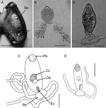
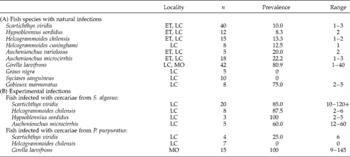
Bucephalid sporocysts were found primarily in the mantle and gonads of both bivalves (fig. 1A), with a prevalence of 2.46% in S. algosus (n= 284) and 0.76–1.40% in P. purpuratus (n= 3198). Certain fish species harboured no more than five metacercarial cysts, with prevalences of 8.3–75%; the exception to this pattern was G. laevifrons, which showed the highest prevalence (80.9%) and digenean intensity (table 1A).
Larval stages of Prosorhynchoides spp. from mussel hosts, showing: (A) sporocysts inside the mantle of P. purpuratus; (B–C) cercariae of P. carvajali; (D–E) cercariae of Prosorhynchoides sp. Sp, sporocysts; Po, penetration organ; Ev, excretory vesicle; Cc, caecum; Os, oral sucker; and Pb, posterior bulb. Scale bars = 0.5 mm (A), 100 μm (B, C, E), 50 μm (D).

The prevalence of infection (%) and range of parasite intensity are shown for (A) natural and (B) experimental infections of intertidal fish species from Las Cruces (LC), El Tabo (ET) and Montemar (MO); n= number of specimens examined.

Experimental infections in fish
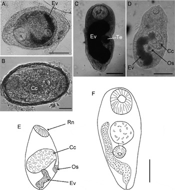
The fish species A. microcirrhis, S. viridis, H. sordidus and H. chilensis that were exposed to infected S. algosus mussels became parasitized with bucephalid cercariae during the experimental period (table 1B, fig. 1B, C); and from the three fish species exposed to infected P. purpuratus mussels, two became parasitized with cercaria larvae, G. laevifrons and S. viridis (table 1B, fig. 1D, E). The metacercariae were mainly found in the muscles of the fish, especially in Auchenionchus spp. and S. viridis, although the fins and body cavities contained several cysts. In G. laevifrons, the metacercariae were found primarily in the caudal fin base. In intense infections, however, cysts were found in the muscles of various parts of the body. The developmental stages of the metacercariae were variable, ranging from small cysts with an embryo occupying the whole capsule and without any distinct organs, to an advanced stage in which the rhynchus, caecum and excretory vesicle were distinguishable (fig. 2A–E). The prevalence of fish infected by digeneans from the mussels S. algosus and P. purpuratus under laboratory conditions was at least 60% and 25%, respectively (table 1B).
Metacercaria of Prosorhynchoides showing: (A) entire specimens of P. carvajali and (B) Prosorhynchoides sp.; (C) excysted metacercaria of P. carvajali in A. microcirrhis from natural infection; (D–E) excysted metacercaria found in S. viridis from experimental infection; (F) naturally excysted metacercaria in a clingfish. Ev, excretory vesicle; Te, testicle; Rn, rhynchus; Os, oral sucker; Cc, caecum. Scale bars = 100 μm.

Clingfish that were fed muscles from infected secondary hosts (S. viridis and A. microcirrhis) did not become infected with the parasite. All parasites were eliminated in the faeces of the clingfish within 24 h after feeding. However, the body length of the trematodes was double that of normally excysted metacercariae (fig. 2E–F).
The prevalence of experimental infections with Prosorhynchoides was higher in S. viridis (Fisher test, P< 0.001), in H. chilensis (Fisher test, P= 0.008) and in H. sordidus (Fisher test, P= 0.009) than for natural infections; however, no difference was found in the prevalence of metacercariae in A. microcirrhis (Fisher test, P= 0.142) between natural and experimental infections when exposed to infected S. algosus. Girella laevifrons had a high prevalence of Prosorhynchoides metacercariae following both the natural and experimental infections, no significant difference was found between the two groups of fish (Fisher test, P= 0.071). There was also no difference in the prevalence in S. viridis between natural infections and exposure to infected P. purpuratus (Fisher test, P= 0.394). However, all fish infected under experimental conditions had a greater abundance of metacercariae than the wild fish. Most naturally infected fish had between 1 and 5 metacercariae per fish, but fish infected in the laboratory contained from 10 to more than 100 metacercariae (table 1B). Naturally infected G. laevifrons had between 1 and 40 metacercariae but contained 9–145 metacercariae per fish following laboratory infections.
Molecular analyses and larval morphology
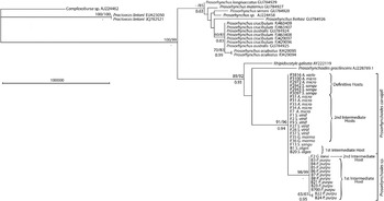
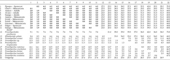
Thirty-four sequences of individual trematodes from various hosts were obtained in this research. The use of different analysis methods (ML, MP, BI) did not substantially influence the results. The molecular analysis showed that all the bucephalids examined, including those from different stages and host species, belonged to the genus Prosorhynchoides (fig. 3). Prosorhynchoides species differed greatly from several Prosorhynchus species (table 2, fig. 3). All analyses of the species in this study clearly revealed two distinct Prosorhynchoides lineages: one group was composed of sporocysts from the bivalve S. algosus, metacercariae from three fish species, and adult trematodes of P. carvajali from another three fish species (fig. 3). The other group was composed of metacercariae from G. laevifrons and sporocysts from P. purpuratus. This trematode group belonged to Prosorhynchoides sp. (designated thus hereafter), which was 1.9–2.0% different from the clade belonging to P. carvajali (table 2, fig. 3). Genetic differences between the two Prosorhynchoides species were related to differences in the morphology of the larval stages, as described below.
Phylogenetic tree to show the relationships between 34 specimens of Prosorhynchoides and other sequences from members of the family Bucephalidae, with GenBank accession numbers based on the SSU rRNA gene. Maximum likelihood (ML), maximum parsimony (MP) and Bayesian inference (BI) analyses to show similar topologies, with values at each branch indicating bootstrap percentages for the ML/MP/BI analyses. The model for the ML tree (HKY+G) had a − lnL score of 1243.57 and an Akaike information criterion (AIC) of 2819.879.

Pairwise sequence divergences for the V4 region of the rRNA gene in adult worms and developing stages of Prosorhynchoides from different hosts; divergence distances are calculated as percentages, shown below the diagonal, using a maximum composite likelihood model with mean numbers of mutations shown above the diagonal for each clade; bold values are those from the present study.

Abbreviations of scientific names of the fish were defined using the first letter of the genus and the first five letters of the specific name.
* Metacercariae in food offered to the clingfish S. sanguineus.
Prosorhynchoides carvajali
Yellow to white sporocysts were found in the mantle and gonads of mussel S. algosus. Measurements of the ramified sporocysts were 4.2–7.0 mm in length between one bifurcation and another and were 102–260 μm in width. Oval-shaped cercariae were of the gasterostome type (fig. 1B, C), showing similar developmental stages inside the sporocysts, measuring 169.8–283.0 μm long × 69–116.9 μm wide; posterior bulb, 30.2–47.1 μm long × 88.7–94.3 μm wide, located at the posterior end of the body with one filament at each side; tegument with small spines; excretory vesicles small and blackish (22.6–43.3 μm long × 9.4–18.8 μm wide); post-equatorial oral sucker followed by a round and large caecum 32.1–56.6 μm long × 32.1–47.1 μm wide with a thick wall (fig. 1C).
Oval-shaped metacercariae were primarily found in muscles and occasionally in the body cavity and on the fins; metacercarial cyst oval (fig. 2A), 262–450 μm long × 135–300 μm wide; tegument with small spines. Freed embryos from cysts were 230–475 μm long × 156–225 μm wide (fig. 2C, D). Certain specimens with a protuberance anterior to the rhynchus (fig. 2D). Excretory vesicle 162.2–226.4 μm long; caecum ranging from 60.4 to 86.8 μm in length, rhynchus ranging from 69.8 to 79.2 μm (fig. 2C, D). Excretory vesicle with blackish, oil-like elements and micro-organisms (possibly bacteria) that were expelled each time the vesicle contracted. Sexual organs poorly developed.
Prosorhynchoides sp.
Yellow to white sporocysts were found in the mantle and gonads of mussel P. purpuratus (fig. 1A). Measurements of the ramified sporocysts were 2.8–6.5 mm in length between one bifurcation and another, and were 95–221 μm in width. Cercariae were the gasterostome type (fig. 1D, E). Body oval shaped, 134.2–197.8 μm long × 58.1–70.7 μm wide. Posterior bulb, 30.1–46.7 μm long × 70.1–95.9 μm wide, located at posterior end of the body with one long filament at each side. Tegument with small spines. Excretory vesicle small, blackish, 40.0–82 μm long × 10–13 μm wide. Post-equatorial oral sucker followed by a round caecum 25–30 μm long × 31–40 μm wide.
Oval-shaped metacercariae found primarily in muscles close to hypural bones or at base of caudal fin rays. Tegument covered with small spines. Metacercarial cysts oval, 300–350 μm long × 200–340 μm wide (fig. 2B). Sexual organs poorly developed.
Discussion
This study identified the life cycle of P. carvajali using the following three methods: experimental infections (from bivalves to fish), morphological characteristics at juvenile stages and molecular analyses (different digenean developmental stages and host species). Sporocysts from the mytilid S. algosus were molecularly identical to P. carvajali adults. Molecular sequences were not obtained for all metacercariae found in eight naturally infected fish species in which Prosorhynchoides cysts were observed, although we confirmed that the metacercariae from A. microcirrhis and G. marmoratus corresponded to P. carvajali. We confirmed from experimental infections using fish exposed to cercariae from S. algosus that the cercariae of this digenean species could infect A. microcirrhis, S. viridis, H. chilensis and H. sordidus. Thus, at least five intertidal fish species serve as intermediate secondary hosts for P. carvajali. Some of these fish species (the labrisomids Auchenionchus spp.) have also been recorded as definitive hosts (Muñoz & Bott, Reference Muñoz and Bott2011).
We also found another Prosorhynchoides species, which has not been recorded in the parasite fauna from Chile (Muñoz & Olmos, Reference Muñoz and Olmos2008). This unidentified species, Prosorhynchoides sp., was present in the mytilid P. purpuratus as a first intermediate host and in the fish G. laevifrons as a secondary intermediate host. A molecular analysis showed that Prosorhynchoides sp. exhibits between 1.9 and 2.0% genetic divergence from P. carvajali; this level of divergence is sufficient to differentiate them as different Prosorhynchoides species (Nolan & Cribb, Reference Nolan and Cribb2005). The cercariae and metacercariae of P. carvajali and those of the newly found Prosorhynchoides sp. were morphologically similar, although they did exhibit differences in body size. Prosorhynchoides carvajali were larger than Prosorhynchoides sp., the posterior bulb was narrower than the body width in P. carvajali but wider than the body width in Prosorhynchoides sp., and the caecum was more conspicuous in P. carvajali, possibly due to the latter's body size and thicker wall. Prosorhynchoides sp. adults are expected to be smaller than those of P. carvajali.
Natural infections with Prosorhynchoides sp. metacercariae were more prevalent in G. laevifrons than in any other fish, and experimental infections were also very successful in this fish species, specifically in terms of the intensity of infections of digeneans (table 1), confirming the intermediate hosts for Prosorhynchoides sp. However, the definitive host for this digenean is still unknown. There are two potential explanations for this uncertainty about the identity of the definitive host: (1) the definitive host for Prosorhynchoides sp. may be a subtidal or demersal fish rather than an intertidal fish; the parasite communities in intertidal fish have been studied extensively, but only P. carvajali has been recorded (e.g. Flores & George-Nascimento, Reference Flores and George-Nascimento2009; Muñoz & Cortés, Reference Muñoz and Cortés2009; Muñoz & Bott, Reference Muñoz and Bott2011; Muñoz & Delorme, Reference Muñoz and Delorme2011), and G. laevifrons spends its juvenile period in the intertidal environment and then lives in the subtidal zone while maturing; (2) Prosorhynchoides sp. and P. carvajali may be cryptic species that co-occur in the same definitive host species (labrisomid fish), as has been demonstrated for many digenean species (e.g. Vilas et al., Reference Vilas, Criscione and Blouin2005; Curran et al., Reference Curran, Tkach and Overstreet2013; McNamara et al., Reference McNamara, Miller and Cribb2013).
Mapping the life cycle of parasites allows us to understand the routes of parasite transmission, predator and prey interactions, and the relationship between a parasite and its environment. Identifying the specific hosts involved in parasite life cycles also allows us to define aspects of parasite population dynamics that are required for epidemiological evaluations. In Chile, however, these characteristics are poorly understood. Therefore, this study has contributed to the knowledge of the life cycle of P. carvajali, the second marine digenean life cycle to be well described in Chile; that of Proctoeces sp. (Fellodistomidae) was the first, although there is a degree of controversy regarding the morphological and molecular analyses applied by different authors (George-Nascimento et al., Reference George-Nascimento, Balboa, Aldana and Olmos1998; Aldana et al., Reference Aldana, González, Loot, Pulgar and Marquet2009; Oliva et al., Reference Oliva, Valdivia, Cárdenas, George-Nascimento, González, Guiñez and Cuello2010; Muñoz et al., Reference Muñoz, López and Cárdenas2013). The present study also revealed the existence of a species of Prosorhynchoides that has not been reported previously. One part of the life cycle of this species was determined for the larval stages in the first and secondary hosts, but the adult stage has yet to be characterized.
Acknowledgements
We thank Dr M.T. Gonzalez for facilitating the use of her laboratory for several molecular analyses and Mr David Jara for his help with the improvement of the figures.
Financial support
This study was supported by the research grant project DIPUV 12-2008 to G.M. and partially funded by Fondecyt 1120868.
Conflict of interest
None.
Ethical standards
The authors assert that all procedures contributing to this work comply with the ethical standards of the relevant national guides on the care and use of laboratory animals (http://investigacion-uv.cl/bioetica-y-bioseguridad/procedimientos-instructivos-y-formularios/) and has been approved by the institutional committee (Facultad de Ciencias del Mar y de Recursos Naturales, Universidad de Valparaíso).