Introduction
Many lichens can establish a new thallus from small pieces (thallus fragmentation) or have evolved special reproductive structures such as soredia or isidia that allow the dispersal of both symbiotic partners simultaneously (Büdel & Scheidegger Reference Büdel and Scheidegger2008). However, for numerous lichens only the fungal partner seems to disperse, either by sexually generated spores or by asexual conidia, and in order to re-establish the symbiosis these species face the challenge of finding a suitable photobiont in the environment (Bowler & Rundel Reference Bowler and Rundel1975; Hestmark Reference Hestmark1990, 1991 Reference Hestmarka , Reference Hestmarkb, Reference Hestmarkc , 1992 Reference Hestmarka , Reference Hestmarkb ; Nash Reference Nash2008). A potential source of such suitable algae is symbiotic propagules dispersed by other lichen species (Beck et al. Reference Beck, Friedl and Rambold1998; Fedrowitz et al. Reference Fedrowitz, Kaasalainen and Rikkinen2011). Many lichen photobionts may not occur frequently in a free-living stage due to their long co-evolution with fungi in lichen symbiosis (Ahmadjian Reference Ahmadjian1988). The degree of specificity in the symbiotic relationship differs substantially between lichen taxa, from rare cases of reciprocal one-to-one specificity (e.g. Otalora et al. Reference Otálora, Martínez, O’Brien, Molina, Aragón and Lutzoni2010) or high specificity of mycobionts for a single or a small number of photobionts (e.g., Paulsrud et al. Reference Paulsrud, Rikkinen and Lindblad2000; Yahr et al. Reference Yahr, Vilgalys and DePriest2004; Lindgren et al. Reference Lindgren, Velmaala, Högnabba, Goward, Holien and Myllys2014; Muggia et al. Reference Muggia, Pérez-Ortega, Kopun, Zellnig and Grube2014; Nyati et al. Reference Nyati, Scherrer, Werth and Honegger2014; O’Brien Reference O’Brien2014; Leavitt et al. Reference Leavitt, Kraichak, Nelsen, Altermann, Divakar, Alors, Esslinger, Crespo and Lumbsch2015) to generalist mycobionts that are more flexible and may associate with multiple partners (Piercey-Normore & DePriest Reference Piercey-Normore and DePriest2001; Piercey-Normore Reference Piercey-Normore2005; Guzow-Krzemińska Reference Guzow-Krzemińska2006; Yahr et al. Reference Yahr, Vilgalys and DePriest2006; Hauk et al. Reference Hauk, Helms and Friedl2007; Nelsen & Gargas Reference Nelsen and Gargas2008; Muggia et al. Reference Muggia, Vancurova, Skaloud, Peksa, Wedin and Grube2013). In general, one would predict that a lichen-forming fungus that needs to re-establish the symbiotic state at every reproductive cycle should be more flexible with regard to the photobionts it associates with than a lichen that propagates the intact symbiosis.
The peltate lichen-forming fungus Umbilicaria spodochroa Hoffm. grows in high abundance on the rocky coasts of southern Norway. It reproduces sexually through ascospores from apothecia on the upper surface, and the number of apothecia is positively correlated with thallus size (Ramstad & Hestmark Reference Ramstad and Hestmark2001), a common trait also observed in several other members of the genus Umbilicaria Hoffm. (Hestmark et al. Reference Hestmark, Skogesal and Skullerud2004; Gregersen et al. Reference Gregersen, Hegseth, Hestmark, Kongsbak and Moe2006). To form a lichen thallus, ascospores of U. spodochroa must re-establish symbiosis de novo with suitable photobionts encountered in the environment. Alternatively, new thalli could be formed through the growth of small thallus fragments containing both symbionts, but neither fragmentation nor erosion of thalli seems common in this particular species (G. Hestmark, unpublished field observations).
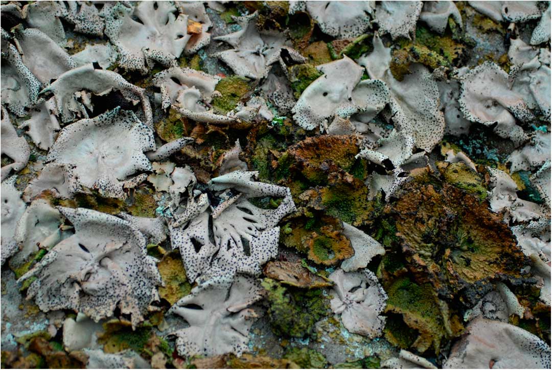
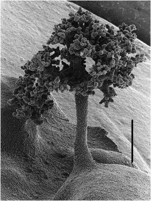
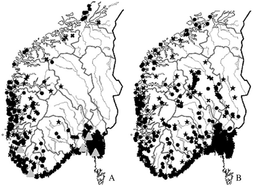
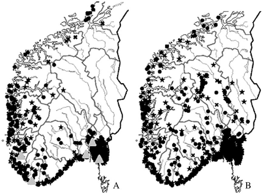
On the coastal cliffs of Scandinavia, U. spodochroa commonly grows in mixed populations with another peltate lichen, Lasallia pustulata (L.) Mérat (Fig. 1), and competitive overgrowth interactions between the two species were demonstrated both in situ and experimentally by Hestmark (1997 Reference Hestmarka ). Although both species belong to the same family, molecular phylogenetic studies indicate that they are not closely related within the Umbilicariaceae (Miadlikowska et al. Reference Miadlikowska, Kauff, Hofstetter, Fraker, Grube, Hafellner, Reeb, Hodkinson, Kukwa and Lücking2006, Reference Miadlikowska, Kauff, Högnabba, Oliver, Molnár, Fraker, Gaya, Hafellner, Hofstetter and Gueidan2014). The two species have almost identical physiological niche responses to light and temperature (Kappen et al. Reference Kappen, Schroeter, Hestmark and Winkler1996, Reference Kappen, Schroeter, Scheidegger, Sommerkorn and Hestmark1997; Hestmark et al. Reference Hestmark, Schroeter and Kappen1997), and very similar population dynamics (Hestmark 1997 Reference Hestmarkb , Reference Hestmarkc , Reference Ramstad and Hestmark2000; Sletvold & Hestmark Reference Sletvold and Hestmark1999; Ramstad & Hestmark Reference Ramstad and Hestmark2000). In contrast to U. spodochroa, L. pustulata reproduces mainly through isidia, that is, packages of fungal hyphae and algae produced as coralloid or tree-like structures on the upper surface of the thallus (Fig. 2). Thalli typically start to produce isidia when they reach a minimum size of 5 cm in diameter and the isidia become more abundant as thalli grow older and larger (Hestmark 1992 Reference Hestmarkb ). Very large thalli may also develop apothecia and reproduce sexually (Hestmark 1992 Reference Hestmarkb ). Compared to ascospores, isidia are heavy and roll onto the rock surface adjacent to the thallus of origin. Because of the common co-occurrence of these two lichens in the same habitats, L. pustulata could serve as a potential ‘photobiont donor’ via its isidia from which newly dispersed ascospores of U. spodochroa could obtain a compatible photobiont. The almost entirely sympatric ranges of the two species in Norway, with that of U. spodochroa being slightly narrower (Fig. 3), could indicate that prior establishment of L. pustulata in any given habitat facilitates the successful colonization by U. spodochroa. The two co-occurring species seem to form an ecological guild, i.e. a group of species that exploit the same class of environmental resources in the same way (Simberloff & Dayan Reference Simberloff and Dayan1991), in this case mediated by a photobiont as has been previously proposed for other groups of lichens (Beck et al. Reference Beck, Friedl and Rambold1998, Reference Beck, Kasalicky and Rambold2002; Beck Reference Beck1999; Rikkinen et al. Reference Rikkinen, Oksanen and Lohtander2002; Rikkinen Reference Rikkinen2003; Werth Reference Werth2012).
Great progress in the identification of lichen photobionts has been made over the past decade due to the application of molecular methods. Large scale phylogenetic analyses of ITS sequence data of the unicellular green algal genus Trebouxia Puymaly, the most common lichen photobiont, have revealed at least 25 main lineages (Muggia et al. Reference Muggia, Pérez-Ortega, Kopun, Zellnig and Grube2014; O’Brien Reference O’Brien2014). Recent studies by Sadowska-Des et al. (Reference Sadowska-Des, Balint, Otte and Schmitt2013, Reference Sadowska-Des, Dal Grande, Lumbsch, Beck, Otte, Hur, Kim and Schmitt2014) demonstrated that the mycobiont of L. pustulata associates with several haplotypes of Trebouxia in Europe. Comparative data for U. spodochroa do not exist. Studies of other Umbilicaria taxa indicate that, overall, members of this genus are generalists and exhibit substantial flexibility (i.e. low specificity) with regard to their choice of Trebouxia photobionts (Romeike et al. Reference Romeike, Fridl, Helms and Ott2002; Jones et al. Reference Jones, Hogg, Wilkins and Green2013). The aims of the present study were to 1) evaluate whether U. spodochroa and L. pustulata share the same algal symbionts (forming a photobiont-mediated guild) through comparative study of ITS sequences of the photobionts associated with these two species growing side by side in several localities in Norway; 2) determine if U. spodochroa, which mostly propagates only the fungal partner, hosts a larger diversity of photobionts than L. pustulata, which propagates the intact symbiosis.
Materials and Methods
Sample collection
Individual thalli (4–5 cm in diam.) of Lasallia pustulata and Umbilicaria spodochroa growing adjacent to each other (<1 cm between the thallus margins) were sampled in pairs from five different localities on the south coast of Norway (Fig. 1 & Table 1): 1) Rogaland, Forsand kommune, Esmark-moraine, 58°54'14"N, 6°8'37"E, leg. et det. G. Hestmark 2007; 2) Vest-Agder, Flekkefjord kommune, Hidre-heiene, W of old farm Håland, 58°15'54"N, 6°34'48"E, leg. et det. G. Hestmark 2011; 3) Vestfold, Nøtterøy kommune, shore at Torød, 59°10'0"N, 10°26'26"E, leg. et det. G. Hestmark 2013; 4) Akershus, Frogn kommune, Drøbak, Torkildstranda, 59°38'46"N, 10°38'5"E, leg. et det. G. Hestmark 2013; and 5) Østfold, Hvaler, Kråkerøy, Ødegården, shore, 59°9'9"N, 10°56'53"E, leg. et det. G. Hestmark 2011. With the exception of the first locality, which is an old moraine located c. 60 m above sea level and 2 km from the sea, the remaining localities were all on sloping coastal cliffs, 20–30 m above sea level. Altogether, photobionts of 19 pairs (38 specimens) of L. pustulata and U. spodochroa were investigated (Table 1). The lichen material was deposited in the Oslo University Herbarium (O).
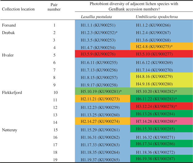
Photobiont diversity found in adjacent specimens (19 pairs) of Lasallia pustulata and Umbilicaria spodochroa in five localities in Norway. GenBank accession numbers are shown in parentheses. Asterisks indicate ITS sequences representing each haplotype in the phylogenetic analysis as shown in Fig. 4
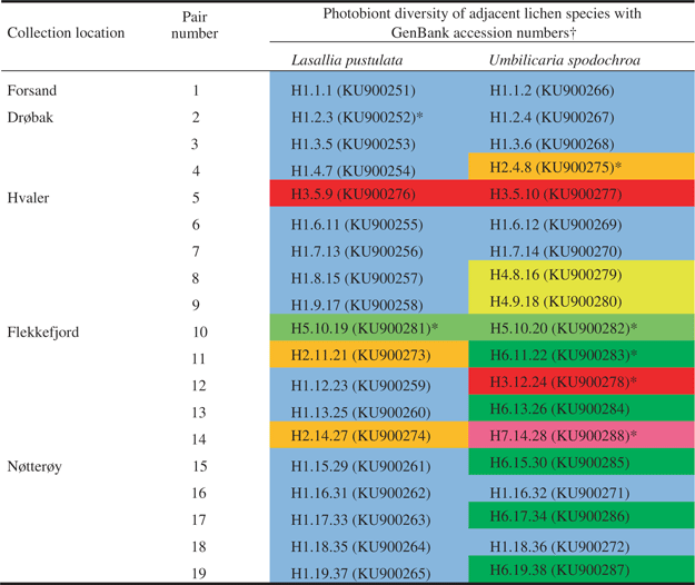
†Key to information given for each photobiont. Three numbers are given for each haplotype; the first = the unique haplotype number also indicated in different colours (H1 -7), the second = the pair number (1 -19), the third = the number of the individual thallus (1 -38). For instance H1.1.1=Haplotype 1, pair number 1, thallus number 1.
DNA isolation, sequencing and sequence alignment
Approximately 0·5 cm2 of each lichen thallus was homogenized with 0·7 mm zirconium beads for 8 s in a mini-beadbeater. Genomic DNA was extracted using a modified protocol from Zolan & Pukkila (Reference Zolan and Pukkila1986) with 2% sodium dodecyl sulphate (SDS) as extraction buffer. Isolated DNA was resuspended in sterile water and stored at −20°C. The ITS region of the photobionts was amplified from the lichen total genomic DNA using the following algal-specific and/or general primer pairs (including newly developed primers shown in parentheses): nrITSaJOFOR2 and nrITSaJOREV2 (Sadowska-Des et al. Reference Sadowska-Des, Balint, Otte and Schmitt2013); nrITSSaJOFOR2 and nrITS4T (Kroken & Taylor Reference Kroken and Taylor2000); nrITSSaJOFOR2 and nrITS4 (White et al. Reference White, Bruns, Lee and Taylor1990); nrITSSaJOFOR2 and Treb_1R (5’ACCTCAGGTCGAAAGCCAAA3’); nrITS1T (Kroken & Taylor Reference Kroken and Taylor2000) and Treb_3R (5’CTGACCTCAGGTCGAAAGCCAA3’). All PCR products were cleaned with ExoSAP (Affymetrix Inc., CA, USA) following the manufacturer’s protocol. Sequencing was carried out in 10 µl reactions using: 1 µl primer (10 µM), 1 µl purified PCR product, 0·75 µl BigDye (BigDye Terminator Cycle sequencing kit, ABIPRISM version 3.1; PerkinElmer, Applied Biosystems, Foster City, CA), 3·25 µl BigDye buffer, and 4 µl double-distilled water. Clean up reactions were performed over Sephadex G-50 DNA grade columns, eluting in water. Samples were then injected directly in an ABI 3730xl DNA analyzer (PE Applied Biosystems, Foster City, CA) utilizing a 22 s injection time and a 50 cm capillary array, at the Sequencing and Genomic Technologies Shared Resource core facility, part of the Duke Center for Genomic and Computational Biology.
Sequences were assembled and edited using the software package Sequencher™ 5.1 (Gene Codes Corporation, Ann Arbor, MI, USA) and subjected to BLAST searches (Wheeler et al. Reference Wheeler, Barrett, Benson, Bryant, Canese, Chetvernin, Church, DiCuccio, Edgar and Federhen2007) to confirm the algal origin of each sequence fragment. GenBank accession numbers for the 38 ITS sequences generated in this study are provided in Table 1. These ITS sequences represent seven haplotypes defined by using a 100% similarity criterion in Sequencher.
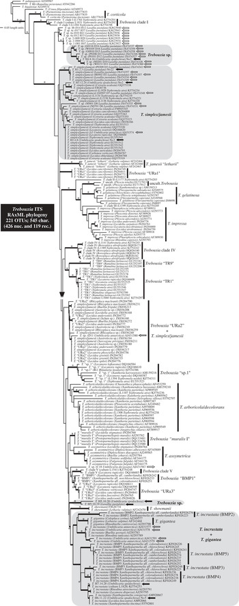
Single representative sequences for each of the seven haplotypes were added to an ITS reference data set from GenBank, that we assembled based on the Trebouxia phylogeny from the study by Muggia et al. (Reference Muggia, Pérez-Ortega, Kopun, Zellnig and Grube2014). The exception was Haplotype 5 which was represented by two sequences due to a few nucleotide uncertainties in one of them. The data matrix included multiple representatives of all major lineages and selected potential species (see also O’Brien Reference O’Brien2014), as well as the existing sequences of photobionts from Lasallia and Umbilicaria (Romeike et al. Reference Romeike, Fridl, Helms and Ott2002; Jones et al. Reference Jones, Hogg, Wilkins and Green2013; Sadowska-Des et al. Reference Sadowska-Des, Balint, Otte and Schmitt2013, Reference Sadowska-Des, Dal Grande, Lumbsch, Beck, Otte, Hur, Kim and Schmitt2014) for a total of 221 OTUs (Fig. 4). Sequences were aligned manually using MacClade 4.08 (Maddison & Maddison Reference Maddison and Maddison2005), and ambiguously aligned regions (sensu Lutzoni et al. Reference Lutzoni, Wagner, Reeb and Zoller2000), were excluded from the first set of phylogenetic analyses. The maximum likelihood analyses on 429 characters were performed using RAxML-HPC2 version 7.2.8 (Stamatakis Reference Stamatakis2006; Stamatakis et al. Reference Stamatakis, Hoover and Rougemont2008) as implemented on the CIPRES portal (Miller et al. Reference Miller, Pfeiffer and Schwartz2010). Optimal tree and bootstrap searches were conducted with the rapid hill-climbing algorithm for 1000 replicates with the GTRGAMMA substitution model (Rodríguez et al. Reference Rodríguez, Oliver, Marin and Medina1990) estimated for two partitions (ITS1+ITS2, and 5.8S). The second set of analyses was conducted on an extended data set with the re-inclusion of four ambiguous regions that were coded using PICS-Ord (Lücking et al. Reference Lücking, Hodkinson, Stamatakis and Cartwright2011), giving a total of 545 characters (426 nucleotides and 119 recoded characters). Trebouxia galapagensis (Hildreth & Ahmadjian) Gärtner (AJ249567) and Trebouxia sp. from Ramalina peruviana Ach. (AY842266) were used to root the phylogenies (Muggia et al. Reference Muggia, Pérez-Ortega, Kopun, Zellnig and Grube2014; O’Brien Reference O’Brien2014). We consider bootstrap values ≥70% as strong support. The NEXUS file and the resulting RAxML phylogeny were deposited in TreeBASE (accession number http://purl.org/phylo/treebase/phylows/study/TB2:S19031).
Results
The photobionts associated with 19 pairs of Lasallia pustulata-Umbilicaria spodochroa from five localities in Norway are represented by seven ITS haplotypes (hereafter referred to as H1–7) of Trebouxia algae (Table 1 & Fig. 4). Most haplotypes differed from each other substantially (i.e. by 10–37 nucleotide substitutions), except for two sets of haplotypes (H6/H7 and H2/H3) which differed only by one and two nucleotide substitutions, respectively. The most common haplotype (H1) was found in 22 of 38 thalli examined (57%), of which 15 were L. pustulata and seven were U. spodochroa (Table 1). This haplotype was present at all five study localities. Three of the remaining haplotypes (H2, H3, and H6) were less common (three to five occurrences) and appeared in two sites only, whereas three others (H4, H5 and H7) were rare and restricted to a single locality (Table 1). The highest diversity of photobionts was found in Flekkefjord, where six of the seven haplotypes were present. In the remaining sites (excluding Forsand because only one pair of lichens was sampled) up to three haplotypes of Trebouxia were detected. In about half of the lichen pairs examined (9 of 19), adjacent thalli of L. pustulata and U. spodochroa shared the same photobiont haplotype, which was H1 in seven of these nine pairs (Table 1). As expected, the mostly sexually reproducing U. spodochroa was found associated with a higher diversity of photobionts (seven haplotypes) than the mostly asexually reproducing L. pustulata (four haplotypes), which was found to be associated with the same haplotype (H1) in 15 of the 19 thalli sampled. Three haplotypes of Trebouxia (H4, H6 and H7) were found only in U. spodochroa thalli.
The ITS phylogeny for Trebouxia (Fig. 4) inferred for this study is largely in agreement with previous published phylogenies (e.g. Muggia et al. Reference Muggia, Pérez-Ortega, Kopun, Zellnig and Grube2014; O’Brien Reference O’Brien2014). The addition of coded characters greatly improved the resolution and support in some parts of the tree, especially for relationships among the most similar sequences. However, as in previous phylogenies, the backbone remained poorly supported (Muggia et al. Reference Muggia, Pérez-Ortega, Kopun, Zellnig and Grube2014; O’Brien Reference O’Brien2014). No conflicting relationship was detected between the tree based on 545 characters (i.e. 426 nucleotides and 119 PICS-Ord characters derived from the recoded ambiguously aligned regions; Fig. 4) and the phylogeny based on 429 characters (nucleotides only and ambiguously aligned regions excluded; tree not shown). Thirty-four internodes weakly supported in the phylogeny based on nucleotide characters only, received bootstrap support above 70% with the addition of PICS-Ord characters, but the opposite was true for five internodes; 41 branches were highly supported by both phylogenetic analyses. Overall, bootstrap support was lower in our phylogeny compared to the results from Muggia et al. (Reference Muggia, Pérez-Ortega, Kopun, Zellnig and Grube2014). This might be due to the more conservative approach in delimiting ambiguously aligned regions, which were excluded from our phylogenetic analyses since only four regions were reintegrated as coded characters. In contrast to Muggia et al. (Reference Muggia, Pérez-Ortega, Kopun, Zellnig and Grube2014), sequences in the T. simplex/jamesii clade were resolved in two groups, one of which also included members of Trebouxia “URa2”. However, none of these clades received strong bootstrap support. We also found that T. gigantea (Hildreth & Ahmadjian) Gärtner was nested within T. incrustata Ahmadjian ex Gärtner, and sequences of those two taxa were recovered within multiple well-supported monophyletic groups (Fig. 4).
Most Trebouxia sequences from our sampling sites are nested within two main clades corresponding to the poorly supported T. simplex/T. jamesii complex (H1, H2, and H3) and the T. incrustata+T. gigantea group (H6 and H7) that received high bootstrap support in our study (Fig. 4). Both clades contain photobionts associated with members of Umbilicaria or Lasallia such as U. antarctica Frey & I. M. Lamb in the T. incrustata clade and L. pustulata in the T. simplex/T. jamesii clade from previous studies (e.g., Sadowska-Des et al. Reference Sadowska-Des, Balint, Otte and Schmitt2013, Reference Sadowska-Des, Dal Grande, Lumbsch, Beck, Otte, Hur, Kim and Schmitt2014; Romeike et al. Reference Romeike, Fridl, Helms and Ott2002). Haplotype 4, found associated only with U. spodochroa in our study, was recovered in a well-supported monophyletic clade of Trebouxia strains associated exclusively with L. pustulata in previous studies. This clade might represent a new Trebouxia species (Trebouxia sp. in Fig. 4). Haplotype 5 was recovered as a distinct, well-supported lineage which could also represent a new Trebouxia species but its relationship to other Trebouxia lineages is poorly supported (Fig. 4). A revised classification of the diversity that has been documented within the genus Trebouxia is urgently needed and has been advocated (e.g. Leavitt et al. Reference Leavitt, Nelsen, Lumbsch, Johnson and St Clair2013, Reference Leavitt, Kraichak, Nelsen, Altermann, Divakar, Alors, Esslinger, Crespo and Lumbsch2015) in order to identify lichen photobionts and better understand patterns of associations among symbionts. Overall, the phylogenetic placements of our isolates were in agreement with nucleotide BLAST results based on the sequences deposited in GenBank. For example, H6 and H7 share an 84-nucleotide insertion (excluded from ML analyses) in ITS1 of T. incrustata, the highest BLAST hit (100% query cover and 99% identity) being in the sequence of a photobiont found in other lichens (e.g., Protoparmeliopsis muralis (Schreb.) M. Choisy and Xanthoparmelia tinctina (Maheu & A. Gillet) Hale).
Discussion
Our study demonstrates that the two co-occurring lichen species Lasallia pustulata and Umbilicaria spodochroa often share the same photobiont within a single site and across different localities, and might therefore form a photobiont-mediated guild sensu Rikkinen (Reference Rikkinen2003). It is also very likely that U. spodochroa ‘pirates’ algal cells from isidia of L. pustulata, as corroborated by the sharing of the rare Haplotypes 3 in Hvaler and 5 in Flekkefjord. However, empirical evidence would be needed from re-lichenization where, for example, isidia of L. pustulata are the only available source of photobiont for U. spodochroa. The fact that in many cases U. spodochroa utilizes photobionts other than those present in adjacent L. pustulata (especially H6; Table 1) suggests that the former species does not entirely depend on the latter for successful colonization and establishment. The overlap in geographical range of the two species is thus less likely to be explained solely by the mutual dependence on the same photobiont, and perhaps involves other environmental and/or reproductive factors. Ecological and evolutionary theory suggests that competitive exclusion should lead to the loss of one or other of these species due to the similarity of their requirements (MacArthur & Levins Reference MacArthur and Levins1967; Abrams Reference Abrams1983; Abrams & Rueffler Reference Abrams and Rueffler2009). It is therefore possible that the co-occurring L. pustulata and U. spodochroa select different photobionts to achieve some degree of niche-separation, which is driven in part by their contrasting mode of reproduction. To what degree some of the detected Trebouxia haplotypes occur in a free-living state or in association with other lichen species in the same habitat has yet to be examined.
Overall, the most widespread, common and shared Trebouxia was represented by Haplotype 1 (H1), which also associates more frequently with L. pustulata (79%) than with U. spodochroa (37%). The same Trebouxia haplotype was found in approximately one third of the 44 specimens of L. pustulata collected across Europe by Sadowska-Des et al. (Reference Sadowska-Des, Balint, Otte and Schmitt2013). Whether an association with a particular photobiont haplotype reflects some degree of physiological adaptation of the fungus to particular local habitats or the availability/ frequency of compatible photobionts, remains unknown. In L. pustulata, Sadowska-Des et al. (Reference Sadowska-Des, Balint, Otte and Schmitt2013) found hardly any genetic variation in several loci of the mycobiont sampled within a broad geographical range in Europe, suggesting that there is no correlation between mycobiont-photobiont haplotypes. Trebouxia Haplotype 1 has only rarely been recorded from other lichen species (e.g. Cetraria aculeata (Schreb.) Fr.; Fernandez-Mendoza et al. Reference Fernandez-Mendoza, Domaschke, Garcia, Jordan, Martin and Printzen2011; Domaschke et al. Reference Domaschke, Vivas, Sancho and Printzen2013). This may, however, indicate the under-sampling of certain environments, including coastal cliff habitats in Norway, rather than its uniqueness. The taxonomic identity of this strain remains unclear as two names, T. angustilobata (del Campo et al. Reference del Campo, Hoyo, Casano, Martinez-Alberola and Barrena2010) and T. jamesii (e.g., Domaschke et al. Reference Domaschke, Vivas, Sancho and Printzen2013), are being used, and the phylogenetic placement of Haplotype 1 within a broadly defined T. simplex/T. jamesii complex is not conclusive as it has been placed where boundaries among species are not well defined and supported.
Our study confirms the capacity of L. pustulata to utilize several different strains of Trebouxia. This was earlier demonstrated by Sadowska-Des et al. (Reference Sadowska-Des, Balint, Otte and Schmitt2013), but the genetic variation we detected among the photobionts was lower, perhaps due to the more limited sampling area restricted to the southern coast of Norway. Umbilicaria spodochroa seems to accept a wider range of algal partners (seven haplotypes) compared with L. pustulata (four haplotypes). This pattern may be explained partly by the greater likelihood of algal switches in U. spodochroa because a new thallus is often re-established de novo through ascospores by the mycobiont encountering a suitable algal partner in the environment, compared to L. pustulata which transmits its photobiont from one generation to the next using isidia. In these circumstances, it might be advantageous for a sexually reproducing species, which is required to re-associate with a new photobiont at each generation, to be a generalist when selecting a ‘suitable photobiont’ especially when competing with a mainly clonally reproducing species that propagates the symbiosis intact. This is the pattern we observed in the two umbilicate lichen species sampled. In the genus Umbilicaria this strategy is not restricted to sexually reproducing species but seems to occur also in about one third of its species that reproduce mainly by specialized asexual fungal propagules called thalloconidia seceded from the lower cortex, which are strictly fungal (Hestmark Reference Hestmark1990, 1991 Reference Hestmarka , Reference Hestmarkb , Reference Hestmarkc , 1992 Reference Hestmarka ). Numerous photobionts (based on the ITS) and diverse sharing patterns were found in four thalloconidia-producing species (U. antarctica, U. decussata (Vill.) Zahlbr., U. kappenii Sancho et al. and U. umbilicarioides (Stein) Krog & Swinscow) in the Antarctic (Romeike et al. Reference Romeike, Fridl, Helms and Ott2002). Similarly, a number of Trebouxia strains associated with U. aprina Nyl. and U. decussata were reported from several localities in Antarctica (Jones et al. Reference Jones, Hogg, Wilkins and Green2013). Somewhat in contrast to these results, another study of U. aprina in Antarctica revealed only a single algal strain in 19 thalli (Pérez-Ortega et al. Reference Pérez-Ortega, Ortiz-Alvarez, Green and de los Rios2012), but this may be due to the restricted geographical sampling in that particular study. The fact that different algae were detected in thalli of L. pustulata supports previous studies reporting that even in a mostly clonally reproducing species with propagules containing both partners, diverse symbiotic associations occur at various spatial scales (e.g. Piercey-Normore Reference Piercey-Normore2009; Dal Grande et al. Reference Dal Grande, Widmer, Wagner and Scheidegger2012; Sadowska-Des et al. Reference Sadowska-Des, Balint, Otte and Schmitt2013).
Our results show that U. spodochroa associates with three Trebouxia haplotypes (H4, H6 and H7) not found in the thalli of the neighbouring L. pustulata. The H4 strain was, however, previously reported from L. pustulata (Sadowska-Des et al. Reference Sadowska-Des, Balint, Otte and Schmitt2013) and H7 is known from other saxicolous lichens such as Lecanora dispersa (Pers.) Sommerf. (UTEX 784), and Tephromela atra (DC.) Hafellner (Muggia et al. Reference Muggia, Pérez-Ortega, Kopun, Zellnig and Grube2014). Overall, Trebouxia strains associated with Umbilicaria/Lasallia are not spread evenly across the ITS phylogeny but cluster in a few lineages (Trebouxia sp., T. simplex/T. jamesii, and T. incrustata+T. gigantea), and might include new Trebouxia species. Having a more stable phylogeny for Trebouxia will not only help in establishing boundaries among potential species and reveal their phylogenetic relationships, but will also facilitate the taxonomic revision of the genus and allow a more reliable identification of the most common lichen photobiont.
Mixed populations of Umbilicaria spodochroa (light grey) and Lasallia pustulata (olive-green) on coastal cliffs of southern Norway. Black dots on U. spodochroa are apothecia.

SEM of isidium on the thallus of L. pustulata: an asexually generated coralloid propagule that includes the fungus and alga in bundles. Scale=0·5 mm.

Distribution maps of U. spodochroa (A) and L. pustulata (B) in Norway. Stars and crosses indicate collections without exact coordinates, localized to county. Grey triangles indicate sample sites in this study, from left to right: Forsand, Flekkefjord, Torød, Drøbak, Hvaler. Maps generated from the lichen database Norsk Lavdatabase of the Natural History Museum, University of Oslo courtesy of E. Timdal.
Phylogenetic placement of seven haplotypes (H1–H7) of photobionts sampled from adjacent thalli of Lasallia pustulata and Umbilicaria spodochroa in Norway, in a broad phylogenetic context of the Trebouxia phylogeny based on ITS sequences (545 characters including 426 nucleotide sites and 119 recoded PICS-Ord characters) for 221 OTUs. See Table 1 for the sampling design and abbreviations used. Reference sequences were selected from Muggia et al. (Reference Muggia, Pérez-Ortega, Kopun, Zellnig and Grube2014), Sadowska-Des et al. (Reference Sadowska-Des, Balint, Otte and Schmitt2013, Reference Sadowska-Des, Dal Grande, Lumbsch, Beck, Otte, Hur, Kim and Schmitt2014) and Romeike et al. (Reference Romeike, Fridl, Helms and Ott2002). The phylogeny was rooted with T. galapagensis AJ249567 and Trebouxia AY842266 following Muggia et al. (Reference Muggia, Pérez-Ortega, Kopun, Zellnig and Grube2014) and O’Brien (Reference O’Brien2014). Arrows indicate sequences of photobionts from L. pustulata, U. spodochroa (published – white arrows pointing to the left; from this study – black arrows), U. antarctica (grey arrows) and U. decussata (white arrow pointing to the right). Clades containing Trebouxia sequences from this study are delimited by grey boxes. Clade annotations follow Muggia et al. (Reference Muggia, Pérez-Ortega, Kopun, Zellnig and Grube2014). Thick branches indicate bootstrap support ≥70%. Stars indicate internodes that received high bootstrap support (>70%) in the maximum likelihood analysis based on nucleotides only (i.e. without PICS-Ord characters). Haplotype 5 (H5) is represented by two sequences due to a few nucleotide uncertainties in one of the sequences.

The authors are grateful to Brendan Hodkinson for his assistance in the installation and implementation of PICS-Ord. Funding for this study came from a research sabbatical grant from the University of Oslo to GH.