Implications
Feeding pigs with reduced protein diets (RPD) has been recognized as a successful nutritional strategy to increase intramuscular fat (IMF) content, with important economic implications in the pig industry, although its consequences on carcass fat deposition are yet to be established. This report sheds light on the events involved in subcutaneous fat deposition by exploiting adipose membrane’s biophysical properties, particularly membrane fluidity and permeability to glycerol in lean and fatty pigs, fed RPD. RPD appeared to promote undesirable increase on total fatty acids in subcutaneous fat, which is likely to be mediated by higher fluidity of adipose membranes.
Introduction
High levels of total fat intake have been shown to increase obesity and cancer risk in industrialized countries (Van Barneveld, Reference Van Barneveld2003). Regarding this, modern pork industry focused their efforts on producing pigs with a lower percentage of fat (Jung et al., Reference Jung, Shim, Na and Choe2015) to satisfy consumers’ preferences (Van Barneveld, Reference Van Barneveld2003). However, leaner meat results not only in reduction of subcutaneous fat, but also of IMF. Indeed, white European breeds (e.g. Large White and Landrace) have been genetically selected toward reduced subcutaneous fat, and, consequently, reduced amount of IMF (Jeremiah et al., Reference Jeremiah, Gibson, Gibsona, Ballc, Akerd and Fortin1999). IMF is a key contributor to important aspects of sensory traits and nutritional value of meat quality (Wood et al., Reference Wood, Enser, Fisher, Nute, Sheard, Richardson, Hughes and Whittington2008).
Over the past decade, our research group has been working on nutritional strategies using RPD supplemented or not with specific amino acids, as lysine or arginine (Madeira et al., Reference Madeira, Costa, Alfaia, Lopes, Bessa, Lemos and Prates2013 and Reference Madeira, Alfaia, Costa, Lopes, Martins, Lemos, Moreira, Santos-Silva, Bessa and Prates2015) to improve the quality of pork. For non-ruminants, lysine is usually the first limiting amino acid in animal feeds. Lysine may affect growth performance by reducing protein synthesis while increasing the proportion of energy available for fat storage (Witte et al., Reference Witte, Ellis, McKeith and Wilson2000; Teye et al., Reference Teye, Sheard, Whittington, Nute, Stewart and Wood2006; D´Souza et al., Reference Doran, Moule, Teye, Whittington, Hallett and Wood2008). Like others (Wood et al., Reference Wood, Nute, Richardson, Whittington, Southwood, Plastow, Mansbridge, da Costa and Chang2004; Doran et al., Reference D´Souza, Pethick, Dunshea, Pluske and Mullan2006; Suárez-Belloch et al., Reference Suárez-Belloch, Guada and Latorre2015), we have reported that reducing the proportion of protein relative to energy in pigs’ diet consistently increases IMF deposition (Madeira et al., Reference Madeira, Costa, Alfaia, Lopes, Bessa, Lemos and Prates2013, Reference Madeira, Alfaia, Costa, Lopes, Lemos, Bessa and Prates2014 and Reference Madeira, Alfaia, Costa, Lopes, Martins, Lemos, Moreira, Santos-Silva, Bessa and Prates2015). However, the effects of these dietary manipulations on subcutaneous fat are not yet completely understood. Although some authors conclude that protein reduction increases backfat thickness (Huff-Lonergan et al., Reference Huff-Lonergan, Bass, Malek, Dekkers, Prusa and Rothschild2002; Lebret, Reference Lebret2008), others report no changes (Wood et al., Reference Wood, Nute, Richardson, Whittington, Southwood, Plastow, Mansbridge, da Costa and Chang2004; Suárez-Belloch et al., Reference Suárez-Belloch, Guada and Latorre2015).
In line with this, in the present work we hypothesized that the effects of RPD and RPD corrected for lysine (RPDL) on carcass fat deposition from crossbred pigs and Alentejana purebred, a lean and fatty genotype, respectively, are mediated by alterations on adipose membrane biophysical properties. Although crossbred pigs result from a genetic heritage of different high-performing pigs, as Large White, Landrace and Pietrain (Jeremiah et al., Reference Jeremiah, Gibson, Gibsona, Ballc, Akerd and Fortin1999), Alentejana breed is genetically pure with a greater propensity to accumulate fat (Ramalho, Reference Ramalho2007). Moreover, Alentejana is an autochthonous Mediterranean breed traditionally reared under extensive conditions, and similarly to the Spanish Iberian pig, has a low capacity for lean tissue deposition (Garcia-Valverde et al., Reference Garcia-Valverde, Barea, Lara, Nieto and Aguilera2008).
Material and methods
Experimental design: animals and diets
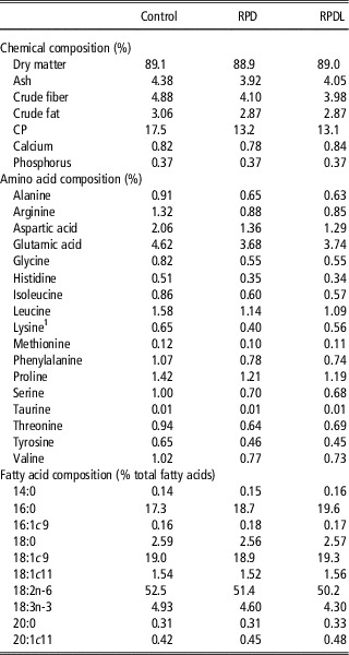
In total, 18 commercial crossbred (50% Large White, 25% Landrace and 25% Pietrain) and 18 Alentejana purebred entire male pigs with an initial body weight (BW) of 60.0±1.67 kg (mean±SE) were used. Pigs were fed a standard concentrate diet from weaning until the beginning of the experiment. Thereafter, pigs from each genotype were randomly assigned to one of three diets (n=6). The experimental diets were iso-energetically formulated (13.5 ME/kg), differing in crude protein (CP) and lysine contents, as follows: 17.5% CP and 0.7% lysine (control diet), 13.1% CP and 0.4% lysine (RPD), or 13.2% CP and 0.6% lysine (RPDL). l-lysine was added to the RPDL diet to equilibrate the level of this amino acid with the control diet. The chemical composition of experimental diets, with amino acid and fatty acid profiles, is presented in Table 1. Analytical methods used to assess diets composition are published in Madeira et al. (Reference Madeira, Costa, Alfaia, Lopes, Bessa, Lemos and Prates2013).
Chemical composition of control and reduced protein diets, with (RPDL) or without (RPD) lysine correction

1 Calculated lysine content: 0.60%, 0.40% and 0.60% for control, RPD and RPDL diets, respectively.
Pigs were slaughtered at an average live BW of 93.1±2.01 kg. After electrical stunning and exsanguination, samples from subcutaneous fat were collected from the right carcass side directly above longissimus lumborum muscle. At 24 h postmortem, the backfat thickness was measured in the left carcass side at the last rib position (P2).
This trial was conducted at the facilities of L-INIA (INRB, Vale de Santarém, Portugal) under the guidelines for the care and use of experimental animals, following EU directive 86/609/EEC.
Total fatty acids in the subcutaneous fat
Subcutaneous fat samples were lyophilized (−60°C and 2.0 hPa) to constant weight, kept dry at −20°C, and analyzed within 2 weeks. Total fatty acids were extracted from the lyophilized samples (Folch et al., Reference Folch, Lees and Sloane Stanley1957) using dichloromethane : methanol 2 : 1 (v : v), converted to methyl esters (FAME) (Raes et al., Reference Raes, Fievez, Chow, Ansorena, Demeyer and De Smet2004) and determined using a gas chromatograph HP6890A (Hewlett-Packard, Avondale, PA, USA), equipped with a flame ionization detector and a CP-Sil 88 capillary column (100 m; 0.25 mm i.d.; 0.20 m film thickness; Chrompack, Varian Inc., Walnut Creek, CA, USA) with the chromatographic conditions described elsewhere (Alves and Bessa, Reference Alves and Bessa2009). The quantification of total FAME was done using nonadecanoic acid (19 : 0) from Supelco (Bellefonte, PA, USA), as internal standard.
Preparation of adipose membrane vesicles from subcutaneous fat
Membrane vesicles were prepared from subcutaneous fat by differential centrifugation, using a previously described method (Martins et al., Reference Martins, Lopes, Costa, Martins, Santos, Prates, Moura and Soveral2011). Crude membranes re-suspended in mannitol-HEPES buffer (100 mM mannitol, 10 mM Tris-HEPES, pH 7.4, 120 mOsM) at a concentration of 0.2 mg protein/ml were immediately used for transport experiments. The protein content was determined by the Bradford technique (Bradford, Reference Bradford1976) using the microassay procedure of Bio-Rad Protein Assay Dye Reagent Concentrate.
Vesicle size of all membrane vesicles was determined by Quasi-Elastic Light Scattering (Brookhaven Instruments, Holtsville, NY, USA) (Soveral et al., Reference Soveral, Macey and Moura1997), revealing homogenous populations with monomodal distributions with a mean hydrodynamic diameter of 310±35 nm.
Lipid composition of adipose membrane vesicles
Membranes were lyophilized and fatty acids were converted to FAME (Christie et al., Reference Christie, Dobson and Adlof2007). The resulting FAME were analyzed by gas chromatography, as described in detail by Martins et al. (Reference Martins, Lopes, Martins, Madeira, Santos, Prates, Moura and Soveral2010). Total cholesterol was extracted from lyophilized membranes (Martins et al., Reference Martins, Lopes, Martins, Madeira, Santos, Prates, Moura and Soveral2010) and quantified, in triplicate, based on the external standard technique, from a standard curve for peak area v. cholesterol concentration.
Membrane fluidity measurements
Membrane fluidity was evaluated by measuring the fluorescence anisotropy of two probes incorporated in the membrane, namely, 1,6-diphenyl-1,3,5-hexatriene (DPH), or 1-(4-(trimethylamino)-phenyl)-6-phenyl-1,3,5-hexatriene (TMA-DPH), from Molecular Probes (Eugene, OR, USA). DPH is incorporated in the membrane at the fatty acyl groups’ level, whereas TMA-DPH is anchored by its cationic moiety at the membrane/water interface, probing the membrane region closer to the phospholipids head groups (Pebay-Peyroula et al., Reference Pebay-Peyroula, Dufourc and Szabo1994). Measurements of fluorescence anisotropy (r) were conducted, as previously described (Martins et al., Reference Martins, Lopes, Martins, Madeira, Santos, Prates, Moura and Soveral2010) and calculated using the equation r=(I VV −GI VH )/(I VV +2 GI VH ), where I VV and I VH are the fluorescence intensities, the subscripts indicate the vertical (V) or horizontal (H) orientations of the excitation and emission polarizers, and G=I HV /I HH is the instrumental factor. The fluorescence intensity data points used for calculations were the average of three identical aliquots (after blank subtraction) measured on a Varian Cary Eclipse fluorescence spectrophotometer (Varian, Mulgrave, Victoria, Australia).
Evaluation of membrane glycerol permeability
Stopped-flow assays were performed on a HI-TECH Scientific PQ/SF-53 (Hi-Tech Scientific, Bradford-on-Avon, UK) apparatus, at temperatures ranging from 12°C to 37°C, as described by Martins et al. (Reference Martins, Lopes, Martins, Madeira, Santos, Prates, Moura and Soveral2010). Membrane vesicles equilibrated in 120 mOsM mannitol-HEPES buffer were confronted to an external solution where the impermeant solute was partially replaced by glycerol (60 mOsM mannitol plus 180 mOsM glycerol, creating an inwardly directed glycerol gradient). After the first fast vesicle shrinkage due to water outflow, glycerol influx in response to its chemical gradient was followed by water influx with subsequent vesicle re-swelling. Glycerol permeability was calculated as P gly =k (V o /A), where k is the single exponential time constant fitted to the light scattering signal of glycerol influx and V o /A is the initial volume to area ratio of the vesicle preparation (Martins et al., Reference Martins, Lopes, Martins, Madeira, Santos, Prates, Moura and Soveral2010). All solution osmolarities were determined from freezing point depression on a semi-micro osmometer (Knauer GmbH, Germany) using standards of 100 and 400 mOsM. The activation energy (E a ) of glycerol transport was calculated from the slope of the Arrhenius plot (lnP gly v. 1/T) multiplied by the gas constant R.
Statistical analysis
Statistical analysis was performed using the Statistics Analysis System (SAS) software package, version 9.1 (SAS Institute, Cary, NC, USA), following a 2×3 factorial arrangement (two genotypes and three diets). Data were checked to normal distribution and variance homogeneity. As variance heterogeneity was detected for most of the variables, the PROCMIXED was used. The model included the fixed effects of genotype, diet and their interaction, and the REPEATED statement considering the GROUP option to accommodate variance heterogeneity. Results are presented as mean and standard error, and considered significantly different when the P-value was inferior to 0.05.
Results
RPD increased total fatty acids in subcutaneous fat but not backfat thickness
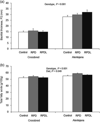
Backfat thickness and total fatty acids of subcutaneous fat were higher in Alentejana than in crossbred pigs (P<0.05, Figure 1). Backfat thickness was not affected by diet (Figure 1a). Total fatty acids were increased by RPD (P<0.05), but RPD and RPDL were similar within the same genotype (Figure 1b).
Backfat thickness at P2 (mm) (a) and total fatty acids (g/100 g) (b) in the subcutaneous adipose tissue from crossbred and Alentejana pigs fed control or reduced protein diets, with (RPDL) or without (RPD) lysine correction. Diet effect on total fatty acids was control<RPDL and RPD, while RPDL=RPD, for both genotypes.

Membrane polyunsaturated fatty acids (PUFA)/saturated fatty acids (SFA) ratio but not cholesterol content was decreased by RPD
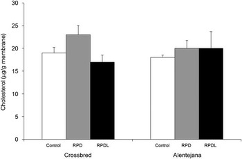
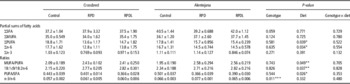
Irrespective of genotype, a lower prevalence of polyunsaturated fatty acids, in particular from the n-6 family, was observed in both RPD and RPDL groups, compared with the control diet (P<0.05, Table 2). In parallel, RPD decreased PUFA/SFA but increased monounsaturated fatty acids (MUFA)/PUFA and 18:1c9/18:2n-6 ratios (P<0.05, Table 2). Adipose membranes from Alentejana were richer in n-3/n-6 ratio than adipose membranes from crossbred pigs (P<0.05, Table 2). Both genotype and diet had no effect on membranes cholesterol content (Figure 2).
Cholesterol concentration (μg/g membrane) of adipose membranes from crossbred and Alentejana pigs fed control or reduced protein diets, with (RPDL) or without (RPD) lysine correction.

Fatty acids (% of total FAME) sums and ratios of adipose membrane vesicles from crossbred and Alentejana pigs fed control or reduced protein diets, with (RPDL) or without (RPD) lysine correction

FAME=fatty acid methyl esters; SFA=saturated fatty acids; MUFA=monounsaturated fatty acids; PUFA=polyunsaturated fatty acids.
ΣSFA=sum of 14:0, 16:0, 17:0, 18:0 and 20:0; ΣMUFA=sum of 16:1c7, 16:1c9, 17:1c9, 18:1c9, 18:1c11 and 20:1c11; ΣPUFA=sum of 18:2n-6, 18:3n-3, 20:2n-6, 20:3n-3, 20:4n-6, 20:5n-3 and 22:5n-3; Σn-6=sum of 18:2n-6, 20:2n-6 and 20:4n-6; Σn-3=sum of 18:3n-3, 20:3n-3, 20:5n-3 and 22:5n-3.
* CTR>RPD and RPDL, RPD=RPDL.
** CTR<RPD and RPDL, RPD=RPDL.
*** Crossbred<Alentejana.
Adipose membranes glycerol permeability was not affected by genotype nor by diet
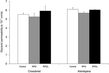
No significant differences were found for glycerol permeability coefficients of adipose membrane vesicles between genotypes or among dietary groups (Figure 3). E a values for glycerol transport were high and similar across all groups ranging from 21.2±0.5 to 25.1±0.8 kcal/mol (88.7±2.0 to 105±3.5 kJ/mol).
Glycerol permeability coefficients (cm/s) of adipose membranes from crossbred and Alentejana pigs fed control or reduced protein diets, with (RPDL) or without (RPD) lysine correction.

RPD increased adipose membranes fluidity
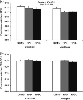
A significant decrease on the fluorescence anisotropy of DPH probe was found in both genotypes of pigs fed RPD (P<0.05, Figure 4a). Bearing in mind that fluorescence anisotropy is inversely related to membrane fluidity, adipose membranes from pigs fed RPD or RPDL are, therefore, more fluid. RPD and RPDL values were identical (Figure 4a). For the fluorescence anisotropy of TMA-DPH probe, no variations were observed across dietary groups (Figure 4b).
Membrane fluidity measurements by fluorescence anisotropy. 1,6-diphenyl-1,3,5-hexatriene (DPH) (a) and 1-(4-(trimethylamino)-phenyl)-6-phenyl-1,3,5-hexatriene (TMA-DPH) (b) probes in adipose membranes from crossbred and Alentejana pigs fed control or reduced protein diets, with (RPDL) or without (RPD) lysine correction. Diet effect on DPH probe was control>RPDL and RPD, while RPDL=RPD, for both genotypes.

Discussion
This study enabled to understand the effect of RPD, corrected or not for lysine, on subcutaneous fat deposition in lean and fatty pigs by exploiting adipose membrane biophysical properties associated with fatty acid content and glycerol transport in adipocytes. Not surprisingly, backfat thickness and total fatty acids of subcutaneous fat were determined by genetic background being higher in Alentejana purebred than in crossbred pigs. In marked contrast to modern highly improved pig breeds selected for low levels of fatness, the Alentejana breed is much more lipogenic (Nieto et al., Reference Nieto, Miranda, Garcia and Aguilera2002). Like us, Huff-Lonergan et al. (Reference Huff-Lonergan, Bass, Malek, Dekkers, Prusa and Rothschild2002) observed that dietary lysine level did not change carcass fat depth and reported a positive correlation between backfat thickness and IMF content. In a companion paper, we reported that RPD increased IMF deposition by ~50% in crossbred pigs, without affecting animal growth rate (Madeira et al., Reference Madeira, Costa, Alfaia, Lopes, Bessa, Lemos and Prates2013). These results clearly show that IMF increment is mediated by lysine restriction (Madeira et al., Reference Madeira, Costa, Alfaia, Lopes, Bessa, Lemos and Prates2013). In our experiment, the protein reduction, corrected or not with lysine, did not affect backfat thickness, but increased total fatty acids on subcutaneous fat in lean and fatty pigs. Doran et al. (Reference D´Souza, Pethick, Dunshea, Pluske and Mullan2006) also observed a large effect on IMF (by ~86.3%) accompanied by an increase in subcutaneous fat lipid content.
In view of the results found for backfat thickness and total fatty acids content, we hypothesize that some biophysical mechanisms of adipose cellular membranes might be held responsible for subcutaneous fat deposition. In particular, we exploited adipose membranes fluidity and permeability to glycerol, which are well known biophysical properties that may facilitate glycerol availability for triacylglycerols (TAG) formation. Glycerol is the backbone molecule of TAG and is crucial for TAG synthesis in the cell. Glycerol release from the adipocyte in response to energy balance may be mediated by a protein glycerol-channel that regulates glycerol accumulation (Kishida et al., Reference Kishida, Kuriyama, Funahashi, Shimomura, Kihara, Ouchi, Nishida, Nishizawa, Matsuda, Takahashi, Hotta, Nakamura, Yamashita, Tochino and Matsuzawa2000). Like other small uncharged molecules, glycerol can cross the cell membrane through lipid bilayer diffusion (i.e. passive pathway) or through a protein channel called aquaglyceroporin (i.e. facilitated diffusion pathway) (Kishida et al., Reference Kishida, Kuriyama, Funahashi, Shimomura, Kihara, Ouchi, Nishida, Nishizawa, Matsuda, Takahashi, Hotta, Nakamura, Yamashita, Tochino and Matsuzawa2000; Kondo et al., Reference Kondo, Shimomura, Kishida, Kuriyama, Makino, Nishizawa, Matsuda, Maeda, Nagaretani, Kihara, Kurachi, Nakamura, Funahashi and Matsuzawa2002). The activation energy values, E a , found for the glycerol transport were consistently high, indicating that the permeation pathway is not protein channel-dependent in pigs and, in fact, is quite stable across experimental groups, revealing dietary treatments independency. Our group also observed high values of E a for glycerol transport in ruminants, as a consequence of lipid diffusion pathway (Campos et al., Reference Campos, Moura, Oliva, Leandro and Soveral2011). In the present work, neither genetics nor low dietary protein levels (with or without lysine correction) had an impact on glycerol permeability of adipose membranes from subcutaneous fat. For comparative purposes, glycerol permeability and E a values available in the literature for adipose tissue isolated membranes are very scarce. An exception is the report by Martins et al. (Reference Martins, Lopes, Costa, Martins, Santos, Prates, Moura and Soveral2011) on mesenteric fat from bovines in which P gly values were nearly fivefold lower than the ones herein observed.
In contrast to glycerol permeability, membrane fluidity was more sensitive to the influence of genetics and dietary treatments. Using the fluorescence polarization technique with DPH probe, higher membrane fluidity (lower fluorescence anisotropy) was observed in fatty pigs compared with lean pigs. This is in accordance to the findings of Lee (Reference Lee1985), who reported an increase in fluidity as characteristic of a wide variety of cellular membranes, not limited to adipocytes, of obese v. lean mice. The TMA-DPH probe was unchanged, reflecting that fluidity varies most certainly due to adjustments at the fatty acyl groups’ level, rather than at the membrane surface.
Besides genetics, membrane fluidity may also be affected by diet due to changes on the degree of saturation of phospholipid fatty acids (York, Reference York1985). Dietary protein reduction increased the fluidity of adipose tissue membranes, regardless of genotype which may be associated with a greater balance between oleic acid (18:1c9) and linoleic acid (18:2n-6) in membranes lipid composition, as previously reported by York (Reference York1985). Although cholesterol content was unchanged, specific phospholipid changes can result on a redistribution of cholesterol within the lipid bilayer, therefore affecting membrane rigidity (Marsh, Reference Marsh2009). Dietary protein reduction increased membrane fluidity which may exert some influence on fatty acids uptake by adipocytes allowing us to validate the research hypothesis previously formulated. The addition of lysine had no impact on membrane fluidity, either assessed by DPH or TMA-DPH. Fatty acids may enter the cell across the plasma membrane by simple diffusion, or facilitated by membrane-associated proteins (Schwenk et al., Reference Schwenk, Holloway, Luiken, Bonen and Glatz2010). The increase of fluidity at the hydrocarbon zone level, as shown by DPH, may facilitate fatty acids diffusion through the membrane lipid bilayer and is in line with the increased fatty acid content on subcutaneous fat from lean and fatty pigs fed RPD.
Conclusions
We acknowledge RPD as a successful nutritional strategy to increase IMF in pigs without affecting backfat thickness. However, the reduction of dietary protein fed to pigs increased total fatty acids from subcutaneous fat and adipose membranes fluidity, regardless of pig’s genotype. This increment on adipose tissue membranes fluidity may be held responsible for facilitating fatty acids uptake from adipocytes, which promotes undesirable increase on total fatty acids in subcutaneous fat.
Acknowledgements
Financial support from Fundação para a Ciência e a Tecnologia – Ministério da Educação e Ciência (FCT-MEC, Portugal) grant (PTDC/CVT/2008/99210), CIISA project (UID/CVT/2013/00276) and individual fellowships to A. P. M. (SFRH/BD/2009/65046), S. V. M. (SFRH/BPD/2009/63019) and M. S. M. (SFRH/BD/2008/48240) are acknowledged. P. A. L. is a researcher from the FCT-MEC program ‘Ciência 2008’ and Incentivo 2014 project (AGR/UI0276/2014).