Case report
A 5-month-old boy was brought by his parents with a history of difficulty in sucking breast milk, failure to thrive, and distressed breathing. The symptoms started from 2 weeks of age. There was no history of cyanosis or cyanotic spells. The weight was 3.5 kg. The baby was in mild distress with a respiratory rate of 44 breaths per minute and a pulse rate of 136 per minute, with a bounding type of pulse. The precordium was hyperdyanamic. The first and second heart sounds were normal. There was an ejection type of systolic murmur of grade three of six, best heard over the left third intercostal space and conducted over the precordium.
The electrocardiogram showed sinus tachycardia with right bundle branch block. The chest X-ray showed cardiomegaly with a cardiothoracic ratio of 65%. Echocardiography revealed situs solitus, atrioventricular and ventriculoarterial concordance, and normal venous drainage. There was a small secundum type of atrial septal defect with left-to-right shunt. The ventricular septum was intact. The left atrium and ventricle were dilated. In the parasternal long-axis view, there was a continuous jet of blood flow to the left atrium, arising from the descending aorta. This was confirmed in the sagital plane of the subcostal view. We could not interrogate the jet with continuous or pulsed wave Doppler because of alignment issue. In the modified apical four-chamber view, we noticed a defect between the aorta and the main pulmonary artery, with systolic left-to-right shunt, diagnostic of aortopulmonary window. There was no significant gradient. There was a high-pressure tricuspid regurgitation with a pressure gradient of about 58 mmHg, suggestive of significant pulmonary hypertension.
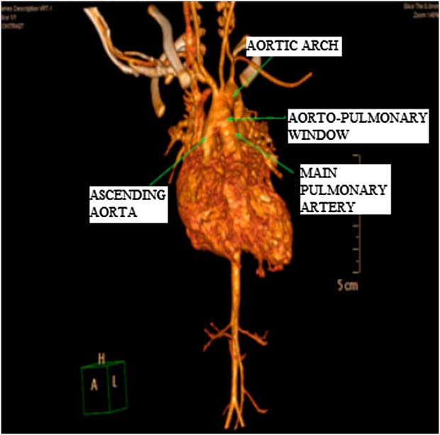
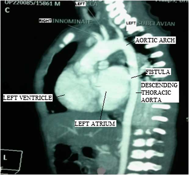
The parents did not give consent for invasive study, and thus to prove these findings a computed tomography was done. Computed tomography confirmed the findings revealed by echocardiography. The three-dimensional reconstructed image shows the heart in the anterolateral view, which clearly demonstrates the aortopulmonary window. The tomographic image of heart in lateral view demonstrates ascending aorta, arch, and descending thoracic aorta. The descending aorta is posterior to the left atrium, and there is a direct fistulous communication between them (Figs 1 and 2). This fistula is very short in length and is smooth. The baby was stabilised medically and referred for corrective surgery, but later lost to follow-up.
A three-dimensional computed tomographic image of the heart in the anterolateral aspect. The aortopulmonary window is seen clearly between the ascending aorta and the main pulmonary artery before branching.

A tomographic image of the heart in the lateral view shows the descending aorta posterior to the left atrium. There is a fistula between the descending thoracic aorta and the left atrium.

Discussion
The communication between the ascending aorta and the left atrium is more commonly reported than that of the descending aorta and left atrium.Reference Jean, Fermin, Velarde, Laired and Silva 1 , Reference Nihoyannopoulos, Sapsford and Oakley 2 , Reference Topcuoglu, Salih, San, Kayhan and Ulus 3 These are more commonly acquired and usually caused by trauma, operation, or infection.Reference Behnam 4 , Reference Shah, Livesey and Rakhit 5 , Reference Ananthasubramaniam 6 The congenital fistulas are usually associated with aneurysmal aortic sinuses, commonly opening in to right-sided chambers. There was no history of any surgical procedures in this baby to suggest an acquired origin of descending aorto–left atrial fistula. There were no aneurysmal dilations of the aorta or its sinuses. All the observed defects were most probably of congenital origin. The presence of the descending aorto–left atrial fistula cannot be explained embryologically.Reference Shebani, Khan and Tofeig 7 In previously reported cases of descending aorta to left atrial fistulas, there was a distinct communicating vessel.Reference Nihoyannopoulos, Sapsford and Oakley 2 , Reference Shebani, Khan and Tofeig 7 In one of the case reports, the abnormal vessel had a connection to a pulmonary vein from the descending aorta.Reference Shebani, Khan and Tofeig 7 In our case, there is a very short communication between the descending aorta and left atrium, apparently more like a window than a distinct vessel. These two types of malformations could be related and may be considered parts of a spectrum. The association of congenital anomalies of descending aorto–left atrial fistula with aortopulmonary window and secundum atrial septal defect has not been reported previously in the literature.
Supplementary material
For supplementary material referred to in this article, please visit http://dx.doi.org/doi:10.1017/S1047951112002156