Introduction
The term ‘phyma’ is the Greek word meaning growth and is used to describe the end stage of rosacea. Typically it affects the nose, rhinophyma, but it can also affect the ears, otophyma, the chin, gnatophyma, forehead, metophyma and eyelids, blepharophyma.Reference Jansen and Plewig1 Unlike rhinophyma, otophyma is rarely seen and therefore there is little on the subject in the English language literature. Consequently, the management of otophyma is largely based on previous experiences with rhinophyma.
Search strategy
A literature search was undertaken using PubMed, The Cochrane Library and Ovid and the following search criteria: ‘otophyma’, ‘rhinophyma’, ‘ear’ and ‘rosacea’ and ‘cancer and rhinophyma’. The above searches were performed with the following limits: English language, humans, and published in the past 20 years.
Case report
A 46-year-old male presented to the ENT department with a four year history of slow growing fleshy growths on both auricles and bilateral, recurrent otitis externa with an associated gradual hearing loss. He denied any trauma to his ears, his only relative past history was that of dermatitis affecting both hands and he had no significant family history.
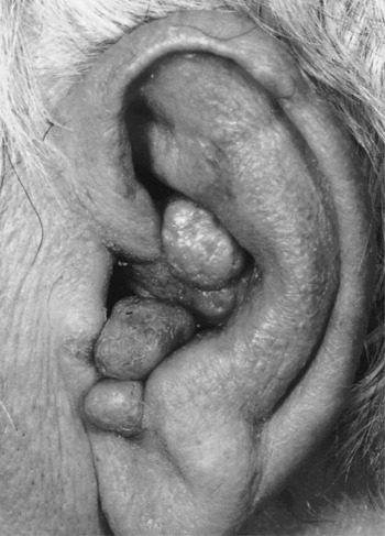
Physical examination demonstrated fleshy growths on both auricles. One mass could be seen on his right auricle in the cavum concha and three masses on his left auricle, one in each of the cymba concha, cavum concha and inferior to the tragus with partial obstruction of both external auditory canals (Figure 1). The skin of his external auditory canals was soft, moist, swollen and inflamed, consistent with otitis externa. No other facial skin changes were noted. His audiogram at presentation confirmed a mild bilateral high frequency sensorineural hearing loss with a superimposed conductive component.
Left ear at presentation.

The physical appearance of these growths did not allow a definitive diagnosis to be made; therefore one lesion from his left pinna was excised with a potassium titanyl phosphate laser for both diagnostic and therapeutic purposes. Pathology results from the sample showed a polypoid portion of skin with patchy chronic inflammation suggestive of a fibroepithelial polyp. Proliferation of atypical melanocytes in the basal layer was also noted although the significance of this was unclear. As lentigo maligna could not be fully excluded, excision of any other changing pigmented lesions in the area was recommended. The remaining three lesions were subsequently excised and the pathology findings from these were also consistent with fibroepithelial skin polyps.
The pathology results did not explain the cause of the fleshy growths and after discussion with the dermatologists a 4 mm punch biopsy from his left ear was undertaken. This specimen showed prominent pilosebaceous units cuffed by an infiltrate predominately composed of lymphocytes, mild vascular telangectasia and scarring of the superficial dermis. Mucin deposits were also noted within the dermis and a congo red stain for amyloid was negative. Findings were not considered specific but suggested that conditions such as lupus needed to be considered. No evidence of autoimmune disease was found.
The case records were taken to a regional dermatology meeting that reached the consensus that otophyma was the most likely diagnosis.
Discussion
Otophyma, unlike rhinophyma, is rarely seen, with only one other case being described in the English language literature.Reference Ekmekci, Koslu and Sakiz2 As a result the diagnosis, investigation and management of it are based upon that of rhinophyma, the final stage of rosacea.
Rosacea is a common skin condition with prevalence ranging between 0.5 and 10 per cent and is described as a four stage process with phyma development being the final stage. Although more common in females, men are more likely to develop rhinophyma resulting from the hyperplasia of sebaceous glands and fibrous tissue of the nose.Reference Rohrich, Griffin and Adams3 The possibility of rosacea affecting the ear canal has previously been raised with the thought that any of the cutaneous markers of rosacea may involve the ear canal. There was, however, no suggestion that it may involve the pinna and no evidence to support the theory at that time.Reference Malak and Hadi4
Rhinophyma is often a diagnosis made on inspection, with the macroscopic appearance of a tuberous enlargement of the lower half of the nose where the skin is irregularly thickened with prominent follicles.Reference Redett, Manson, Goldberg, Girotto and Spence5 However, diagnosis is complicated by the fact that other pathologies can mimic or be obscured by rhinophyma. Granuloma eosinophilicum, sarcoidosis and lymphoma may mimic itReference Rohrich, Griffin and Adams3 and various forms of malignancy have been found within rhinophyma including basal cell carcinoma,Reference McKenna and McKenna6 squamous cell carcinomaReference Lutz and Otley7 and angiosarcoma.Reference Gallardo, Bosch, Vidal, Cabra, Rodrigo and De Galvez8 It is for this reason that any tissue samples from surgical excisions should be submitted for histological examination. Recent or rapid changes in a rhinophymatous nose warrant surgery.Reference Rohrich, Griffin and Adams3
Histological findings in rhinophyma are characterised by sebaceous hyperplasia, inflammation and acanthosis. In severe cases the histology has not always shown this characteristic pattern and cases have been described where the sebaceous units have been destroyed and replaced with persistent oedema and fibrosis.Reference Rohrich, Griffin and Adams3 Pathological findings from the lesions excised in this case of otophyma did describe chronic inflammatory changes and prominent pilosebaceous units similar to those in rhinophyma.
• Otophyma is a rare condition with only one other case report in the English literature
• Otophyma is equivalent to rhinophyma occuring on the ear and is part of the rosacea spectrum
• Management is based on experiences with rhinophyma
• Medical and surgical options exist but surgical intervention is more successful
• Here we demonstrate that laser excision is a successful intervention and results in a good cosmetic appearance
The severity of rhinophyma can vary greatly and therefore management options also vary. Only the early stages of rhinophyma, minimal skin thickening and no nasal deformity, may benefit from medical treatment with steroids, antibiotics or retinoids, but this will not cause substantial regression of existing disease.Reference Gupta and Chaudhury9 Consequently, surgical management is the mainstay of treatment,Reference Rohrich, Griffin and Adams3, Reference Gupta and Chaudhury9 with the aim of restoring the normal contour of the nose whilst minimising scar formation.Reference Gupta and Chaudhury9 Surgical methods used can be divided into two groups according to the extent of excision, either complete or incomplete. Incomplete excision, or debulking, using methods such as dermaplaning, dermabrasion, cryosurgery, electrosurgery, sharp blade excision and laser excision are now the favoured methods as they result in better cosmetic appearance.Reference Rohrich, Griffin and Adams3, Reference Gupta and Chaudhury9 Re-epithelialisation is achieved from the remaining pilosebaceous units. Complete excision is reserved for cases with a focus of malignancy which needs to be completely excised, those with deep infiltration or when the presence of scar tissue makes partial excision difficult.Reference Redett, Manson, Goldberg, Girotto and Spence5 Cosmetic outcomes achieved with complete excision are not as good as incomplete excision and therefore the latter is the preferred surgical option.Reference Gupta and Chaudhury9
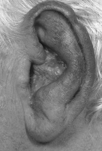
Surgical treatment for rhinophyma is often curativeReference Redett, Manson, Goldberg, Girotto and Spence5 and to date, in this patient, the excision of otophyma using a potassium titanyl phosphate laser has achieved a good cosmetic appearance (Figure 2) and there has been no recurrence.
Left ear post-excision of lesions.

Only one other case of otophyma is described in the English language literature and that case presented with a six-year history of enlargement, redness and oiliness of the right earReference Ekmekci, Koslu and Sakiz2 rather than the discrete fleshy growths as seen in this patient. Similarly though, neither individual had other signs of rosacea at presentation.
To our knowledge, this is the first case of otophyma presented in the otolaryngological literature. Here we have described the difficulties faced in diagnosing this rare condition and our successful management of this case.