INTRODUCTION
Blastocystis spp. is a protozoan parasite commonly found in the human gastrointestinal tract. The symptoms attributed to infection with Blastocystis spp. include diarrhoea, abdominal pain or discomfort, etc. Blastocystis spp. from humans and animals exhibit similar morphological features. Extensive genetic variability has been reported in Blastocystis spp. of both human and animal origin. A number of molecular techniques used to study the genetic diversity of Blastocystis spp. include PCR-restriction fragment length polymorphism (RFLP) (Abe et al. Reference Abe, Wu and Yoshikawa2003a , Reference Abe, Wu and Yoshikawa b , Reference Abe, Wu and Yoshikawa c ), PCR followed by dideoxy sequencing (Abe, Reference Abe2004; Yoshikawa et al. Reference Yoshikawa, Wu, Kimata, Iseki, Ali, Hossain, Zaman, Haque and Takahashi2004; Stensvold et al. Reference Stensvold, Brillowska-Dabrowska, Nielsen and Arendrup2006), and PCR with subtype-specific (sequence-tagged site [STS]) primers (Abe et al. Reference Abe, Wu and Yoshikawa2003a , Reference Abe, Wu and Yoshikawa b , Reference Abe, Wu and Yoshikawa c ; Khan and Alkhalife, Reference Khan and Alkhalife2005; Yakoob et al. Reference Yakoob, Jafri, Beg, Abbas, Naz, Islam and Khan2010).
Oral inoculation with 1×10⁵ cysts of Blastocystis sp. strain RN94-9 in rats resulted in chronic infection in the caecum for 4 weeks after infection (Iguchi et al. Reference Iguchi, Yoshikawa, Yamada, Kimata and Arizono2009). Histological examination revealed only a slight increase in goblet cells in the caecal mucosa 1–3 weeks postinfection but no inflammatory cell infiltration nor mucosal sloughing. Significant upregulation of the expression of interferon-γ, IL-12 and TNF-α, but not IL-6 or granulocyte–macrophage colony stimulating factor (GM-CSF) was demonstrated in the caecal mucosa at 2 weeks post-infection. The induction of local host responses, including mild goblet cell hyperplasia, and significant upregulation of type-1 and proinflammatory cytokines, suggests that Blastocystis sp. strain RN94-9 is a weakly pathogenic organism that could elicit proinflammatory as well as protective responses in local tissues (Iguchi et al. Reference Iguchi, Yoshikawa, Yamada, Kimata and Arizono2009). The cysteine proteases of Blastocystis ratti WR1, a zoonotic isolate, can activate IL-8 gene expression in human colonic epithelial cells. The molecular mechanism by which Blastocystis activates IL-8 gene expression in human colonic epithelial T84 cells and the production of IL-8 is dependent on NF-κB activation (Puthia et al. Reference Puthia, Lu and Tan2008). Previously, an experimental in vitro study demonstrated Blastocystis spp. is able to trigger inflammatory cytokine response in colonic epithelial cells (Long et al. Reference Long, Handschack, König and Ambrosch2001). After 24 h incubation of Blastocystis spp. with the cell lines HT-29 and T-84, B. hominis cells were not able to cause cytopathic effects, but significantly increased the release of IL-8 and GM-CSF. However, after the first 6 h of co-incubation, the production of IL-8 was not increased in HT-29 cells and even reduced when Escherichia coli (bacteria or lipopolysaccharide) were present during co-incubation. Similar effects were observed using supernatants of Blastocystis spp. culture. These data indicate that Blastocystis spp. induces as well as modulates the immune response in intestinal epithelial cells (Long et al. Reference Long, Handschack, König and Ambrosch2001).
Irritable bowel syndrome (IBS) is common and has a significant medical and socioeconomic impact. It is considered to be a biopsychosocial disorder resulting from an interaction among many factors that included diet, gut microflora, visceral hyperalgesia, genetic and environmental factors, infection, inflammation, gut motility and psychological factors (Drossman et al. Reference Drossman, Camileri, Mayer and Whitehead2002). IBS is associated with abdominal pain or discomfort and an alteration in bowel habits (Drossman et al. Reference Drossman, Camileri, Mayer and Whitehead2002). There has been a suggestion that some patients with IBS may harbour bacterial overgrowth leading to low-grade inflammation (O'Leary and Quigley, Reference O'Leary and Quigley2003) and immune activation (O'Sullivan et al. Reference O'Sullivan, Clayton, Breslin, Harman, Bountra, McLaren and O'Morain2000; Chadwick et al. Reference Chadwick, Paulus, Bethwaite, Tie and Wilson2002), and their symptoms may be ameliorated by its eradication (Pimentel et al. Reference Pimentel, Chow and Lin2000). Imbalances in pro- and anti-inflammatory cytokine production may promote ongoing low-grade inflammation after an acute gastroenteritis, and subsequently lead to IBS (van der Veek et al. Reference van der Veek, van den Berg, de Kroon, Verspaget and Masclee2005). Thus abnormal neuroimmune interactions may contribute to the altered gastrointestinal physiology and hypersensitivity that underlies IBS (Barbara et al. Reference Barbara, De Giorgio, Stanghellini, Cremon and Corinaldesi2002). Previously, we demonstrated that stool culture for Blastocystis spp. was positive in 46% of the patients with D-IBS (Yakoob et al. Reference Yakoob, Jafri, Jafri, Khan, Islam, Beg and Zaman2004). In the present study, we determined the cytokine profile in patients with D-IBS and in those with concomitant Blastocystis sp. type 1 and 3 infection. We evaluated cytokine production in vitro from PBMC and determined tissue cytokine levels of IL-8, IL-10, IL-12 and TNF-α. IL-12 is a marker of Th1 response and IL-10 of T regulatory response. The outcome of an immune response depends on the balance between pro- and anti-inflammatory responses. We aimed for a comprehensive overview of cytokine profiles including the anti-inflammatory cytokine IL-10.
MATERIALS AND METHODS
Study population
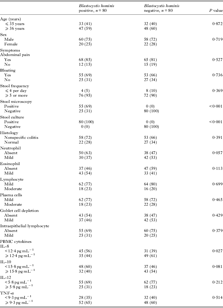
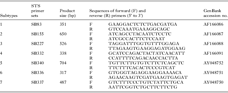
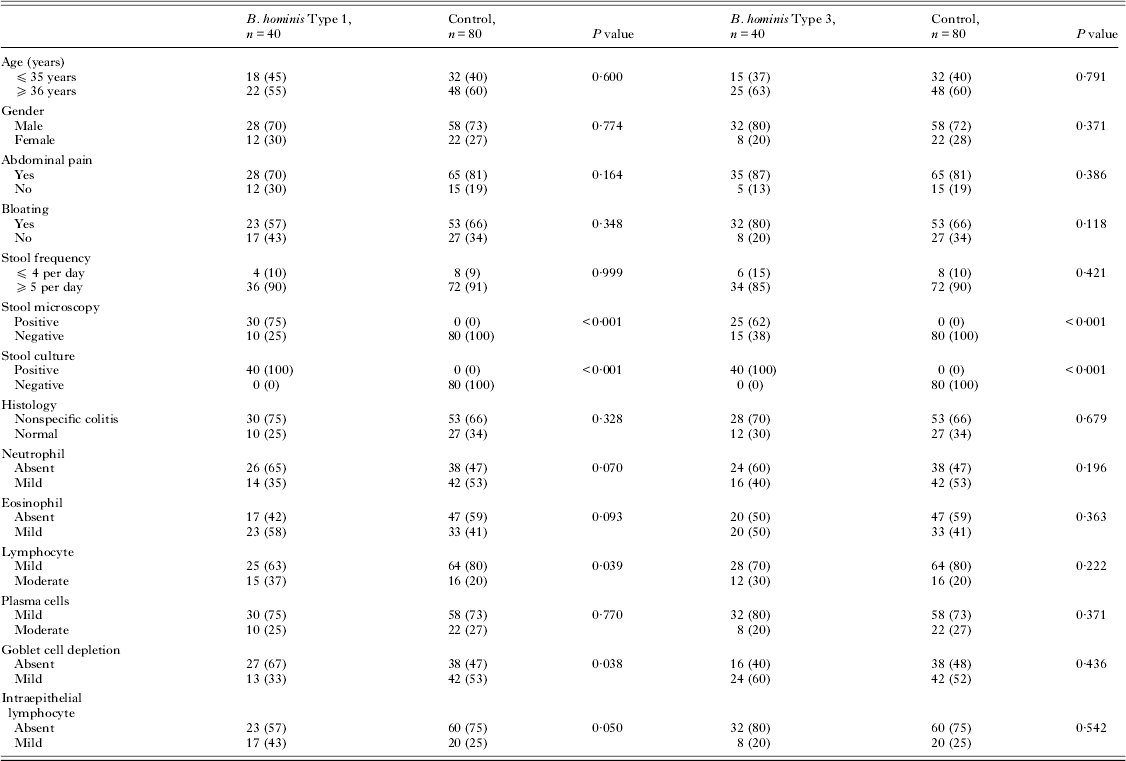
This prospective study was conducted at the Aga Khan University in Karachi, Pakistan. One hundred and seventy patients with symptoms suggestive of D-IBS, according to the Rome III criteria, who attended the gastroenterology clinic from December 2009 to December 2011, were enrolled. Rome III criteria define IBS as recurrent abdominal pain or discomfort at least 3 days per month in the last 3 months that started at least 6 months before diagnosis, cannot be explained by a structural or biochemical abnormality, and is associated with at least two of the following: improvement with defecation, onset associated with a change in frequency of stool, and onset associated with a change in form (appearance) of stool (Drossman and Dumitrascu, Reference Drossman and Dumitrascu2006). Other symptoms that support the diagnosis but are not part of the criteria include abnormal stool frequency (⩽3 bowel movements per week or >3 bowel movements per day), abnormal stool form (lumpy/hard or loose/watery), defecation straining, urgency, or feeling of incomplete bowel movement, passing mucus and bloating. Four possible IBS subtypes include IBS with constipation (C-IBS), IBS with diarrhoea (D-IBS), mixed IBS (M-IBS) and un-subtyped IBS depending on the predominant stool pattern (Drossman and Dumitrascu, Reference Drossman and Dumitrascu2006). Of these ten patients were excluded; three had coinfection of Giardia lamblia and one Entameba coli with Blastocystis spp. while there were 2 each with Blastocystis sp. types 2 and 4 and 1 each with Blastocystis sp. types 5 and 6, respectively. Forty (25%) were infected with Blastocystis sp. type 1 and 40 (25%) with Blastocystis sp. type 3, respectively (Table 2). Eighty patients with D-IBS and Blastocystis spp. infection were defined as ‘cases’ and 80 D-IBS patients without Blastocystis spp. infection were ‘control’ (Table 1). Cases were subdivided into patients with D-IBS and Blastocystis spp. infection type 1 or type 3. Standardized subtype-specific (STS) primers for Blastocystis sp. subtype 1 (SB83) and subtype 3 (SB227) defined Blastocystis sp. type 1 and 3. The study was approved by the Ethics Committee of the Aga Khan University and written informed consent was obtained from all patients. After enrolment of eligible patients in the study, a detailed history and physical examination was carried out. Colonoscopy of the patient was performed to rule out any organic disease and four rectal biopsies were taken, two each for histopathology and cytokine levels. Blood samples were obtained for cytokines analysis. Three stool samples collected from each patient on alternate days were examined by microscopy and cultured for Blastocystis spp. Coeliac disease, small bowel bacterial overgrowth and lactose intolerance were excluded by measuring tissue transglutaminase antibodies (IgA and IgG) and lactose intolerance by hydrogen breath test, respectively. The inclusion criteria were adults from 18 to 61 years of age with D-IBS, satisfying ROME III criteria (Drossman and Dumitrascu, Reference Drossman and Dumitrascu2006). Exclusion criteria were pregnant and lactating females, inflammatory bowel disease and other systemic disease, patients on laxatives or anti-diarrhoeal drugs that could influence the motility of gut, patients on antibiotics that could alter the enteric flora, patients with any ongoing infection, not willing to participate and those with Blastocystis spp. infection of more than one genotype. We chose to study only D-IBS patients as they were more frequently documented to be positive for Blastocystis spp. in our patients previously as compared with other IBS subtypes (Yakoob et al. Reference Yakoob, Jafri, Jafri, Khan, Islam, Beg and Zaman2004). The stool samples from D-IBS patients were screened with the seven kinds of STS primers 1, 2, 3, 4, 5, 6 and 7. However, we studied only genotypes of Blastocystis sp. type 1 and 3 which are the predominant types of Blastocystis spp. identified among our D-IBS patients (Yakoob et al. Reference Yakoob, Jafri, Beg, Abbas, Naz, Islam and Khan2010).
Comparison of characteristics of controls with and without B. hominis infection

Histology
Colonic biopsy specimens were obtained from rectum at the recto-sigmoid junction and were fixed immediately in 10% formalin in sodium phosphate buffer and sent to the Department of Pathology for processing. Biopsies were embedded in paraffin, and histological sections were stained with haematoxylin and eosin for evaluation. Histological inflammation was graded as 0–3 (0 – nil or without evidence of histopathology, 1 – mild, 2 – moderate, 3 – severe) according to the infiltration by neutrophils, eosinophils, lymphocytes, plasma cells and epithelial cell damage e.g. goblet cell depletion, intraepithelial cell lymphocytes (IEL). The pathologist was blinded to cytokine data.
Microscopy of fecal smear
Fecal sample microscopy was done as described before (Zaman and Khan, Reference Zaman and Khan1994). Briefly, approximately 2 mg of feces was thoroughly emulsified on a glass slide in one drop of physiological saline and covered with a cover slip. A similar preparation was made on another slide using Lugol's iodine. These preparations were examined under both the low power (×10) and high dry (×40) objectives. Three fecal samples were examined on alternate days before giving a negative diagnosis of infection with an intestinal protozoan.
Culture of feces
Blastocystis spp. culture was done by inoculating approximately 50 mg of feces into Jones’ medium. Blastocystis spp. culture was performed using Jones medium without starch (Zaman and Khan, Reference Zaman and Khan1994). The cultures were incubated at 37 °C and examined after 2–3 days for one week to exclude slow-growing parasites. The sediment was examined under both the low power (×10) and high dry (×40) objectives. The positive samples were subcultured for another 3 days using fresh media.
Extraction of genomic DNA
Genomic DNA of Blastocystis spp. was extracted by using a Stool DNA Extraction kit (Qiagen) according to the manufacturer's protocol. Extracted DNA was stored at −20 °C until PCR was carried out for Blastocystis spp. genotyping.
Blastocystis spp. genotyping by PCR with STS primers
Seven kinds of STS primers developed for typing the Blastocystis spp. isolates were used as described previously (Table 2A) (Yoshikawa et al. Reference Yoshikawa, Nagano, Wu, Yap, Singh and Takahashi1998, Reference Yoshikawa, Abe, Iwasawa, Kitano, Nagano and Takahashi2000, Reference Yoshikawa, Abe and Wu2003; Abe et al. Reference Abe, Wu and Yoshikawa2003a , Reference Abe, Wu and Yoshikawa b ; Yan et al. Reference Yan, Su, Ye, Liao, Ye, Li, Luo and Chen2006; Li et al. Reference Li, Zhou, Du, Wang, Wang, Jiang, Yoshikawa, Steinmann, Utzinger, Wu, Chen, Chen and Zhang2007a , Reference Li, Zhang, Lv, Zhang, Yoshikawa, Wu, Steinmann, Utzinger, Tong, Chen and Zhou b ). Seven standardized subtype-specific STS primers were used, namely SB83 (351 bp) for subtype 1, SB340 (704 bp) for subtype 2, SB227 (526 bp) for subtype 3, SB337 (487 bp) for subtype 4, SB336 (317 bp) for subtype 5, SB332 (338 bp) for subtype 6, and SB155 (650 bp) for subtype 7 according to a recent classification terminology (Stensvold et al. Reference Stensvold, Suresh, Tan, Thompson, Traub, Viscogliosi, Yoshikawa and Clark2007). Typing of the Blastocystis spp. isolates was conducted through PCR amplification on the basis of the presence or absence of the products with parallel control. The PCR conditions consisted of one cycle denaturing at 94 °C for 3 min, 30 cycles including annealing at 59 °C for 30 s, extending at 72 °C for 60 s, denaturing at 94 °C for 30 s, and additional cycle with a 5-min chain elongation at 72 °C (PCR System 9700, Perkin Elmer, USA). The PCR products and molecular markers were electrophoresed in 2% agarose gel with Tris-acetate-EDTA electrophoresis buffer. The size markers were 100-bp ladder (Promega, USA). The PCR amplification for each primer pair was repeated at least thrice. Bands were visualized by the imaging system (Gel Doc 2000, Gel Documentation System, Bio-Rad, UK) after being stained with ethidium bromide.
The sequence-tagged site (STS) primer used

Isolation of peripheral blood mononuclear cells
Blood samples from patients with D-IBS (approximately 4 mL) were collected in sterile ethylene diaminetetra-acetate (EDTA) containing tubes. Peripheral blood mononuclear cells were isolated using Histopaque (Sigma-Aldrich, USA) according to the density gradient centrifugation method (Chomczynski and Sacchi, Reference Chomczynski and Sacchi1987). The collected PBMCs were washed twice with Roswell Park Memorial Institute (RPM 1640) culture medium and resuspended in growth medium of RPMI supplemented with 10% foetal bovine serum (FBS), 100 U mL−1 penicillin-streptomycin and 2·5 μg mL−1 of fungizone at 37 °C in a CO2 incubator containing 5% CO2, 95% air and 100% humidity. The supernatant was removed after 3 days and stored at −70 °C until further testing.
Cytokine assays
Cell culture supernatants were harvested and analysed for cytokines by ELISA techniques with commercially available kits. Human IL-8, 10, 12 and TNF-α kits were obtained from BD OptEIA. All cytokine assays were calibrated against the World Health Organization international standards by the kit manufacturer. The lower limits of detection for the individual assays are as follows: IL-8, 0·8 pg mL−1; IL-10, 2 pg mL−1; IL-12, 4 pg mL−1 and TNF-α, 2 pg mL−1.
Biopsy cytokines using real-time quantitative PCR with SYBR Green
TRIzol
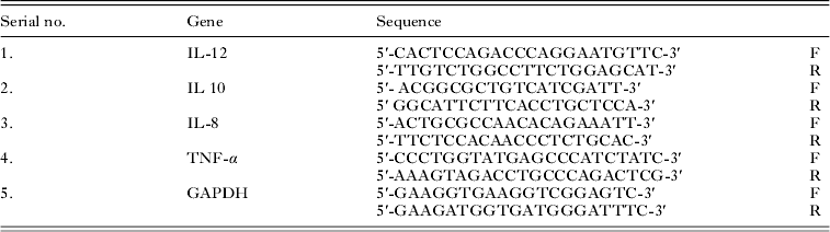
Intestinal biopsy specimens obtained for RNA extraction were collected in an Eppendorf vial containing TRIzols Reagent (Invitrogen Corporation, USA) and stored in a liquid nitrogen container for transport to the laboratory and stored at −70 °C until further use. Total RNA was extracted from endoscopic biopsy samples of colonic mucosa with TRIzol method described previously. Reverse transcription of the extracted RNA was performed using RNase H-deficient reverse transcriptase (Superscript II, Life Technologies) and oligo (dT) primers (Life Technologies). Aliquots (2 μL) of reverse transcription reaction mixture (20 μL) were used for quantitation of IL-12, IL 10, IL-8 TNF-α and GAPDH gene expression by real-time PCR assays (Table 2B) (Tsukada et al. Reference Tsukada, Nakamura, Iimura, Iizuka and Hayashi2002). The SYBR Green QRT PCR was used to quantify IL-12, IL-10, IL-8, TNF-α and GAPDH gene expression (PE Applied Biosystems, Foster City, CA). The PCR reactions were performed using the SYBR Green QRT PCR kit (PE Applied Biosystems) as described previously (Heid et al. Reference Heid, Stevens, Livak and Williams1996). After activation for 10 min at 95 °C, 40 cycles of 15 s at 95 °C and 1 min at 62 °C was carried out in model icycler (Biorad). Real-time fluorescence measurements was recorded and the threshold cycle (Ct) value for each sample calculated by the above sequence detector (Heid et al. Reference Heid, Stevens, Livak and Williams1996). For IL-12, TNF-α and GAPDH standard curves of Ct values were obtained from real-time PCR of pMFGhTNF, pBSKIhIL-12 and PCRII GAPDH (Reference plasmids). The relative fold change in mRNA expression of the target gene was calculated with 2−∆∆CT method using the software GENEX. The 2−∆∆CT method presents the data as fold change in mRNA expression of the target gene, normalized to the mRNA expression of the housekeeping gene. Glyceraldehyde-3-phosphate dehydrogenase was used as the housekeeping gene in all the QRT-PCR experiment. Each biopsy sample obtained from the same patient was tested in duplicate, and the average of two Ct values was used in this study.
Cytokines primer sequence

F = Forward primer; R = Reverse primer.
Tsukada et al. (Reference Tsukada, Nakamura, Iimura, Iizuka and Hayashi2002).
Statistical methods
Results are expressed as mean±s.d. for continuous normally distributed variables whereas median with IQR (interquartile range) for non-normal variables and number (percentage) for categorical data. Differences in mean comparison of characteristics of IBS patients with and without Blastocystis sp. infection type-I and 3 with control group for normally distributed variables were performed by using the analysis of variance (ANOVA) and Tukey's HSD test was used for multiple comparison, however for non-normal variables a non-parametric Kruskal–Wallis test and Mann–Whitney test were used for within-group comparison. Differences in proportion were compared by Pearson Chi-square or Fisher exact where appropriate. A P value of <0·05 was considered as statistically significant. All P values were two sided. Statistical interpretation of data was performed by using the computerized software program SPSS version 19.
RESULTS
Ninety-five (59%) of the patients were over 35 years of age. Abdominal pain, bloating and frequency of stool were equally common in both groups (Table 1). Fecal smear microscopy for Blastocystis spp. was positive in 55 (69%) and Blastocystis spp. culture was positive in 80 (50%) (Table 1).
Blastocystis spp. subtypes and histology
Non-specific colitis was present in 58 (72%) with Blastocystis spp. infection and in 53 (66%) without (P = 0·391) (Table 1). Of the 58 Blastocystis positive D-IBS with non-specific colitis, Blastocystis sp. subtype 1 was 30 (75%) compared with control of 53 (66%) (P = 0·328) and Blastocystis sp. subtype 3 was 28 (70%) compared with control of 53 (66%) (P = 0·679). There was no significant difference in the distribution of neutrophils, eosinophils, mononuclear cells or evidence of epithelial cell damage in the colonic mucosal biopsies in the two groups (Table 1).
Blastocystis spp. types and PBMC cytokines
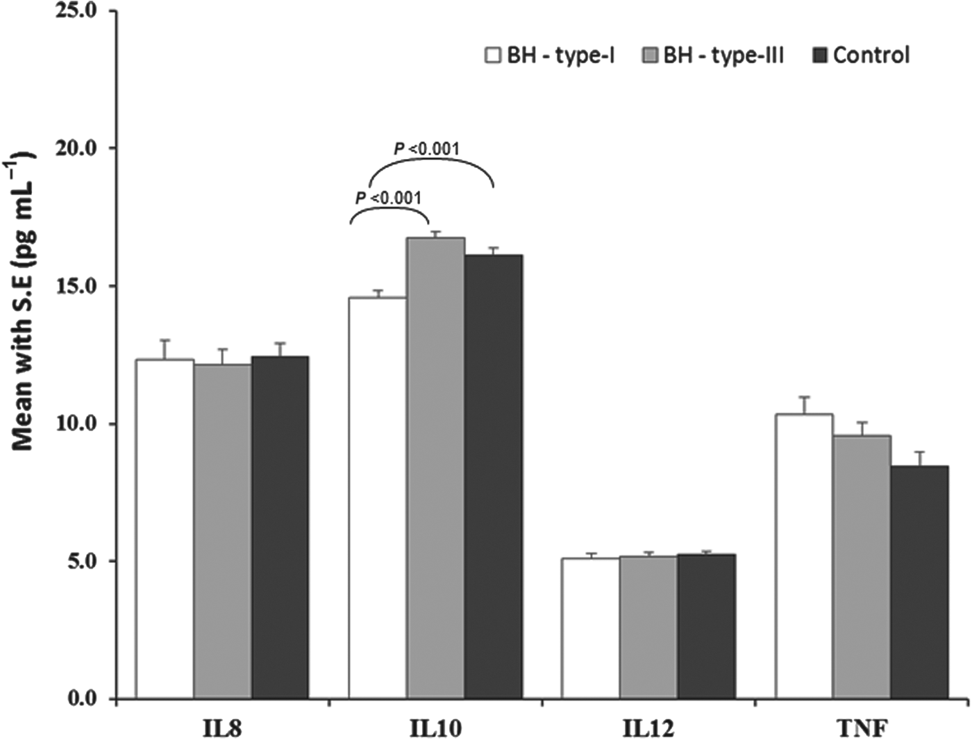
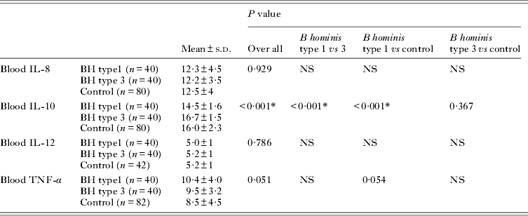
Patients with D-IBS infected with Blastocystis spp. demonstrated low PBMC production of IL-8 in 45 (56%) (P = 0·027) compared with control 31 (39%) (Table 1). There was no difference in the PBMC production of IL-10, IL-12 and TNF-α by PBMCs in vitro in the Blastocystis spp. infected and control groups (Table 1). Patients with D-IBS infected with Blastocystis sp. type 1 demonstrated low PBMC production of IL-10 in 36 (90%) compared with control 37 (46%) (P<0·001) (Fig. 1). The mean concentration of IL-10 (14·5±1·6 pg mL−1, P<0·001) was low in Blastocystis sp. type 1 compared with those with Blastocystis sp. type 3 (16·7±1·5 pg mL−1) and controls (16±2·3 pg mL−1) (Table 4), and these differences remained significant after adjustment for multiple comparisons, P<0·001 and P<0·001, respectively (Table 4).
Comparison of PBMC cytokines with Blastocystis sp. type 1 and 3 and control.

Comparison between Blastocystis spp. types with PBMC expressed cytokines and histology
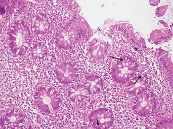
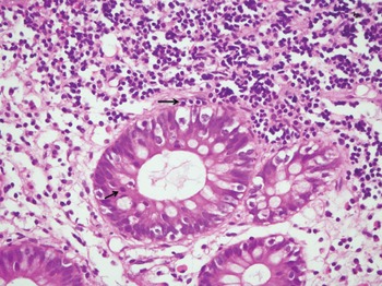
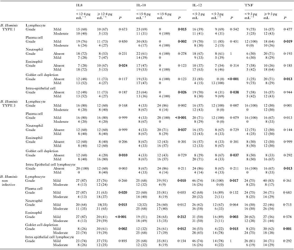
In Blastocystis sp. type 1, mucosal lymphocyte and IEL infiltration were 15 (37%) (P = 0·039) and 17 (43) (P = 0·050), respectively compared with control of 16 (20%) and 20 (25%) (Table 3). In Blastocystis sp. type 1, IL-8 was associated with mild eosinophil infiltration in 18 (72%) (P = 0·024); IL-10 with lymphocyte in 25 (69%) (P = 0·015), plasma cell infiltration in 30 (83%) (P = 0·002) and IEL in 23 (64%) (P = 0·026) (Figs 3–5); IL-12 was associated with goblet cell depletion in 13 (100%) (P<0·001) and IEL in 9 (69%) (P = 0·038) and TNF-α also with plasma cell infiltration in 12 (100%) (P = 0·019) (Table 5). In Blastocystis sp. type 3, IL-8 was associated with goblet cell depletion in 16 (80%) (P = 0·010) and IEL in 8 (40%) (P = 0·003); IL-10 in above mean values with mild lymphocyte in 24 (86%) (P = 0·002) and plasma cell infiltration in 28 (100%) (P<0·001); IL-12 was associated with goblet cell depletion in 20 (71%) (P = 0·037) and TNF-α was associated with lymphocyte in 12 (50%) (P = 0·001) and plasma cell infiltration in 8 (33%) (P = 0·013) (Table 5).
Comparison of characteristics of controls with B. hominis type genotype

Comparison between Blastocystis spp. types and cytokine mRNA expression in colonic mucosa
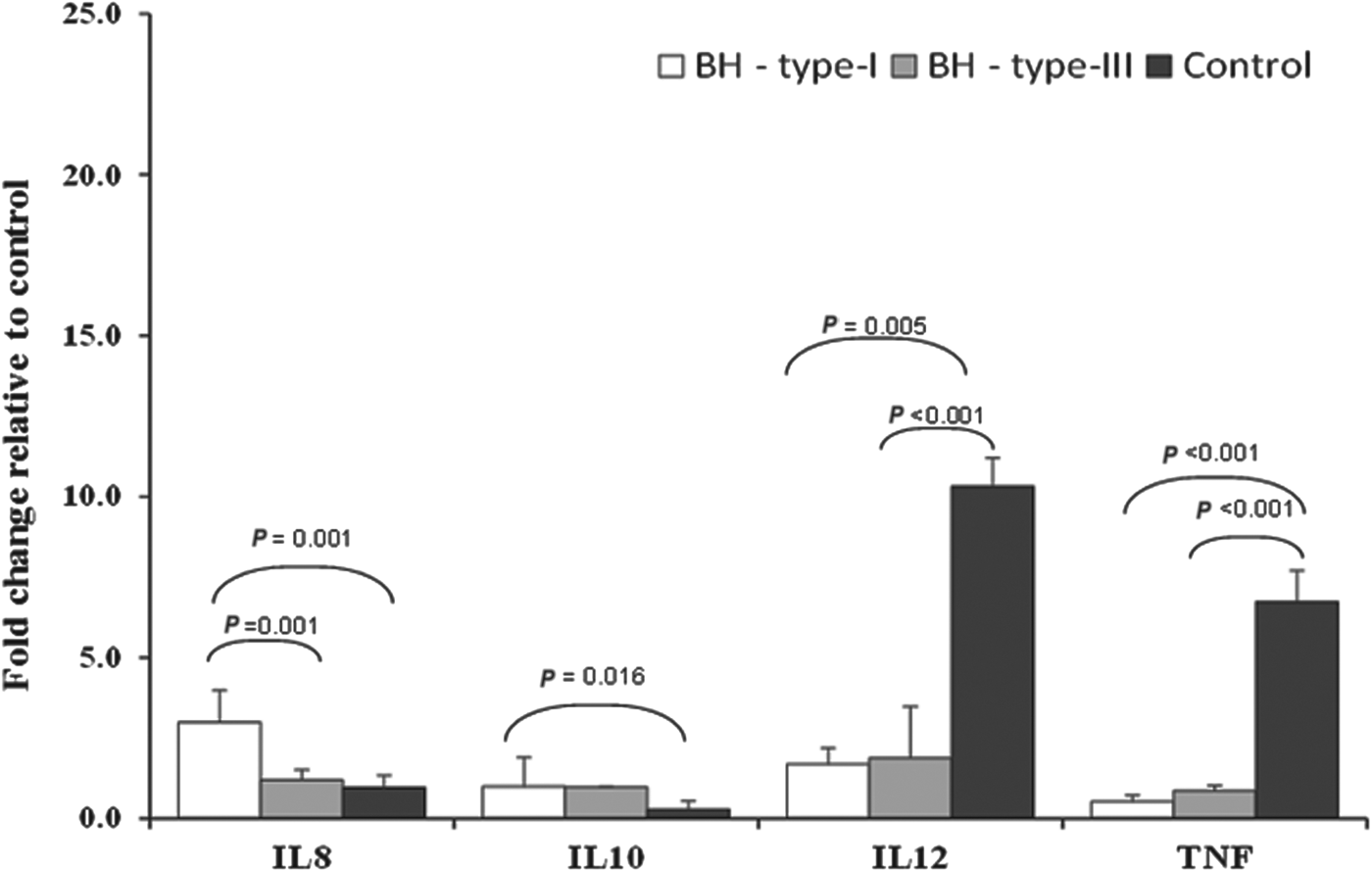
The expression of IL-8 mRNA (pg mL−1) in colonic mucosa in Blastocystis sp. type 1 (median with interquartile range) was 3·4 (1·04–7·30), type 3 was 1·2 (0·21–2·14) and for control was 1 (0·27–9·8) (P = 0·016). The expression of IL-10 mRNA (pg mL−1) in colonic mucosa in Blastocystis sp. type 1was 0·78 (0·34–5·7), type 3 was 1 (0·97–0·99) and for control was 0·27 (0·04–3·6) (P = 0·153). The expression of IL-12 mRNA (pg mL−1) in colonic mucosa in Blastocystis sp. type 1 was 1·5 (1·2–5·15), type 3 was 2 (0·22–7·06) and for control was 1 (2·1–13·8) (P = 0·005). The expression of TNF-α mRNA (pg mL−1) in colonic mucosa in Blastocystis sp. type 1 was 0·54 (0·05–1·5), type 3 was 0·87 (0·53–1·8) and for control was 6·7 (2·03–14·7) (P<0·001). The IL-8mRNA expression was increased in Blastocystis sp. type 1 compared with type 3 (P = 0·001) and control (P = 0·001) (Fig. 2). The expression of mRNA of IL-10 was low in the control group and Blastocystis sp. type 3 and type 1 (Fig. 2). The IL-10 mRNA expression was increased in Blastocystis sp. type 1 compared with control (P = 0·016) (Fig. 2). The expression of mRNA of IL-12 was higher in the control group compared with Blastocystis sp. type 3 and type 1 (P<0·001 and 0·005, respectively) (Fig. 2). The expression of mRNA of TNF-α was higher in the control group compared with Blastocystis sp. type 3 and type 1 (P = <0·001 and 0·001, respectively) (Fig. 2).
Comparison of cytokine mRNA expression in colonic mucosa between B. hominis subtypes and control group.

Colonic glands showing intraepithelial lymphocytes in the glandular lining epithelium. H&E ×10.

Colonic glands showing intraepithelial lymphocytes in the glandular lining epithelium H&E ×20.

Colonic glands showing mucin (goblet cell) depletion. H&E ×20.

DISCUSSION
Blastocystis spp. is an extracellular luminal organism. It is able to evade the host immune response by suppressing iNoS production and cleaving immunoglobulin (Puthia et al. Reference Puthia, Vaithilingam, Lu and Tan2005; Mirza et al. Reference Mirza, Wu, Kidwai and Tan2011). This may also compromise these protective responses to other coinfecting pathogens allowing them to infect host epithelia and cause disease. There have been studies investigating the priming of human immune responses by intestinal protozoa (Djuardi et al. Reference Djuardi, Sartono, Wibowo, Supali and Yazdanbakhsh2010). The presence of Blastocystis spp. as the predominant species in pregnant mothers was shown to dampen the innate and adaptive responses to purified protein derivative (PPD) of mycobacterium tuberculosis (Djuardi et al. Reference Djuardi, Sartono, Wibowo, Supali and Yazdanbakhsh2010). Earlier studies have shown that Blastocystis spp. infection can be associated with impaired intestinal permeability (Mirza et al. Reference Mirza, Wu, Teo and Tan2012) as well as lower total leucocyte and neutrophil count (Reiman et al. Reference Reiman, Thompson, Feng, Hari, Knight, Cheever, Rosenberg and Wynn2006).
In this study, colonoscopic examination of D-IBS patients with Blastocystis spp. demonstrated no pathological changes or only non-specific inflammation without evidence of invasion with Blastocystis spp., in keeping with previous studies (Chen et al. Reference Chen, Louie, McCormick, Walker and Shi2005). Also, Blastocystis strain RN94-9 induced neither epithelial injury nor inflammatory cell infiltration in rat caecal mucosa (Iguchi et al. Reference Iguchi, Yoshikawa, Yamada, Kimata and Arizono2009). The non-invasive nature of strain RN94-9 was further confirmed by normal expression levels of epithelial tight junction proteins. In D-IBS with Blastocystis spp. infection, PBMCs IL-8 and IL-10 responses were less marked in Blastocystis sp. type 1 compared with control (Table 3). The mean IL-10 was significantly lower in Blastocystis sp. type 1 compared with Blastocystis sp. type 3 and control (Table 4). In Blastocystis sp. type 1, there was colonic mucosal eosinophil infiltration associated with IL-8 compared with Blastocystis sp. type 3 (Table 5) and lymphocyte infiltration and goblet cell depletion compared with control (Table 3). In contrast, in a mouse model of Blastocystis spp. infection (Iguchi et al. Reference Iguchi, Yoshikawa, Yamada, Kimata and Arizono2009) goblet cell hyperplasia was reported. Moreover, goblet cell hyperplasia has also been reported in Giardia infections as well (Ponce-Macotela et al. Reference Ponce-Macotela, González-Maciel, Reynoso-Robles and Martínez-Gordillo2008). It is known that T helper type 2 cytokines (IL-13 and IL-4) regulate the development of goblet cell hyperplasia in the gut during infection and an increased number of goblet cells plays an important role in host protective immunity against infection (Khan and Alkhalife, Reference Khan and Alkhalife2005). Goblet cell hyperplasia, increased mucin and fluid secretion and enhanced intestinal propulsive activity results in the eviction of noxious agents from the gut lumen. However, a single administration of recombinant adenovirus vector expressing IL-12 (Ad5IL-12) in Trichinella spiralis-infected mice inhibited infection-induced muscle hypercontractility and goblet cell hyperplasia. This also correlated with upregulated interferon-γ (IFN-γ) expression and downregulated IL-13 expression in the muscularis externa layer (Khan et al. Reference Khan, Blennerhasset, Ma, Matthaei and Collins2001). These results indicate that transfer and overexpression of the IL-12 gene during Th2-based infection shifts the immune response towards Th1 and abrogates the physiological responses to infection, attenuating both muscle hypercontractility and goblet cell hyperplasia (Khan et al. Reference Khan, Blennerhasset, Ma, Matthaei and Collins2001). O'Malley et al. (Reference O'Malley, Dinan and Cryan2010) reported goblet cell hyperplasia in a rat model of anxiety and depression. The finding of goblet cell depletion in both case and control in this study is surprising and may be explained on the basis that Th2 reaction when replaced by a proinflammatory reaction leads to goblet cell depletion in D-IBS in humans.
Comparison of peripheral cytokines with B. hominis types and control

* P value <0·05 was considered as statistically significant; NS = not significant; s.d. = Std. deviation.
Comparison between B. hominis types, peripheral blood mononuclear cells cytokines and histological parameters

Histological inflammation was graded as 0 = nil, 1 = mild, 2 = moderate, 3 = severe.
IL-12 plays a key role in induction of Th1 immune responses by stimulating the production of IFN-γ and TNF-α from T and natural killer (NK) cells. In this study, IL 12 was associated with goblet cell depletion in both cases and control (Table 5). Long et al. (Reference Long, Handschack, König and Ambrosch2001) in an in vitro study reported Blastocystis sp. type 1 elicited a significant increase in proinflammatory IL-8 in HT-29 and T-84 IEC human colonic cell lines. This was observable after 24 h exposure to parasites (Long et al. Reference Long, Handschack, König and Ambrosch2001). However, the PBMCs culture supernatants harvested after 72 h of incubation did not show significant cytokine variation among Blastocystis spp. types and control (Tables 4 and 5). It has been previously reported that cytokine changes presented within 6 h (Long et al. Reference Long, Handschack, König and Ambrosch2001) and normalized after that period. IL-10, having a regulatory role, is produced by activated lymphocytes in later phases of the immune response (Schröder et al. Reference Schröder, Mrowietz, Morita and Christophers1987; De Waal Malefyt et al. Reference De Waal Malefyt, Abrams, Bennett, Figdor and de Vries1991). The anti-inflammatory cytokine IL-10 inhibits monocytes production of pro-inflammatory cytokines including TNF-α which subsequently regulates both early neutrophilic infiltration and eosinophil recruitment (Asseman et al. Reference Asseman, Mauze, Leach, Coffman and Powrie1999). Secretion of IL-10 is also induced by TNF-α linking its production to inflammation (Powrie et al. Reference Powrie, Leach, Mauze, Menon, Caddle and Coffman1994). In Blastocystis spp. type 1 and type 3, IL-12 and TNF-α mRNAs expression were also low compared with in control (Fig. 2). The low IL-12 and TNF-α in coinfection with Blastocystis spp. may be attributed to the immune modifying effect of Blastocystis spp. Previously, the downregulation of IFN-γ and TNF-α together with the upregulation of IL-6, IL-8, as well as NF-κB gene expressions were seen in the PBMCs stimulated with 1 μg mL−1 of Blastocystis antigen. This suggested that Blastocystis antigen stimulated the humoral immune responses in PBMCs, which may lead to inflammatory reactions and propagation of the cells to combat the infection (Chandramathi et al. Reference Chandramathi, Suresh and Kuppusamy2010).
IL-10 is important in regulating inflammatory response as it reduces the production of chemotactic factors, such as IL-8 (Turner et al. Reference Turner, Williams, Sankaran, Lazarus, Sinnott and Hutchinson1997; Olivo-Diaz et al. Reference Olivo-Diaz, Romero-Valdovinos, Gudiño-Ramirez, Reyes-Gordillo, Jimenez-Gonzalez, Ramirez-Miranda, Martinez-Flores, Martinez-Hernandez, Flisser and Maravilla2012). Previously in animal models, gene transcription of type 1 and proinflammatory cytokines IFN-γ, IL-12 and TNF-α was significantly upregulated in the caecal mucosa. These results suggest that Blastocystis infection in rats induces local host responses to exposed antigens (Iguchi et al. Reference Iguchi, Yoshikawa, Yamada, Kimata and Arizono2009). In D-IBS with Blastocystis spp., the IL-8mRNA expression was increased in Blastocystis sp. type 1 compared with type 3 (P = 0·001) and control (P = 0·001) (Fig. 2). In keeping with the previous studies that showed Blastocystis strains N and II that belong to subtype 1 and WR1 belonging to subtype 4 stimulated the release of chemokine IL-8 from a human colonic epithelial cell line in vitro, possibly mediated by the organism-derived cysteine protease (Long et al. Reference Long, Handschack, König and Ambrosch2001; Puthia et al. Reference Puthia, Vaithilingam, Lu and Tan2005).
Blastocystosis may not be a highly inflammatory disease (at least in the context of these isolates) as evidenced by low levels of cytokine among Blastocystis spp. types and control. The severity of Blastocystosis spp. infection may be mediated by host factors. Secretion of high levels of enteric anti-Blastocystis IgA has been found to be a common factor in all symptomatic, but not asymptomatic, cases of Blastocystis (Mahmoud and Saleh, Reference Mahmoud and Saleh2003). This would also explain why some individuals can be carriers of the organism without expressing symptoms (Markell and Udkow, Reference Markell and Udkow1986).
The limitation of this study is that the case-to-control patient ratio of 1:1 is too small to draw any firm conclusion. A case-to-control ratio of 2:1 or 3:1 would have been preferable. The size of the case group was limited by the number of eligible patients fulfilling the Rome III criteria for D-IBS infected with Blastocystis sp. subtype 1 or 3 and the resources required for experiments on this sample size. In conclusion, in patients with D-IBS and concomitant Blastocystis sp. type 1 infection, there is a low IL10 response compared with type 3 and D-IBS without Blastocystis spp. and this may be attributable to an immune modifying effect of Blastocystis sp. infection in D-IBS.
ACKNOWLEDGEMENT
We are grateful to the staff of the JUMA research building for their help during the completion of this work.
CONFLICT OF INTERESTS
All authors declare they have no conflict of interest.