Chronic subdural hematoma (CSDH) is a common disabling neurosurgical disease which is increasing in prevalence.Reference Balser, Farooq and Mehmood1 CSDH is traditionally treated with surgical drainage which is complicated by a high rate of recurrence of approximately 20–30%.Reference Dian, Linton and Shankar2 In the last few years, embolization of middle meningeal artery (EMMA) is emerging as a novel treatment modality for the treatment of CSDH and has been reported in several retrospective studies, systematic reviews, and prospective studies.Reference Dian, Linton and Shankar2–Reference Fiorella and Arthur4 EMMA is a minimally invasive, technically straightforward, and relatively safe procedure, which could potentially obviate the need of surgical drainage while significantly reducing the rate of recurrence to a low single-digit percentage. Moreover, it has been proposed that the effect size for reduction of recurrence is as high as 20%.Reference Dian, Linton and Shankar2
Multiple prospective randomized control trials are being conducted in different parts of the world to test the safety and efficacy of EMMA. Prospective randomized controlled trials are essential for EMMA to be accepted as the universal standard of care in treating CSDH. Several pertinent technical points have been raised for the conduct and reporting of these clinical trials. One of the most important points is the development of a standardized grading system to report the clinical and technical efficacy off the treatment.Reference Fiorella, Hirsch and Arthur5 While several clinical scores are in use for reporting clinical neurological outcome, there is no technical grading system available to grade the technical success of EMMA for the treatment of CSDH. The purpose of this letter is to propose a simple, feasible, and reliable grading system for scoring the technical success of EMMA in the treatment of CSDH.
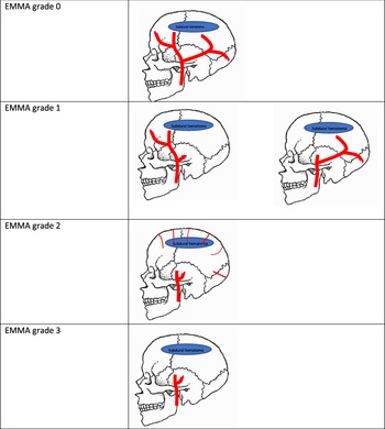
The grading system is predominantly based on the extent of embolization of the middle meningeal artery (MMA) territory. This is based on the hypothesis that embolization of both anterior and posterior branches of MMA leads to more complete resolution of the CSDH compared to single-branch embolization. Figure 1 illustrates a schematic diagram of the grading system ranging from EMMA grade 0 to EMMA grade 3.
Schema for the grading of embolization of middle meningeal artery (EMMA) for management of chronic subdural hematoma. The grading assessment is done on the final proximal trunk external carotid angiogram done at the end of the EMMA.

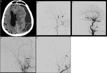
EMMA grade 0 (Figure 2b, c) – simply means no embolization of the MMA. This could be due to difficult access issues, iatrogenic spasm, or dissection of the proximal trunk of the MMA by the micro-catheter or microguidewire. Due to the rich meningeal collateral, the continued filling of major branches of the MMA is seen on the proximal external carotid artery (ECA) trunk angiogram.
Chronic subdural hematoma on CT head (a). External carotid artery (ECA) angiogram showing classic anterior and posterior divisions (arrows) of the middle meningeal artery (MMA) (b). (c) Common carotid angiogram showed MMA arising from the ophthalmic artery and EMMA could not be achieved (grade 0). (d) Post-EMMA, ECA angiogram shows continued filling of the anterior division territory of MMA by the ipsilateral accessary meningeal artery (arrow) filling 50% of the MMA territory (grade 1). (e) Post-EMMA, ECA angiogram shows embolization of most of MMA territory except small filling (arrow) of the anterior division (grade 2). (f) Post-embolization of the middle meningeal artery (EMMA), there is no residual filling of the MMA territory (grade 3).

EMMA grade 1 (Figure 2d) represents approximately 50% of MMA territory embolization. It simply means embolization of either anterior or posterior branch of the MMA. The other branch of the MMA is either not embolized or could not be embolized for technical reasons similar to that described in EMMA grade 0.
EMMA grade 2 (Figure 2e) represents the case where most of the MMA territory is embolized through both anterior and posterior branch. However, due to the proximal occlusion/embolization of the main trunk of the MMA, the distal MMA territory can be seen filling through the collateral meningeal supply with meningeal parenchymal blush seen on the proximal ECA trunk angiogram.
EMMA grade 3 (Figure 2f) represents the most ideal situation where both anterior and posterior branch of the MMA were completely embolized with no obvious meningeal parenchymal blush seen on the proximal ECA trunk angiogram at the end of embolization.
Our grading system proposes a simple and easy to interpret system which can effectively communicate the technical efficacy of EMMA. A standardized grading system is greatly needed to communicate the results of multiple clinical trials and future studies on EMMA for CSDH treatment. Whether the type, distribution, and volume of embolysate affect the outcome can be debatable; however, it is intuitive that the more extensive the embolization, the better the outcome. The best evidence in support of this has been shown by Catapano et al.Reference Catapano, Nguyen and Wakim6,Reference Catapano, Ducruet and Nguyen7 Importantly and of great clinical significance is the fact that that our grading system can be applied to both polyvinyl alcohol particle embolization and liquid embolic agent.
While this grading system may continue to evolve in the future, it certainly fills a significant void in clinical practice where a consistent grading system to report the results of embolization is highly warranted.
This proposed grading system is simple and follows parallel principles that have been shown in the thrombolysis in cerebral infarction grading systemReference Higashida, Furlan and Roberts8 and the aneurysm occlusion grading system.Reference Roy, Milot and Raymond9
Hopefully, future EMMA trials will adopt our grading system for reporting technical outcomes as this would increase the comparability and interpretability of study findings.
Disclosures
No financial disclosures or conflicts of interest to declare except Jai Shankar is the principal investigator of EMMA-Can study in Canada.
Statement of Authorship
Jai Shankar conceptualized and wrote the first draft of the manuscript.
Zul Kaderali developed the idea and edited the manuscript.