Introduction
Anthropogenic impacts, such as pollution, dam construction and predatory fishing, are wellknown deleterious for the maintenance of fish biodiversity (Botta et al., Reference Botta, Sciara, Arranz, Murgas, Pereira and Oberlender2010; Reis et al., Reference Reis, Albert, Di Dario, Mincarone, Petry and Rocha2016; Pelicice et al., Reference Pelicice, Azevedo-Santos, Vitule, Orsi, Lima Junior, Magalhães, Pompeu, Petrere and Agostinho2017; Ilha et al., Reference Ilha, Rosso and Schiesari2019; Bem et al., Reference Bem, Ribolli, Röpke, Winemiller and Zaniboni-Filho2021). These impacts can generate a decline in the taxonomic and functional diversity of fish fauna, in addition to negatively impacting the reproductive aspects of wild populations (Keppeler et al., Reference Keppeler, Andrade, Trindade, Sousa, Arantes, Winemiller, Jensen and Giarrizzo2022). Artificial reproduction may be considered a viable alternative to reduce the pressure on fishing stocks synergically with conservation efforts for native species (Oliveira et al., Reference Oliveira, Ashikaga, Foresti and Senhorini2015; Hilsdorf et al., Reference Hilsdorf, Hallerman, Valladão, Zaminhan-Hassemer, Hashimoto, Dairiki, Takahashi, Albergaria, Gomes, Venturieri, Moreira and Cyrino2022).
Previous knowledge related with oocyte characteristics and post-fertilization events provides important information that explains reproduction mechanisms, and permits embryonic manipulation such as post-larval management (Santos et al., Reference Santos, Yasui, Xavier and Nakaghi2016; Arashiro et al., Reference Arashiro, Yasui, Calado, Nascimento, Santos, Santos, Levy-Pereira, Monzani, Siqueira-Silva and Senhorini2018). This information is considered an essential tool and serves as support for new studies with the application of biotechnologies used in fish reproduction (Silva et al., Reference Silva, Santos, Senhorini, Paes, Valentin, Fujimoto, Nascimento, Yasui and Nakaghi2017), such as germ cell transplantation (Coelho et al., Reference Coelho, Yo, Mira-López, Monzani, Arashiro, Fujimoto, Senhorini and Yasui2019) and chromosome set manipulation (Bertolini et al., Reference Bertolini, Lopez, Nascimento, Arashiro, Siqueira Silva, Santos, Senhorini and Yasui2019). Some species of South American Serrasalmidae, popularly known as ‘pacus’ are considered one of the most produced group in aquaculture (Peixe BR,. Reference Peixe2024). In this group, the subfamily Myleinae are an important fishing resource for commercial and subsistence fisheries as a source of protein for riverside populations in the Amazon region (Francesco and Carneiro Reference Francesco and Carneiro2015).
Prosomyleus rhomboidalis is a medium-sized species distributed in rapid rivers that drain the Brazilian and Guiana shields (Kolmann et al., Reference Kolmann, Hughes, Hernandez, Arcila, Betancur-R, Sabaj, López-Fernández and Ortí2021). The species is important for the Amazonian ecosystem since it may widespread seeds from important trees (Santos, Reference Santos2019; Pereyra et al., Reference Pereyra, Hallwass, Begossi, Giacomin and Silvano2023). In the Xingu River it is popularly known as ‘pacu-de-seringa,’ and stands out among the species exploited in both commercial and artisanal fisheries, representing an important food resource for riverside communities, in which some areas were impacted by hydroelectric dams (Pagezy and Jégu, Reference Pagezy and Jégu2002; Francesco, Reference Francesco and Carneiro2015; Choueri and Nascimento, Reference Choueri and Nascimento2019; Fricke, Eschmeyer, Reference Fricke, Eschmeyer and Van der Laan2023). Such a panorama suggests research efforts for conservation and maintenance of natural populations, but on the other hand, a substantial gap in knowledge regarding the ecology and early morphology exists in this species. Recently, a critical decline in natural populations has been reported by fishermen, which may be related to increasing anthropogenic impacts, compromise the dynamics and maintenance of fish biodiversity (Keppeler et al., Reference Keppeler, Andrade, Trindade, Sousa, Arantes, Winemiller, Jensen and Giarrizzo2022).
The reproduction of fish species in loco is then an important initial action to reduce impacts on natural fishing stocks and minimize the risk and threat for the species. In addition, description of gamete characteristics and early development are important to understand the reproductive characteristics of the species for future conservation and aquaculture purposes. Therefore, this study aims to characterize macroscopically the female gametes, evaluate the survival rate of embryos generated in vitro and describe in detail the main stages and post-fertilization phases of the embryonic development in this species.
Materials and methods
The study was conducted at the Laboratory of the Center for Environmental Studies, Vitoria do Xingu – PA, Brazil. Adult P. rhomboidalis used in the experiments were obtained by collecting in the Xingu River according with the rules Ethics Committee on the Use of Animals (CEUA #4751220822) and with the licence permission SISBIO #79124-2 from the Ministry of Environment of Brazil.
Origin of broodstock and fertilization
The adult P. rhomboidalis was collected by local fishermen during the rainy season spawning season, from November 2022 to January 2023. The specimens were caught using a 18-mm gillnets in the Xingu River, an area known as Volta Grande and transported by boat in 250 L tanks to river margin (–3.3755S, –51.9398W). During fish collecting, some females presented spontaneous ovulation and were used for fertilization trials.
For in vitro fertilization, six individuals used in this experiment were previously anaesthetized with eugenol 100 mg/L (Phytoterápica, Guarulhos/SP, Brasil), sexed based on the morphology of the anal fin (females presents a single lobe, males present double lobes, Figure 1). Firstly, three males (1.604 ± 0.11 g and 33.16 ± 0.32 cm) did not release the sperm by stripping, and then they were euthanized using an overdose of eugenol 300 mg/L–1. Sperm was collected by testis mincing in a 15 mL tube containing 5 mL of modified Ringer’s solution (128.3 mM NaCl, 23.6 mM KCl, 3.6 mM CaCl2, 2.1 mM MgCl2) and then maintained at ∼ 2.5°C (Yasui et al., Reference Yasui, Senhorini, Shimoda, Pereira-Santos, Nakaghi, Fujimoto, Arias-Rodriguez and Silva2015). Three sampled females (1.784 ± 0.13 g and 33.26 ± 0.72 cm) presented spontaneous ovulation during capturing and were used for fertilization trials. As the females release few oocytes with high amount of urine, a new procedure for in vitro fertilization was developed. Firstly, 5 mL of diluted sperm was added to a 1-L plastic bowl, and then the oocytes were stripped on the diluted sperm.
Morphology of the anal fin of Prosomyleus rhomboidalis. (A) females have a single lobe; (B) males have double lobes.

Immediately after stripping, 50 mL of water was added to complete the gamete activation, followed by gently mixing by hand. After stripping of the gametes, the fish were immediately released into the Xingu River.
A small aliquot of oocytes (∼15 oocytes) was taken at the time of stripping (non-fertilized) and immediately after fertilization. The incubation was performed in floating apparatus, in 30-litre aquariums with a recirculation system and the temperature set at 27.5°C. For the embryonic development study, three couples were used and each spawning that reached hatching was considered a replication.
Collection and embryonic development
After fertilization, the embryos were collected according to cell development. Aliquots of 10 to 20 embryos were randomly selected and observed under a trinocular stereomicroscope (Nikon SMZ 800N, Tokyo, Japan). Each developmental stage was considered when about 50% of the embryos had the same morphological characteristics.
Embryos were observed at the following intervals: each fifteen min until 60 min post-fertilization (mpf); each 5 min at 60 mpf until 5 h post-fertilization (hpf); each 20 min from 5 hpf to 7 hpf; each 30 min from 7 hpf to 11 hpf; each 30 min from 11 hpf to 24 hpf; and each 60 min from 24 hpf until hatching, with adaptations from our previous study (Coelho et al., Reference Coelho, Arashiro, Disselli, Pereira-Santos, Mira-López, Monzani, Senhorini, Fujimoto and Yasui2021). A small aliquot was separated and kept under the same conditions obtain the survival rates at cleavage, blastula, gastrula, segmentation and hatching when the percentages of normal and abnormal larvae were also measured. Larvae hatched with patterns that allowed them to swim freely and without malformations in the distribution of the yolk, development of the tail or head was classified as normal. Those that hatched with difficulty getting rid of chorion residue, with swimming problems due to malformations in the tail, head, or even the size and conformation of the yolk, were classified as abnormal.
During development, three embryos were selected to obtain digital images, considering the individuals that best represented development at each stage (zygote, initial cleavage, blastula, gastrula, segmentation and hatching) (Kimmel et al., Reference Kimmel, Ballard, Kimmel, Ullmann and Schilling1995; Fujimoto et al., Reference Fujimoto, Kataoka, Sakao, Saito, Yamaha and Arai2006), fixed in 2.5% glutaraldehyde in Dulbecco’s phosphate-buffered saline (Sigma # D5773, St. Louis, EUA) (D-PBS) in 2 mL cryogenic tubes, for later description of the development phases. Digital images were obtained using a charge-coupled device camera (CCD) (PrimeCam, INTERVISION SNZ12, DC 12V3A, China), connected to the stereomicroscope using Prime Life Science software. For the characterization of the oocytes, the following variables were measured: total diameter (EGD) before and after hydration; yolk diameter (YD) and perivitelline space (PS), all in millimetres (Nakatani et al., Reference Nakatani, Agostinho, Bialetzki, Baumgartner, Sanches, Makrakis and Pavanelli2001).
Statiscal analysis
Data are presented as mean ± standard error (SE). All values were obtained from triplicate assessments using gametes from independent parental sources. Embryonic survival at each developmental stage, under the same incubation temperature, was calculated and compiled using Microsoft Excel (2016).
Results
Macroscopic and morphometric analysis of oocytes
The oocytes are yellowish in colour, spherical in shape, non-adhesive, with a small and translucent PS. The eggs are demersal, remaining at the bottom of the incubator.
Non-hydrated oocytes of the species had an average of 2.15 ± 0.02 mm, increasing to 2.51 ± 0.02 mm in diameter after hydration, resulting in an increase of 0.36 ± 0.03 mm corresponding to the PS (Figure 2).
Unhydrated Prosomyleus rhomboidalis oocyte observed fresh under a stereomicroscope (A); View of the newly fertilized egg with emphasis on the perivitelline space formed, represented by the arrow, after hydration making visible the structure that provides mechanical and chemical protection to the embryo, called chorion (*) (B).

Figure 2 Long description
The image consists of two parts. Part A shows an unhydrated Prosomyleus rhomboidalis oocyte observed fresh under a stereomicroscope. Part B displays a newly fertilized egg with emphasis on the perivitelline space formed after hydration, indicated by an arrow. The structure providing mechanical and chemical protection to the embryo, called chorion, is marked with an asterisk.
Survival rates and embryo development of Prosomyleus rhomboidalis
The percentages of unfertilized eggs are shown in Table 1. In general, embryos incubated at 27.5°C showed satisfactory survival rates during development with, hatching above 96%, although higher mortality rates were observed in the gastrula phase.
Developmental stages of Prosomyleus rhomboidalis maintained at 27.5°C including the percentages of each stage (n = 3). Development was successful and presented high survival and normal larvae at hatching stage. Data are shown as mean ± standard error

Table 1 Long description
The table presents data on the developmental stages of Prosomyleus rhomboidalis eggs maintained at 27.5 degrees Celsius. It includes the total number of eggs, the percentage of unfertilized eggs, and the percentages of eggs at various developmental stages: 2 to 64 cells, blastula, gastrula, segmentation, hatching, normal, and abnormal. The table has 1 row and 8 columns. Row 1: Total eggs, 439; Unfertilized, 1.83 plus or minus 0.61 percentage; 2 to 64 Cells, 98.17 plus or minus 0.61 percentage; Blastula, 98.17 plus or minus 4.91 percentage; Gastrula, 97.71 plus or minus 3.85 percentage; Segmentation, 96.76 plus or minus 9.52 percentage; Hatching, 96.79 plus or minus 9.52 percentage; Normal, 96.21 plus or minus 2.40 percentage; Abnormal, 3.79 plus or minus 2.40 percentage. The data are shown as mean standard error.
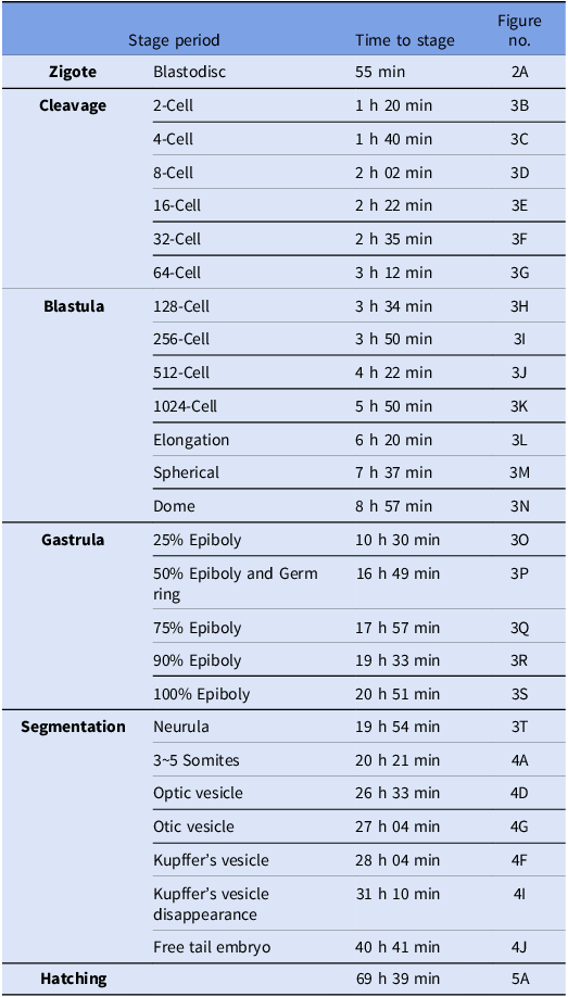
Embryonic development took 69 hours (h) and 39 minutes (min) from fertilization to hatching. In the following topics, a detailed description of the development stages, as shown in Table 2
Post-fertilization intervals of embryonic development of Prosomyleus rhomboidalis, incubated at 27.5°C. After in vitro fertilization, the embryos were incubated and visualized microscopically to describe the developmental stages. Figures on the table refers to the images described on Fig. 2, 3, 4 and 5

Table 2 Long description
The table outlines the embryonic development stages of Prosomyleus rhomboidalis, detailing the time intervals from fertilization to hatching. It includes stages such as Zigote, Cleavage, Blastula, Gastrula, Segmentation, and Hatching, with specific time periods for each stage. The table has three columns: Stage period, Time to stage, and Figure no. It lists the developmental stages in rows, with corresponding times and figure references. For example, the Zigote stage lasts 55 minutes and is referenced in Figure 2A. The Cleavage stage includes various cell divisions, such as 2-Cell, 4-Cell, 8-Cell, 16-Cell, 32-Cell, and 64-Cell, with times ranging from 1 hour 20 minutes to 3 hours 12 minutes. The Blastula stage includes 128-Cell, 256-Cell, 512-Cell, and 1024-Cell, with times from 3 hours 34 minutes to 5 hours 50 minutes. The Gastrula stage includes stages like 25 percentage Epiboly, 50 percentage Epiboly and Germ ring, 75 percentage Epiboly, 90 percentage Epiboly, and 100 percentage Epiboly, with times from 10 hours 30 minutes to 20 hours 51 minutes. The Segmentation stage includes Neurula, 3-5 Somites, Optic vesicle, Otic vesicle, Kupffer's vesicle, Kupffer's vesicle disappearance, and Free tail embryo, with times from 19 hours 54 minutes to 40 hours 41 minutes. The Hatching stage takes 69 hours and 39 minutes. Each stage is linked to specific figures for visual reference.
Zygote stage
After fertilization and hydration, determined as h zero post-fertilization, the chorion begins to expand, inducing an increase to 2.51 mm in total diameter, corresponding to the PS. At this stage, the formation of the cytoplasmic disc, known as the blastodisc, begins. It is located over a yolk-rich portion at the vegetal pole. The blastodisc corresponds to the first blastomere and covers the animal pole, being observed at 55 min post-fertilization (Table 2) (Figures 2B and 3A).
Early development of Prosomyleus rhomboidalis, during the zygote, cleavage, blastula, gastrula and initial segmentation stages. (A) zygote; (B) 2-cell stage; (C) 4-cell stage; (D) 8-cell stage; (E) 16-cell stage; (F) 32-cell stage; (G) 64-cell stege; (H) early blástula stage, with 128 blastomeres; (I) 256-blastomeres; (J) 512-blastomeres, (K) 1k blastomeres; (L) stretching; (M) sphere; (N) dome; (O) gástrula stage, 25% epiboly; (P) 50% epiboly, germ ring (arrows indicate the germ ring); (Q) 75% de epiboly; (R) 90% of epiboly (arrow indicates the embryonic Shield initial); (S) fase com 100% de epibolia (asterisk indicates the neural tube formation); (T) initial segmentation stage. Scale bar indicates 1 mm.

Figure 3 Long description
The image contains twenty images showing the early development stages of Prosomyleus rhomboidalis. The stages include zygote, 2-cell stage, 4-cell stage, 8-cell stage, 16-cell stage, 32-cell stage, 64-cell stage, early blastula stage with 128 blastomeres, 256-blastomeres, 512-blastomeres, 1k blastomeres, stretching, sphere, dome, gastrula stage with 25 percent epiboly, 50 percent epiboly with germ ring indicated by arrows, 75 percent epiboly, 90 percent epiboly with embryonic shield initial indicated by an arrow, 100 percent epiboly with neural tube formation indicated by an asterisk, and initial segmentation stage. Each stage is depicted in a circular format with a scale bar indicating 1 millimeter.
Clivage period (2 to 64-cell stage)
After the first cleavage, the blastomeres divided at approximately 20 min intervals (Table 2). Cytoplasmic divisions are meroblastic, with divisions exclusively at the animal pole. The cleavage stage comprises six phases (Figure 3B, G), in which the blastomeres divided simultaneously, almost synchronously, with a decrease in cell diameter, ending 3 h and 12 min after fertilization (Table 2).
2-Cell—At this stage, the first mitotic division occurred in the vertical plane, approximately 1 h and 20 min post-fertilization, giving rise to two blastomeres of similar sizes (Figure 3B).
4-Cell—The second cleavage gives rise to four similar blastomeres that divide transversely, forming right angles to the plane of the first cleavage. At this point, it is possible to observe a 2 × 2 matrix on the yolk (Figure 3C).
8-Cell—The third cleavage, characterized by the eight-cell phase, formed vertically to the second cleavage furrow in the four blastomeres (Figure 3D), assuming a 2 × 4 matrix format.
16-Cell—The fourth cell division gives rise to sixteen blastomeres, 02 h and 22 min post-fertilization, arranged in a 4 × 4 matrix pattern (Figure 3E).
32-Cell—The fifth cycle of cleavages produces thirty-two cells, generally presenting an irregular monolayer of blastomeres in the horizontal plane between the margin and the animal pole (Figure 3F).
64-Cell—The cleavage stage ends with the sixth division, giving rise to two layers of overlapping cells, totalling sixty-four blastomeres, 3 h and 12 min post-fertilization, Table 2, (Figure 3G). The arrangement of blastomeres may appear similar to the 32-cell stage, although they are smaller in diameter.
Blastula period (128 to 1024-cell stage)
The blastula stage is characterized by important processes occurring such as the transition in synchrony in the division of blastomeres, formation of the yolk syncytial layer (YSL), followed by the beginning of epiboly. The blastula was defined as initial and final according to the characteristics presented in each phase, according to the methodology described by Fujimoto et al. (Reference Fujimoto, Kataoka, Sakao, Saito, Yamaha and Arai2006) and Santos et al. (Reference Santos, Yasui, Xavier and Nakaghi2016), comprising the phases of 128, 256, 512 and 1024 blastomeres, including the elongation, spherical and dome phases. The morphological characteristics of the embryo are described in the following stages.
128-Blastomeres—At this stage, the blastodisc tends to assume a taller shape compared to previous cleavages, as cell layers are formed with the constant division of blastomeres (Figure 3H).
256 and 512-Blastomeres—The diameter of the blastomeres becomes visibly smaller compared to the previous phase, the blastodisc has an even more elevated and smooth shape, due to the overlapping of cell layers and the reduction of spaces between them (Figures 3I, J).
1024-Blastomeres—The tenth cleavage produced 1024 blastomeres (Figure 3K).
Elongation stage—At this stage, the embryos acquire an elliptical shape due to constant cell division and an increase in the number of blastomeres (Figure 3L).
Sphere stage—The surface of the blastomeres takes on a flatter appearance, giving the embryos a spherical appearance (Figure 3M).
Dome stage—The blastoderm cells assumed a dome shape that covered the yolk towards the animal pole (Figure 3N), beginning the gastrula stage. The embryos developed up to 25 to 30% of the epiboly stage, covering a quarter of the yolk (Figure 3O).
Gastrula period (epiboly)
At this stage, the morphogenetic movements that will initiate the germ ring and head-tail embryonic axis occur, with increased epiboly movement, ending when epiboly is 100% complete with the closure of the blastopore.
50% de epiboly stage and Germ ring phase—Half of the yolk cell (50%) is covered by the blastoderm, as shown in Figure 3P, after 16 h and 49 min post-fertilization, with the involution movements it is possible to observe the beginning of the formation of the germ ring, it is possible to observe different layers, being composed of a superficial layer, the epiblast and an internal layer, the hypoblast.
75% de epiboly stage—Three-quarters of the yolk was covered by the movement of epiboly by the blastoderm (Figure 3Q).
90% de epiboly and formation of the embryonic Shield—The movement of the blastoderm has advanced and covered approximately 90% of the yolk. At this stage, the head-tail embryonic axis is visible (Figure 3R), 19 h and 33 min post-fertilization.
100% de epiboly—The blastoderm completely covered the vitellus with the closure of the blastopore by means of the epiboly movement (100% epiboly), and thus the formation of the neural tube can be observed (Figure 3S, T).
Organogenesis periodsegmentation stage
In the segmentation stage, important events occurred, such as neurulation and somitogenesis phases, classified as early and late. The first somites were observed 20 h after fertilization (Table 2). The somites consist of blocks of mesodermal cells that, when developing, are separated by a myoseptum made up of connective tissue, also known as muscle segments or myomeres. As the number of somites increased, the visual and auditory organs of the embryo became visible, constituting useful morphological features for identifying stages of development.
Initial segmentation—neurula phase—In the neurulation phase, the neural tube could be seen when the embryos were analysed dorsally, characterizing this phase, with the emergence of the first somites, right after gastrulation (one to three somites) (Figures 3S and 4A).
Prosomyleus rhomboidalis embryos, during the segmentation stages, incubated at 27.5°C. (A) Early neurulation stage (arrow indicates the region of formation of the first somites); (B) 5-somites stage; (C) 10-somites stege; (D) 14-somites stage (arrow indicates the formation of the optic vesicle); (E) 18-somites satage, (asterisk indicates the morphological change in the shape of the somites); (F) 20-somites stage (asterisk indicates the formation of the Kupffer’s vesicle); (G) 24-somites stege, (arrow indicates the formation of the otic vesicle); (H) tail begins to detach from the yolk; (I) 27-somites stage, Kupffer’s vesicle is last seen; (J) 28-somites stage, (asterisk indicates the location where the tail is attached to the yolk); (K) 28-somites stage, it is possible to observe the heart (location indicated by arrow); (L) 30-somites stage, embryo in the late segmentation period, with an increase in the number of somites; (M) it is possible to observe a decrease in the perivitelline space; (N) fully formed embryo, tail completely detached, with intense movement within the chorion and ample yolk reserve. Scale bar indicates 1 mm.

Figure 4 Long description
The image contains fourteen separate images of Prosomyleus rhomboidalis embryos at various stages of segmentation, each labeled from A to N. Each image shows the embryo at a different stage of development, with specific features highlighted by arrows or asterisks. Image A shows the early neurulation stage with an arrow indicating the region of formation of the first somites. Image B depicts the 5-somites stage. Image C illustrates the 10-somites stage. Image D shows the 14-somites stage with an arrow indicating the formation of the optic vesicle. Image E represents the 18-somites stage, with an asterisk indicating morphological changes in the shape of the somites. Image F shows the 20-somites stage with an asterisk indicating the formation of the Kupffers vesicle. Image G depicts the 24-somites stage with an arrow indicating the formation of the otic vesicle. Image H shows the stage where the tail begins to detach from the yolk. Image I illustrates the 27-somites stage, where the Kupffers vesicle is last seen. Image J shows the 28-somites stage with an asterisk indicating the location where the tail is attached to the yolk. Image K depicts the 28-somites stage, with an arrow indicating the observable heart. Image L shows the 30-somites stage, representing the late segmentation period with an increase in the number of somites. Image M illustrates a decrease in the perivitelline space. Image N shows a fully formed embryo with the tail completely detached, intense movement within the chorion, and ample yolk reserve. The scale bar indicates 1 millimeter.
5-somites stage—A pair of rudimentary optic vesicles were visible in the brain region (Figure 4B).
14 to 18-somites stage—The optic vesicle becomes evident (Figure 4D), somites now form at a rate of about two per h and the pioneers have a V-shaped appearance that indicates the future division of the myomeres into dorsal (epaxial) muscle bundles and ventral (hypaxial) (Figure 4E). At these stages, all embryos had tails attached to the yolk.
20 a 30-somites stage—beginning of this phase, it was possible to identify the Kupffer vesicle, a prominent structure at the end of the tail (Figure 4F), the otic vesicle becomes visible when the embryo has 24-somites (Figure 4G), tail begins to detach from the yolk (Figure 4H), 27-somites for the Kupffer vesicle is no longer visible (Figure 4I), 28-somites the release of the caudal is evident (Figure 4J), it was possible to observe the emergence of the heart (Figure 4K).
30-somites stage—This phase, known as late segmentation, is the period in which there is an increase in the final number of somites (Figure 4L), and decreasing the PS (Figure 4M), the embryo presents a completely detached tail, intense movement within the chorion and ample yolk reserve (Figure 4N), contractions become stronger, more coordinated and more frequent until the chorion ruptures.
Hatching
The embryos had an average incubation period of 69 h and 39 min. Normal newly hatched larvae were transparent and had between 36 and 39 myomeres, with length measurements of the larvae 5,98 mm. They had a distended posture, closed mouth and anus, with the anus positioned in the median region of the body (Figures 5A and 6A), non-pigmented eyes, otic and optic vesicle (Figure 5B (8), (9)), embryonic fin covering the entire caudal region (Figure 5A (4)). The head was positioned ventrally and adhered to the anterior portion of the yolk sac, which had a circular shape (Figures 5A (2)). Furthermore, the newly hatched larvae remained at the bottom of the incubator, with little vertical movement, probably due to the size of the yolk and the absence of fins. The rates of normal and abnormal larvae are shown in Table 1.
Morphological structures of normal larvae of de Prosomyleus rhomboidalis after hatching. (A) Larvae with central nervous system (notochord indicated by asterisk), pericardial cavity with developing heart (1) indicated by arrow, circular yolk sac (2), presence of myomeres (3), embryonic membrane is present along the caudal region (4); (B) presence of the central nervous system, forebrain (5), midbrain (6) and hindbrain (7), completely depigmented optic vesicle (8), otic vesicle (9).

Figure 5 Long description
The image presents a cross-sectional view of a larva of Prosomyleus rhomboidalis, highlighting various internal structures. In panel A, the larva displays the central nervous system with the notochord marked by an asterisk. An arrow points to the developing heart within the pericardial cavity, labeled as part 1. The circular yolk sac is labeled as part 2, and the presence of myomeres is indicated as part 3. The embryonic membrane extends along the caudal region, labeled as part 4. Panel B focuses on the central nervous system, showing the forebrain labeled as part 5, the midbrain as part 6, and the hindbrain as part 7. Additionally, a completely depigmented optic vesicle is labeled as part 8, and the otic vesicle is labeled as part 9.
Newly hatched larvae of Prosomyleus rhomboidalis, demonstrating morphological patterns for normal and abnormal larvae. (A) Normal Larvae 38 somites (asterisk indicates the formation and position of the anus); (B) Larvae containing an abnormality in the caudal region (arrow indicates the region at the end of the damaged notochord); (C) and (D) Larvae containing abnormality in caudal region (arrow indicates the region with abnormalities in the upward curved tail and down, respectively); (E) Larvae presents several abnormalities, in the cephalic region (indicated by arrow) and a completely curved tail (region indicated by the asterisk); (F) Larvae presents several morphological abnormalities in the caudal region and yolk region (indicated by arrows).

Figure 6 Long description
The image displays six different larvae of Prosomyleus rhomboidalis, each exhibiting various morphological patterns. Image A shows a normal larva with 38 somites, with an asterisk marking the formation and position of the anus. Image B highlights a larva with an abnormality in the caudal region, indicated by an arrow pointing to the damaged notochord. Images C and D depict larvae with abnormalities in the caudal region, with arrows indicating upward and downward curved tails, respectively. Image E presents a larva with multiple abnormalities, including a damaged cephalic region marked by an arrow and a completely curved tail indicated by an asterisk. Image F shows a larva with several morphological abnormalities in the caudal and yolk regions, marked by arrows.
Hatched larvae presenting free swimming and absence of malformations were classified as normal (Figure 6A). Various patterns of abnormalities were found including malformations in the tail, head, or even in size (Figure 6B–F).
Discussion
The characteristics of eggs from Prosomyleus rhomboidalis are different from most freshwater fish, especially in Serrasalmidae, attributes such as low fecundity and long duration of embryogenesis with more of 69 h, (see comparison on Table 3 below) suggest that this species has a life history strategy that combines comparatively low fertility with some degree of parental care, according to Nakatani et al., (Reference Nakatani, Agostinho, Bialetzki, Baumgartner, Sanches, Makrakis and Pavanelli2001) and Rizzo and Bazzoli (Reference Rizzo and Bazzoli2020). This is not a common strategy for the Pacus group, where the pattern is medium to very high fertility, with unprotected pelagic eggs (Romagosa et al., Reference Romagosa, De Paiva and Godinho1990; Leite et al., Reference Leite, Melo, Oliveira, Pinheiro, Campello, Nunes and Salmito-Vanderley2013).
Comparison of some reproductive aspects and embryo parameters of other serrasalmid species

Table 3 Long description
The table compares reproductive aspects and embryo parameters of various serrasalmid species, focusing on fecundity, egg diameter, and incubation period. It includes data for four species: Prosomyleus rhomboidalis, Colossoma macropomum, Piaractus mesopotamicus, and Piaractus brachypomus. The table has four rows and four columns, with headers for Serrasalmid species, Fecundity, Egg diameter (millimeters), and Incubation period (hours). Notable trends include the low fecundity and long incubation period of Prosomyleus rhomboidalis compared to other species. Colossoma macropomum has the highest fecundity at 1,800,000 eggs, while Piaractus brachypomus has the shortest incubation period at 12 hours. The data suggests varying reproductive strategies among serrasalmid species.
Knowledge of the early development stages observed in P. rhomboidalis is similar to that of most freshwater teleosts in terms of post-fertilization events as the case of Danio rerio (Kimmel et al., Reference Kimmel, Ballard, Kimmel, Ullmann and Schilling1995), Misgurnus anguillicaudatus (Fujimoto et al., Reference Fujimoto, Kataoka, Sakao, Saito, Yamaha and Arai2006), Brycon insignis (Isaú et al., Reference Isaú, Rizzo, Amaral, Mourad and Viveiros2013), Astyanax altiparanae (Santos et al., Reference Santos, Yasui, Xavier and Nakaghi2016), Brycon amazonicus (Silva et al., Reference Silva, Santos, Senhorini, Paes, Valentin, Fujimoto, Nascimento, Yasui and Nakaghi2017) and Piaractus mesopotamicus (Coelho et al., Reference Coelho, Arashiro, Disselli, Pereira-Santos, Mira-López, Monzani, Senhorini, Fujimoto and Yasui2021). It was observed a very long period of embryonic greater than 69 h, compared with other pacus species such as P. mesopotamicus 20 h and 35 min at 26°C (Coelho et al., Reference Coelho, Arashiro, Disselli, Pereira-Santos, Mira-López, Monzani, Senhorini, Fujimoto and Yasui2021). This may be related to some factors as a reproductive strategy of the species, since those characteristics of low fecundity, large amount of yolk and diameter, long incubation period are characteristics of intertidal k-strategist spawners (Table 3). Those characteristics suggest some strategies for progeny protection, probably passive protection such as hiding in the rocky bottom of the Xingu River. The parameters discussed above suggest that P. rhomboidalis does not spawn in open waters, as occurs in other migratory species.
The incubation temperature is known to be longer at higher temperatures and vice versa, influencing embryo development time and survival percentages and larvae with malformations (Santos et al., Reference Santos, Yasui, Xavier and Nakaghi2016; Silva et al., Reference Silva, Santos, Senhorini, Paes, Valentin, Fujimoto, Nascimento, Yasui and Nakaghi2017; Arashiro et al., Reference Arashiro, Yasui, Calado, Nascimento, Santos, Santos, Levy-Pereira, Monzani, Siqueira-Silva and Senhorini2018; Coelho et al., Reference Coelho, Yo, Mira-López, Monzani, Arashiro, Fujimoto, Senhorini and Yasui2019, Reference Coelho, Arashiro, Disselli, Pereira-Santos, Mira-López, Monzani, Senhorini, Fujimoto and Yasui2021). In this study, regarding incubation of the embryos of P. rhomboidalis, the temperature was set at 27.5°C, based on previous studies involving Neotropical teleost, since increased temperature at 30°C gave rise to poor hatching rates and an increased percentage of deformed larvae. Such a phenomenon was observed in P. mesopotamicus (Coelho et. al., Reference Coelho, Arashiro, Disselli, Pereira-Santos, Mira-López, Monzani, Senhorini, Fujimoto and Yasui2021), Astyanax altiparanae (Santos et al., Reference Santos, Yasui, Xavier and Nakaghi2016), Brycon amazonicus (Silva et al., Reference Silva, Santos, Senhorini, Paes, Valentin, Fujimoto, Nascimento, Yasui and Nakaghi2017), Pimelodus maculatus and Pseudopimelodus mangurus (Arashiro et al., Reference Arashiro, Yasui, Calado, Nascimento, Santos, Santos, Levy-Pereira, Monzani, Siqueira-Silva and Senhorini2018). In P. rhomboidalis the average temperature of the Xingu River varies from 28 to 32, but during spawning period the temperature is near 30°C (Barreiros et al., Reference Barreiros, Giarrizzo and Godoy2023). However, it is well known that most fish species including Amazonian species, the reproduction is triggered by rain which synchronize spawn in a wide range of fish species, and also cause a decrease on temperature and an increase in dissolved oxygen that are important for incubation and larval maintenance.
Successive cell divisions during the cleavage period, characterized by an increase in the number of cells and a decrease in the size of blastomeres, is commonly observed in teleosts, (Faustino et al., Reference Faustino, Nakaghi and Neumann2011; Nakauth et al., Reference Nakauth, Villacorta-Correa, Figueiredo, Bernardino and França2016; Ganeco-Kirschnik et al., Reference Ganeco-Kirschnik, Franceschini-Vicentini, Faria Paes and Nakaghi2018; Santos et al., Reference Santos, Soares and Bialetzki2020), as well as in the species in this study, P. rhomboidalis.
During the blastula stage, the formation of the YSL begins an extraembryonic structure that, according to Faustino et al., (Reference Faustino, Nakaghi and Neumann2011), plays a fundamental role in early embryonic development. Prior to the transition to exogenous feeding sources, the embryo relies exclusively on the yolk, which is continuously absorbed through the YSL. The presence of this structure during the blastula stage has also been reported by several authors (Fujimoto et al., Reference Fujimoto, Kataoka, Sakao, Saito, Yamaha and Arai2006; Silva et al., Reference Silva, Santos, Senhorini, Paes, Valentin, Fujimoto, Nascimento, Yasui and Nakaghi2017; Coelho et al., Reference Coelho, Yo, Mira-López, Monzani, Arashiro, Fujimoto, Senhorini and Yasui2019).
The cells move convergently during the gastrula stage, a characteristic movement known as epiboly occurring in the direction of the animal pole to the vegetative pole, originating structures such as the germ ring and embryonic axis, as observed by Leite et al. (Reference Leite, Melo, Oliveira, Pinheiro, Campello, Nunes and Salmito-Vanderley2013), for C. macropomum and Nakauth et al. (Reference Nakauth, Villacorta-Correa, Figueiredo, Bernardino and França2016) for B. amazonicus, ending gastrulation when the blastoderm completely covers the yolk.
Several organs develop during somitogenesis, such as optic vesicles, otic vesicles and Kupffer’s vesicle, which comprise developmental milestones, which differentiate into different numbers of somites in different fish species (Essner et al., Reference Essner, Amack, Nyholm, Harris and Yost2005; Faustino et al., Reference Faustino, Nakaghi, Marques, Makino and Senhorini2007; Coelho et al., Reference Coelho, Arashiro, Disselli, Pereira-Santos, Mira-López, Monzani, Senhorini, Fujimoto and Yasui2021). With the development of the embryo, there was a progressive increase in the number of somites, expansion of the cephalic region, increase in the length of the tail and intensification of body movements until the rupture of the chorion, (Kimmel et al., Reference Kimmel, Ballard, Kimmel, Ullmann and Schilling1995; Faustino et al., Reference Faustino, Nakaghi and Neumann2011; Silva et al., Reference Silva, Santos, Senhorini, Paes, Valentin, Fujimoto, Nascimento, Yasui and Nakaghi2017) was observed with P. rhomboidalis.
After hatching, P. rhomboidalis larvae had a large yolk sac, developed eyes but without pigmentation and a mouth in a subterminal and non-functional position. The gills were absent, attributes similar to those found for species with altricial characteristics (Nakatani et al., Reference Nakatani, Agostinho, Bialetzki, Baumgartner, Sanches, Makrakis and Pavanelli2001; Santos et al., Reference Santos, Soares and Bialetzki2020; Coelho et al., Reference Coelho, Arashiro, Disselli, Pereira-Santos, Mira-López, Monzani, Senhorini, Fujimoto and Yasui2021).
In this study, oocytes were characterized in detail for description of developmental stages from fertilization to hatched larvae. Our results provide valuable information for future biotechnological applications, including chromosome set manipulation and the traceability of primordial germ cells, as well as the establishment of stem cell cryobanks as an initial step toward the application of surrogate technologies in serrasalmids.
In conclusion, this work describes in detail, for the first time in this species, the developmental stages and incubation management used in loco reproduction. Different reproductive patterns were found in the Serrasalmidae group. This information is important for basic and applied sciences in both conservation and sustainable aquaculture.
Acknowledgments
The authors thank the Center for Environmental Studies and its collaborators, the reproduction site of the species; the Federal University of Pará, represented by the Xingu Ornamental Fish Aquaculture Laboratory. We are especially grateful to Zaqueu Santos for the scientific illustrations. This study is part of the first author’s master’s thesis in the Graduate Programme in Biodiversity and Conservation at the Federal University of Pará.
Funding
We also thank Norte Energia S/A for the financial support through the scholarship granted under the Research and Development Project ANEEL (PandD-07427-0121/2021), which was essential for the execution of this research.
Competing interests
The authors declare no conflict of interest.
Ethical standards
Ethical approval all applicable international, national and/or institutional guidelines for the care and use of animals were followed by the authors.