

Introduction
The Gentianaceae is the third largest family of the order Gentianales, comprising about 1700 species (Molina & Struwe, Reference Molina and Struwe2009), with two-thirds of the genera being distributed in the tropics (Meszaros & De Laet, Reference Meszaros and De Laet1996). Phylogenetic studies on Gentianaceae suggest six tribes should be recognised: Exaceae, Chironieae, Gentianaceae, Helieae, Potalieae and Saccifolieae (Struwe et al., Reference Struwe, Kadereit, Klackenberg, Nilsson, Thiv, Von-Hagen, Albert, Struwe and Albert2002). The tribe Helieae is strictly neotropical, and comprises 22 genera and 184 species. Phylogenetic studies on Helieae, combining molecular and morphological data, strongly support the monophyly of several genera and two major subclades: the Macrocarpaea (Griseb.) Gilg and Symbolanthus G.Don subclades. The phylogeny of the Symbolanthus subclade, however, has not been completely clarified (Struwe et al., Reference Struwe, Albert, CaliÓ, Frasier, Lepis, Mathews and Grant2009). The genus Calolisianthus (Griseb.) Gilg, in the Symbolanthus subclade (Struwe et al., Reference Struwe, Albert, CaliÓ, Frasier, Lepis, Mathews and Grant2009), is comprised of largely unbranched perennial herbs with sessile oblong-elliptic leaves and 5-merous flowers with large pink corollas in terminal few-flowered cymes (Struwe et al., Reference Struwe, Kadereit, Klackenberg, Nilsson, Thiv, Von-Hagen, Albert, Struwe and Albert2002). This genus consists of 6–10 species distributed in Bolivia and southeastern Brazil, occurring in Brazilian savannas (cerrado) and rocky field (campo rupestre) formations (Struwe et al., Reference Struwe, Kadereit, Klackenberg, Nilsson, Thiv, Von-Hagen, Albert, Struwe and Albert2002).
The cerrado is a savanna physiognomy characterised by strong rainfall seasonality, coupled with constantly high diurnal air temperatures and deep and well-drained soils (Furley, Reference Furley1999). The nutrient-poor, acid soils are an additional limiting factor for plant growth in this region (Haridasan, Reference Haridasan2000). Campo rupestre vegetation is found on the slopes of hills in rocky outcrops, being characterised by a predominant physiognomy of grassland and shrubs (Gottsberger & Silberbauer-Gottsberger, Reference Gottsberger and Silberbauer-Gottsberger2006). The campo rupestre is also characterised by seasonal rainfall, high temperatures during the day and low temperatures during the night, frequent winds (Scarano et al., Reference Scarano, Duarte, Ribeiro, Rodrigues, Barcellos and Franco2001), low soil fertility, and soil with sandstone and quartz (Giulietti et al., Reference Giulietti, Menezes, Pirani, Meguro and Wanderley1987). Because of low fertility and water deficits (Dickison, Reference Dickison2000), morpho-anatomical adaptations, such as root-mycorrhizal associations that increase the water and nutrient absorption (Herrman et al., Reference Herrman, Oelmméller and Buscot2004), may facilitate, or even be essential to, the survival of plants in these two environments.
The comparative anatomy of vegetative organs of Calolisianthus species may be important in our understanding of anatomical evolution and variation in the family. Leaf anatomy has always played an important role in plant taxonomy and may be useful for infrageneric delimitation (Metcalfe & Chalk, Reference Metcalfe and Chalk1950; Kocsis et al., Reference Kocsis, Darók and Borhidi2004; Matias et al., Reference Matias, Soares and Scatena2008; Erxu et al., Reference Erxu, Qiufa, Hongfei, Jingbo, Yueqiang, Feilai and Hui2009; Moon et al., Reference Moon, Hong, Smets and Huysmans2009), as well as being important in our interpretation of adaptation of plant species to ecological constraints. Such detailed anatomical analysis is necessary for an understanding of the effects of environmental constraints on Calolisianthus ecology in cerrado and campo rupestre in Brazil.
Materials and Methods
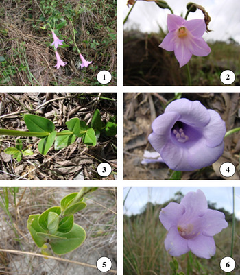
The anatomy of vegetative organs was studied in three species: Calolisianthus pendulus (Mart.) Gilg (Figs 1–2), C. amplissimus (Mart.) Gilg (Figs 3–4), and C. speciosus (Cham. & Schltdl.) Gilg (Figs 5–6). Voucher specimens are housed at VIC (Index Herbariorum: http://sweetgum.nybg.org/ih/): Calolisianthus pendulus (M.N. Delgado, A.A. Azevedo & G.E. Valente VIC 31.361), C. amplissimus (M.N. Delgado, A.A. Azevedo & G.E. Valente VIC 31.366), C. speciosus (M.N. Delgado, A.A. Azevedo & G.E. Valente VIC 31.362).
Morphological diversity in species of Calolisianthus studied. 1–2: Calolisianthus pendulus. 3–4: C. amplissimus. 5–6: C. speciosus.

Samples of Calolisianthus speciosus and C. pendulus for the anatomical study were collected in campo rupestre areas of the Ouro Branco Mountain Range (Giulietti & Pirani, Reference Giulietti, Pirani, Heyer and Vanzolini1988; Giulietti et al., Reference Giulietti, Pirani, Harley, Davis, Heywood, Herrera-MacBryde, Villa-Lobos and Hamilton1997) and samples of C. amplissimus in cerrado areas of the Paraopeba National Forest (Balduino et al., Reference Balduino, Souza, Meira-Neto, Silva and Silva JÚnior2005; Souza et al., Reference Souza, Alves, Silva and Souza2008), in Minas Gerais, Brazil. The campo rupestre area where Calolisianthus pendulus specimens were collected is a slope, located at 20°29′35.5″–20°29′35.7″S and 43°42′36.1″–43°42′36.8″W, with altitudes ranging from 1517 to 1540 m. The area is sunny, windy and rocky, but the soil is soft. Specimens of Calolisianthus speciosus were collected at 20°30′07.6″–20°30′20.02″S and 43°38′39.9″–43°38′21.6″W, with altitudes ranging from 1321 to 1352 m. The area is sunny and dominated by grasses and the soil is hard and sandy. Calolisianthus amplissimus specimens were collected at 19°15′15.13″–19°15′21.4″S and 44°24′04.4″–44°24′06.3″W, with altitudes ranging from 763 to 769 m. This area is a cerrado with some trees and shrubs and the soil is soft and clayey.
Leaf, stem and root samples were fixed in FAA50 and then stored in 70% ethanol (Johansen, Reference Johansen1940). Samples were collected from the basal, medial and apical portions and margins of leaf blades. Stems were sampled at the second and third internodes and roots were sampled in the area of secondary growth.
Samples of root, stem and leaf were processed and embedded in Historesin (Leica Embedding Kit), following standard procedures. Longitudinal and cross-sections, 6–8 μm thick, were obtained using an automatic rotary microtome (model RM2155, Leica Microsystems Inc., Deerfield, USA) and stained with Toluidine Blue in MacIlvaine buffer for 15 minutes (O’Brien & McCully, Reference O'Brien and McCully1981). In extrafloral nectaries and colleters the histochemical test PAS was utilised to detect carbohydrates (Maia, Reference Maia1979), Ruthenium Red for acid polysaccharides, and Sudan Red B for total lipids (Johansen, Reference Johansen1940). Histological slides were prepared to study epidermal characteristics. Entire leaves were cleared by immersion in a 10% NaOH solution for 1 hour and transferred to 20% sodium hypochlorite for approximately 72 hours until they became translucent. Samples were stained in Basic Fuchsin for 1 or 2 hours, dehydrated in an ethanol-xylol series and mounted in Permount (Berlyn & Miksche, Reference Berlyn and Miksche1976). Photographs were taken using an Olympus AX70 microscope equipped with a U-Photo photographic system.
Leaf samples of the species studied were also collected for scanning electron microscopy (SEM) analysis, fixed in Karnovsky’s solution (Karnovsky, Reference Karnovsky1965) in the field, dehydrated in an ethyl alcohol series, and dried to the critical point with CO2. The samples were gold coated as described by Bozzola & Russel (Reference Bozzola and Russel1992) and examined by scanning electron microscope SEM 1430 LEO VP (Cambridge, UK) at 5–15 kV.
Results
Anatomical description
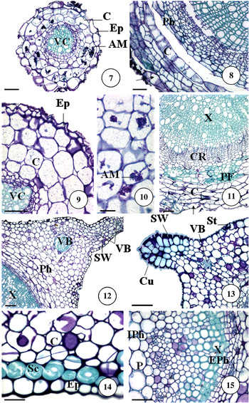
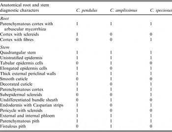
Roots (Table 1). All the roots studied exhibited secondary growth (Figs 7–9, 11). However, we observed that a uniseriate epidermis remained in thin roots. This epidermis consisted of irregularly shaped cells in cross-section (Figs 7, 9). In all species, the cortex is parenchymatous and colonised by Arum-type arbuscular mycorrhiza (AM) (Figs 7, 10). Only cortical cells of Calolisianthus amplissimus contain starch.
Transverse sections of root and stem. 7–8: Root of Calolisianthus pendulus. 9–10: Root of C. amplissimus. 11: Root of C. speciosus. 12: Stem of C. amplissimus. 13: Stem of C. pendulus. 14–15: Stem of C. speciosus. Ep – epidermis, C – cortex, AM – Arum-type arbuscular mycorrhiza, VC – vascular cylinder, X – xylem, Sc – sclereids, PF – perivascular fibre, CR – cambial region, Arrow – cortical cells that replace epidermis, SW – stem wing, VB – vascular bundle, Ph – phloem, Cu – cuticle, St – stomata, EPh – external phloem, IPh – internal phloem, P – pith. Scale bars = 70 μm (7, 8, 9, 11), 50 μm (10, 14, 15), 100 μm (12, 13).

Root and stem characters of Calolisianthus pendulus, C. amplissimus and C. speciosus

1 = presence; 0 = absence.
In thicker roots of Calolisianthus pendulus and C. speciosus the epidermis and some layers of cortical parenchyma with Arum-type AM are discarded and are replaced by elongate cortical cells in the periclinal direction and irregular-shaped cells in the anticlinal direction (Figs 8, 11). Phellogen does not occur in secondary roots of the species studied. Sclereids were found scattered in the cortex of Calolisianthus pendulus, primarily in the innermost layers. In Calolisianthus speciosus there are several layers of fibres arranged between the cortical parenchyma and the secondary phloem (Fig. 11). We found two types of vessel perforation in the root secondary xylem: in Calolisianthus pendulus plates are foraminate and simple, while in C. speciosus and C. amplissimus we observed only simple plates.
Stems (Table 1). The epidermis is unistratified with thick external periclinal walls that are cutinised. Calolisianthus amplissimus has a smooth and fine cuticle, whereas in C. pendulus (Fig. 13) and C. speciosus the cuticle is decorated and thick. Epidermal stem cells in the cross-section are elongated in Calolisianthus pendulus (Fig. 13) and C. speciosus (Fig. 14) and tabular in C. amplissimus (Fig. 12). Stem cortex is parenchymatous and chlorophyllous with subepidermal sclereids in Calolisianthus speciosus (Fig. 14). Three to four cortical bundles are observed towards the stem wings in Calolisianthus amplissimus (Fig. 12) and C. speciosus, but only one is present in C. pendulus (Fig. 13). The endodermis of Calolisianthus pendulus and C. speciosus has clear Casparian strips that are not observed in the material of C. amplissimus that was analysed. The stem vascular system has external and internal phloem in the second and third internodes (Fig. 15). There is an initial secondary growth with the addition of secondary xylem. The vascular cylinder consists of secondary xylem, secondary phloem, vascular cambium and pith. The vessels found in the stem are the same as those in the roots. Parenchymatous pith is fistulous only in Calolisianthus amplissimus.
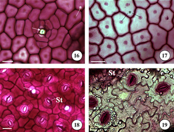
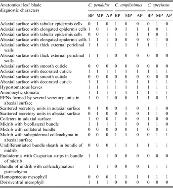
Leaves (Tables 2and 3). In Calolisianthus pendulus the epidermis is unistratified with cells that have straight anticlinal walls (Figs 16, 18). Calolisianthus amplissimus and C. speciosus have straight anticlinal walls on the adaxial surface (Fig. 17) and sinuous walls on the abaxial surface (Fig. 19). All species have cuticular ornamentation visible under light microscopy (Figs 16–17) which is rather thick in Calolisianthus pendulus (Fig. 27) and C. speciosus (Fig. 31) but thinner in C. amplissimus (Fig. 29). All species of Calolisianthus studied have hypostomatous leaves (Figs 27, 29, 31), often with anomocytic stomata (Figs 18–19). We have observed variation in the shape of epidermis cells in cross-section at the basal, medial and apical portions of the same leaf.
Leaf epidermis. 16: Adaxial surface of a Calolisianthus pendulus leaf. 17: Adaxial surface of a C. speciosus leaf. 18: Abaxial surface of a C. pendulus leaf. 19: Adaxial surface of a C. amplissimus leaf. Arrow – secretory unit of nectar, * – cuticular ornamentation, St – stomata. Scale bars = 50 μm (16, 17, 18, 19).

Leaf blade characters of Calolisianthus pendulus, C. amplissimus and C. speciosus

1 = presence; 0 = absence; EFNs = extrafloral nectaries; BP = basal portion; MP = medial portion; AP = apical portion.
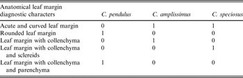
Leaf margin characters of Calolisianthus pendulus, C. amplissimus and C. speciosus

1 = presence; 0 = absence.
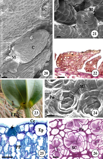
A variable number of non-vascularised colleters are found on the adaxial surface and close to the leaf axis in all three species (Figs 20–22). The colleters are very small and glossy and can only be observed using a stereomicroscope. Micromorphological studies on leaf colleters show that the colleter heads are undulate and uneven (Figs 20–21), possessing a large quantity of mucilage between the secretory structures (Fig. 21). Colleters are formed from large cells with a prominent nucleus, dense cytoplasm and thin cell walls covered with a thin cuticle (Fig. 22). Colleters involved in secretion have the central region separated from the most peripheral cell layers by a space filled with a secretion (Fig. 22) that reacts intensely to the PAS test. The peripheral layer cells are not different from inner cells in shape, but stained more intensively with Ruthenium Red (Fig. 22) and PAS. The Sudan Red B test is negative for the secreted substance and the neck region, staining red only in the thin cuticle that covers the colleter. In Calolisianthus speciosus and C. pendulus, two conspicuous extrafloral nectaries (EFNs) have been observed at the base of the abaxial surface of the leaves. These large extrafloral nectaries, which are described for the first time, are formed by the adhesion of several secretory units. Each secretory unit is a very small nectary that is formed by a few cells (Figs 23–24, 26). Furthermore, a set of secretory units forms an apical nectary at the leaf apex of the three species. These secretory units react positively with PAS (Fig. 26), suggesting a secretion of soluble sugars, and are scattered throughout the leaf on both adaxial (Fig. 25) and abaxial (Figs 24, 26) surfaces.
Secretory structures. 20: Colleters, by scanning electron microscopy, on the adaxial surface of the leaf base of Calolisianthus pendulus. 21–22: Colleters on the adaxial surface of the leaf base of C. speciosus: 21: Colleters under scanning electron microscopy. 22: Colleters under light microscopy, stained with Ruthenium Red. 23: Two conspicuous extrafloral nectaries on the abaxial surface of the leaf base of C. speciosus. 24: Nectar secretory unit of C. speciosus on the abaxial surface of the leaf base, under scanning electron microscopy. 25: Nectar secretory unit (arrow) of C. pendulus at the apex of the leaf, adaxial surface. 26: Nectar secretory unit of C. speciosus at the base of the leaf, abaxial surface, stained with PAS. C – colleter, M – mucilage, BC – broken colleter, * – mucilage accumulated in apoplastic space of colleter head, EFN – extrafloral nectary, SU – secretory unit, Po – secretory pore, Arrow – nectar secretory unit, Cc – cuticle, Ep – epidermis, PP – palisade parenchyma, SC – secretory cell. Scale bars = 10μm (20), 50μm (21, 22), 20μm (24, 25, 26).

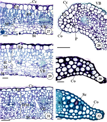
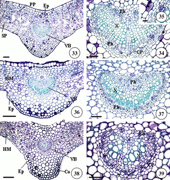
The laminas are dorsiventral with a mesophyll that has three layers of palisade parenchyma in Calolisianthus pendulus (Figs 27, 33) but is homogeneous in C. amplissimus (Figs 29, 36) and C. speciosus (Figs 31, 38). The leaf margin structure varies between the three species: in Calolisianthus pendulus (Fig. 28) the leaf margin is rounded and formed from parenchymatous and collenchymatous cells, in C. amplissimus (Fig. 30) it is acute, curved, and formed only from collenchyma, and in C. speciosus it is curved and formed from collenchyma containing some sclereids (Fig. 32).
Mesophyll and margin. 27: Mesophyll of Calolisianthus pendulus. 28: Leaf margin of C. pendulus. 29: Mesophyll of C. amplissimus. 30: Leaf margin of C. amplissimus. 31: Mesophyll of C. speciosus. 32: Leaf margin of C. speciosus. Cc – cuticle, Ep – epidermis, PP – palisade parenchyma, SP – spongy parenchyma, St – stomata, VB – vascular bundle, Arrow – nectar secretory unit, P – parenchyma, Co – collenchyma, HM – homogeneous mesophyll, Sc – sclereids. Scale bars = 50μm (27, 29, 30, 32), 150μm (28), 100μm (31).

Differences in leaf midrib anatomy have been found between the three species. The vascular bundle of the midrib is bicollateral at the base and in the middle of the leaf (Figs 34, 37, 39) but is always collateral at the leaf apex in all three species. In Calolisianthus pendulus, the bicollateral bundle initiates secondary growth (Fig. 34), with an endodermis with Casparian strips around the midrib (Figs 34–35). Calolisianthus pendulus (Fig. 34) and C. speciosus (Fig. 39) have collenchymatous parenchyma associated with the midrib bundle. Calolisianthus amplissimus (Fig. 36) and C. speciosus (Fig. 38) have subepidermal collenchyma on the abaxial surface.
Leaf midrib. 33–35: Calolisianthus pendulus. 36–37: C. amplissimus. 38–39: C. speciosus. Ep – epidermis, PP – palisade parenchyma, SP – spongy parenchyma, VB – vascular bundle, HM – homogeneous mesophyll, Co – collenchyma, Ph – phloem, X – xylem, CP – collenchymatous parenchyma, Arrow – endodermis. Scale bars = 150μm (33, 36, 38), 50μm (34, 37, 39), 20μm (35).

Discussion
A comparison of the anatomy of vegetative organs in Calolisianthus pendulus, C. speciosus and C. amplissimus shows that C. amplissimus and C. speciosus share a great number of characters, although there are also unique characters in each species. Calolisianthus pendulus has more unique diagnostic characters than C. amplissimus and C. speciosus. Some of the characters shared by Calolisianthus amplissimus and C. speciosus are: leaf midrib with collateral bundle and subepidermal collenchyma in the abaxial surface, homogeneous mesophyll, and an acute and curved leaf margin. In Calolisianthus amplissimus the diagnostic characters are: roots with tabular epidermis cells, stem epidermis cells with smooth cuticle, presence of fistulous pith in the stem, the lack of EFNs at the base of the leaf, and the leaf margin formed from collenchyma. In Calolisianthus speciosus the diagnostic characters are: root cortex with fibres, stem with subepidermal sclereids, and leaf margin formed from parenchyma and sclereids. In Calolisianthus pendulus the unique characters are: root cortex with sclereids, stem pericycle with sclereids, leaf epidermis cells with thick external periclinal walls on the adaxial surface, endodermis with Casparian strips around the midrib, dorsiventral mesophyll, and a rounded leaf margin formed from parenchyma.
Some root traits are diagnostic and may be used to identify the species studied, such as the presence of sclereids or fibres or absence of both. Other characters, like the presence of mycorrhizae, may be closely related to ecological conditions and are not diagnostic. Mycorrhizae are found in many cerrado herbs (Detmann et al., Reference Detmann, Delgado, Rebello, Leite, Azevedo, Kasuya and Almeida2008), these being crucial for increased water absorption in the dry season. Furthermore, the soil of cerrado and campo rupestre is poor in some nutrients, especially phosphorus (Giulietti et al., Reference Giulietti, Menezes, Pirani, Meguro and Wanderley1987; Furley & Ratter, Reference Furley and Ratter1988; Furley, Reference Furley1999), and mycorrhizae can decrease the nutritional stress (Siqueira et al., Reference Siqueira, Lambais and Stumer2002).
Arum-type arbuscular mycorrhizae (AMs) are found in the parenchymatous cortex of the roots of all three species. Arum-type AM consists of hyphae that grow across the cortex apoplast before forming arbuscules within cells (Peterson et al., Reference Peterson, Massicotte and Melville2004). Although we observed Arum-type AM in Calolisianthus species, intracellular hyphal coils that are named Paris-type AM have already been described for several Gentianaceae species (Demuth & Weber, Reference Demuth and Weber1990).
An important and interesting observation is the absence of hairs on the root epidermis of the three species studied. This absence could be an artefact of the root collection method or be due to the fast secondary growth that induces hair senescence. However, this absence could also be due to the intense mycorrhizal colonisation in roots (Imhof, Reference Imhof1999). These mycorrhizae increase access to water and nutrients, being more efficient than root hairs (Herrman et al., Reference Herrman, Oelmméller and Buscot2004; Peterson et al., Reference Peterson, Massicotte and Melville2004).
Anatomical data can contribute to our understanding of adaptive strategies in plants (Dickison, Reference Dickison2000; Matias et al., Reference Matias, Soares and Scatena2008; Delgado et al., Reference Delgado, Azevedo, Valente and Kasuya2009). For example, anatomical xeromorphic characteristics, such as adaptive responses to high luminosity, a stress factor in areas of campo rupestre and cerrado (Gottsberger & Silberbauer-Gottsberger, Reference Gottsberger and Silberbauer-Gottsberger2006), have been identified in stems and leaves of the species studied (e.g. Calolisianthus pendulus and C. speciosus that were collected in sunny areas). There is a thick cuticle and the epidermal cells are elongated in the stems of Calolisianthus pendulus and C. speciosus. Calolisianthus pendulus occurs in the Ouro Branco Mountain Range, on a windward cliff, where strong winds and high light intensities predominate. Calolisianthus speciosus occurs in the Ouro Branco Range in an open field area, consisting mainly of tall grasses that generally shade plants of C. speciosus which are shorter than them. Calolisianthus amplissimus is found in shaded areas of cerrado and has a thinner cuticle, whilst C. pendulus and C. speciosus that occur in full sun in the Ouro Branco Range have a thicker cuticle. The thickness of the cuticle might be influenced by solar radiation (Ashton & Berlyn, Reference Ashton and Berlyn1992). Since the cuticle can reflect intense sunlight (Morais et al., Reference Morais, Medri, Marur, Caramori, Ribeiro and Gomes2004) thicker cuticles prevent excessive water loss (Sarmiento et al., Reference Sarmiento, Goldstein and Meinzer1985; Oliveira et al., Reference Oliveira, Meirelles and Salatino2003), thereby avoiding excessive increases in temperature within the mesophyll.
The shape of epidermal cell walls may also be an environmental adaptation in these species. Plants growing in more shaded areas, such as those of Calolisianthus amplissimus, have more sinuous epidermal cells, whereas plants that grow in full sunlight have straight epidermal cells on the adaxial surface (C. speciosus) or on both leaf surfaces (C. pendulus). Straight or curved walls are generally characteristic of species growing in drier or brighter conditions, whereas undulating walls are found in species growing in more humid or shaded conditions (Stace, Reference Stace1965).
The anatomical study of secretory structures is of fundamental importance for solving taxonomic problems (Solereder, Reference Solereder1908; Metcalfe & Chalk, Reference Metcalfe and Chalk1979). For example, some species of Rubiaceae can be distinguished by the arrangement of colleters (Klein et al., Reference Klein, Gomes, Silva-Neto and Da Cunha2004). Furthermore, secretory structures can also help in our understanding of phylogenetic relationships between related species (Pascal et al., Reference Pascal, Motte-Flora and McKey2000).
Colleters are found on the adaxial basal part of the leaf of all three Calolisianthus species and their presence is a general trait in the Gentianaceae (Struwe & Albert, Reference Struwe and Albert2002). Colleters are likely to be associated with protection of vegetative meristems against dehydration and herbivory (Fahn, Reference Fahn1979; Evert, Reference Evert2006). They are an adaptive strategy to xeric conditions since the secretion of mucilage helps to decrease water loss by cuticle transpiration (Dell, Reference Dell1977). The environmental conditions found in cerrado and campo rupestre, where we sampled Calolisianthus species, could select for colleters in these species, although these colleters occur throughout the tribe Helieae (Struwe et al., Reference Struwe, Kadereit, Klackenberg, Nilsson, Thiv, Von-Hagen, Albert, Struwe and Albert2002).
Conspicuous extrafloral nectaries on the abaxial surface at the leaf base may be a useful taxonomic attribute, as it is present in Calolisianthus pendulus and C. speciosus but absent in C. amplissimus. Extrafloral nectaries at the abaxial leaf base can easily be seen in the field without a magnifying glass which makes its use practical and fast in field surveys. The EFNs found at the abaxial leaf base of Calolisianthus pendulus and C. speciosus consist of several small and inconspicuous secretory units called nectarioles which together form a conspicuous nectary. This character was first identified in sepals of Calolisianthus pendulus (under the synonym Irlbachia pendula (Mart.) Maas) by Vogel (Reference Vogel1998). Nectarioles spread throughout both surfaces of the leaf blade and are observed in all three species. At the leaf apex of all three species a protuberance consisting of an isolated EFN formed from several nectarioles can be viewed without a hand lens. This is the first time that these EFNs have been described.
Some mesophyll anatomical characteristics are also taxonomically informative. The consistently dorsiventral mesophyll in Calolisianthus pendulus differentiates it from C. amplissimus and C. speciosus. A bicollateral bundle is always present in all three species. Only Calolisianthus pendulus has a sheath with Casparian strips in the midrib bundle, constituting typical endodermis. The presence of an endodermis in leaves of Calolisianthus pendulus may be a diagnostic attribute for this species. The leaf margin structure also differentiates the three species. The presence of collenchyma and sclereids in the margin adds stability and support against high winds in open areas (Castro & Menezes, Reference Castro and Menezes1995).
Anatomical characters such as subepidermal sclereids and a hollow stem, conspicuous nectaries at the leaf base, an endodermal layer in the midrib bundle of the leaf blade, and the margin structure are all characters that could be useful to distinguish between Calolisianthus species and could contribute substantially to taxonomic and phylogenetic studies in the Gentianaceae, especially in the Symbolanthus clade. Our results show that the anatomy of vegetative organs is useful for the identification of Calolisianthus species and might play a role in elucidating evolutionary relationships in Calolisianthus. Further research is necessary.
Acknowledgements
The authors thank Maria Fernanda Calió for the identification of Calolisianthus plant material, the officers of Paraopeba National Forest for allowing the collection of samples, and the National Research Council (CNPq) for the Masters degree grant. The authors are indebted to Ms Cristina Ribeiro Marinho and Ms Davi Rodrigo Rossatto for their valuable suggestions.