Introduction
Anxiety disorders are common psychiatric disorders characterized by impaired processing of negative emotions.1, Reference McKay and Tolin2 Functional brain imaging studies have demonstrated abnormal activation toward stimuli eliciting negative emotions in multiple neocortical and subcortical regions in patients with anxiety disorders.Reference Frick, Howner, Fischer, Kristiansson and Furmark3–Reference Padgaonkar, Phuong Uy, DePasque, Galvan and Peris6 However, these studies are frequently limited by small sample sizes, sample heterogeneity, differences in prior treatments, age of patients, and differences in the tasks used, which contribute to inconsistent findings. In this context, meta-analyses can integrate individual studies and enhance our understanding of emotional processing in anxiety disorders—and how it differs from healthy individuals. This synthesis may identify neurofunctional risk and resiliency biomarkers and elucidate the neural basis for cognitive aspects of anxiety (eg, threat perception, fear generalization). Understanding emotion processing in anxiety disorders is critical to developing therapeutic targets in these highly prevalent conditions with variable responses to traditional treatment. Indeed, there has been increased enthusiasm for translational treatment studies incorporating target engagement.Reference Bruckl, Spoormaker and Samann7, Reference Williams, Goldstein-Piekarski and Chowdhry8 Yet, these targets (ie, emotional processing circuitry) are poorly understood in anxiety disorders. Its study is mired by the heterogeneity of prior attempts to understand these targets.Reference Strawn and Levine9
Several earlier meta-analyses reveal that patients with anxiety disorders have abnormal activation during emotion processing in widespread brain regions, including the amygdala, fusiform gyrus, prefrontal cortex (PFC), anterior cingulate cortex (ACC), insula, superior temporal gyrus (STG), parietal cortex, occipital regions, and cerebellum.Reference Bruhl, Delsignore, Komossa and Weidt10–Reference Yu, Ruan and Zhang15 However, these meta-analyses differ in inclusion criteria, especially for types of task contrasts, which produce discrepancies. Specifically, they include contrasts of negative stimuli versus baseline (eg, resting state, fixation cross); negative stimuli versus neutral stimuli; negative stimuli versus positive stimuli; and negative, positive, and neutral stimuli versus baseline. Such methodological differences can meaningfully impact not only the neurofunctional processes probed by the tasks but also the magnitude of the apparent effect and the actual disorder-specific effects. Not surprisingly, prior studies comparing negative emotion stimuli to a central fixation cross instead of neutral stimuli with similar perceptual demands yield different effects. For example, Padgaonkar et al. found that aversive scenes, compared to fixation cross, increased activation in the middle frontal gyrus and precentral gyrus in youth with anxiety disorders, but comparing aversive scenes with neutral scenes was not associated with differences in activation.Reference Padgaonkar, Phuong Uy, DePasque, Galvan and Peris6 This likely relates to the involvement of regions that subserve visual and cognitive processes being recruited in processing negative faces/scenes and fixation crosses differently. Further, some studies suggest distinct brain responses to negative and positive emotional stimuli in both psychiatrically ill and healthy individuals.Reference Fusar-Poli, Placentino and Carletti16, Reference Zhang, Huang and Li17 These findings indicate that comparing negative stimuli to positive stimuli is not equivalent to comparing negative stimuli to neutral stimuli. Combining results from previous meta-analyses, which included studies with significantly different contrasts, may have resulted in a less clear and possibly less meaningful summary of previously published findings.
In addition, including studies contrasting brain responses to different types of visual stimuli (eg, negative faces versus neutral scenes, negative scenes versus neutral faces, and negative faces/scenes versus geometric shapes) that require different perceptional demands in a meta-analysis makes it harder to understand the brain activation during negative emotional processing. Goldin and colleagues found that adults with social anxiety disorder (SAD) compared to controls had altered activation in 14 clusters when comparing the faces expressing negative affect to neutral scenes, but no differences when negative and neutral scenes were compared.Reference Goldin, Manber, Hakimi, Canli and Gross18 The results of negative emotion-related brain activation become more discrepant when comparing negative emotion stimuli to geometric shapes. For example, Gaebler et al. found no differences in activation between patients with SAD and healthy controls when comparing negative faces to geometric shapes.Reference Gaebler, Daniels, Lamke, Fydrich and Walter19 However, among the same group of participants, individuals with SAD exhibited increased activation in the STG when assessing negative scenes in comparison with neutral scenes.Reference Gaebler, Daniels, Lamke, Fydrich and Walter20 Moreover, activation in dorsomedial PFC (dmPFC) was increased in patients with SAD when comparing angry faces to neutral facesReference Evans, Wright, Wedig, Gold, Pollack and Rauch21 but decreased when comparing angry faces to circular images.Reference Binelli, Muniz and Subira22 These conflicting findings highlight the substantial impact of stimulus type being used in the contrasts. Thus, to prevent potential non-emotion-related confounding effects, it is crucial to ensure a high similarity between the task stimuli being compared, with the only difference being the affective valence of the stimuli.
Moreover, in healthy individuals, faces and scenes—the two most commonly used stimuli—recruit separate neural networks during emotion processing.Reference Britton, Taylor, Sudheimer and Liberzon23, Reference Sabatinelli, Fortune and Li24 Consistent with this, different or even opposite activation patterns can be seen in response to emotional faces and scenes in patients with anxiety disorders. For instance, patients with anxiety disorders have increased ventromedial PFC activation to negative scenes compared to neutral scenesReference Strawn, Bitter and Weber25 but decreased ventromedial PFC activation to negative faces relative to neutral faces.Reference Carlisi, Hilbert, Guyer and Ernst26 Given this conflicting evidence, it is necessary to clarify negative scene- and face-specific activation patterns in anxiety disorders separately and quantitatively compare the activation pattern differences between negative scenes (versus neutral scenes) and negative faces (versus neutral faces) meta-analytically.
With these considerations in mind, we examined abnormal regional brain activation during negative emotion processing in patients with anxiety disorders by performing a meta-analysis of studies contrasting negative and neutral emotion stimuli and using stimuli that differ only in emotional valence between the two conditions. Further, we restricted this meta-analysis to generalized anxiety disorder (GAD), SAD, and separation anxiety disorder because they are highly comorbid and more characterized by anxiety symptoms than other anxiety disorders that are more characterized by episodic and discrete fear (eg, panic disorder and specific phobia).Reference Duval, Javanbakht and Liberzon27 We aimed to (1) characterize regional brain activation abnormalities during the processing of negative emotion in these anxiety disorders, (2) examine stimulus-specific activation differences (eg, faces and scenes), and (3) explore demographic features related to alterations in activation. Such an exploration could enhance our understanding of the neural mechanisms of negative emotion processing in anxiety disorders and provide insight into the neurobiological differences between processing negative emotions related to faces and scenes.
Methods
Search strategy and selection criteria
We conducted a systematic search in the PubMed, Web of Science, and EMBASE databases to retrieve relevant studies published prior to September 5, 2022, with the following search terms: (“social anxiety disorder” OR “social phobia” OR “GAD” OR “generalized anxiety disorder” OR “separation anxiety disorder” OR “anxiety”) AND (“fMRI” OR “MRI” OR “functional magnetic resonance imaging” OR “functional” OR “magnetic resonance imaging” OR “task”) AND (“emotion” OR “emotional” OR “emotion regulation” OR “emotional regulation” OR “reappraisal”) AND (“IAPS” OR “picture” OR “faces” OR “words” OR “scripts”). Additional publications were identified by manual search in the reference lists of included studies and previous meta-analyses.
Original task-based functional magnetic resonance imaging (fMRI) studies were included if they (1) were published in a peer-reviewed journal in English; (2) included patients with a primary diagnosis of GAD, SAD, or separation anxiety disorders based on the Diagnostic and Statistical Manual or International Classification of Disease criteria (no restrictions on edition used) and controls; (3) compared negative emotion stimuli with neutral emotion stimuli in the same type (ie, negative faces versus neutral faces, negative scenes versus neutral scenes); (4) directly compared brain activation between patients and controls at the whole-brain level; and (5) reported coordinates of group comparisons in stereotactic space (ie, Montreal Neurological Institute or Talairach). For studies using overlapping samples, we only recruited the one with the largest sample. Studies of patients with neurological disorders (eg, Parkinson’s disorders) with comorbid anxiety were excluded. Studies employing contrasts of negative stimuli versus baseline; negative and positive stimuli versus baseline; negative, positive, and neutral stimuli together versus baseline; negative stimuli versus positive stimuli; negative and positive stimuli versus neutral stimuli; negative scenes versus neutral faces; negative faces versus neutral scenes; negative scenes versus geometric shapes; or negative faces versus geometric shapes were excluded because of unmatched perceptual characteristics.
Two authors (R.W. and L.L.) independently conducted the literature search and selection to ensure comprehensiveness, with discrepancies resolved via discussions. The meta-analysis followed the Preferred Reporting Items for Systematic Reviews and Meta-Analyses (PRISMA) guidelines.Reference Moher, Shamseer and Clarke28 The protocol was registered on the International Prospective Register of Systemic Reviews (PROSPERO) (CRD42022358749).
Voxel-wise meta-analysis
This meta-analysis used the anisotropic effect size version of the signed differential mapping (AES-SDM) software package (version 5.15; http://www.sdmproject.com/software), a statistical tool for meta-analyzing neuroimaging studies.Reference Radua and Mataix-Cols29 Details of the seed-based d mapping (SDM) method have been described elsewhere.Reference Radua, Mataix-Cols and Phillips30, Reference Radua, van den Heuvel, Surguladze and Mataix-Cols31 Briefly, the peak coordinates and corresponding statistical values (ie, t, z, or p values) from each study were extracted and used to recreate effect-size maps of activation differences between the patients with anxiety disorders and controls. Both positive and negative coordinates were reconstructed in the same map so that both positive and negative effects in regions were considered simultaneously.Reference Radua and Mataix-Cols32 Finally, the mean map was obtained by combining individual maps from each study using meta-analytic calculations, weighted by intra-study variance and inter-study heterogeneity.
Statistical significance was determined using standard permutation tests with an uncorrected p < 0.005 and |Z| > 1 as the main threshold. This default threshold has been found to balance sensitivity and specificity optimally and to be an approximate equivalent to a corrected p < 0.05 in AES-SDM.Reference Radua, Mataix-Cols and Phillips30 Besides, a default AES-SDM threshold of cluster size >10 voxels was used to report significant findings.Reference Radua, Mataix-Cols and Phillips30 We also validated our primary meta-analytic findings of activation abnormalities in anxiety disorders using the latest Seed-based d Mapping with Permutation of Subject Images (SDM-PSI) toolbox (version 6.22).Reference Albajes-Eizagirre, Solanes, Vieta and Radua33 More details about the application of the SDM-PSI toolbox are available in the Supplementary materials.
Subgroup analysis and group comparisons
We conducted scene-specific and face-specific subgroup analyses separately to evaluate stimulus-specific alterations in functional activation between patients and controls. Then, we quantitatively compared altered neural activations between the two stimulus types (ie, scene-specific and face-specific) in each voxel to examine the brain response differences between these two common stimulus types. The statistical significance of task effect differences was determined using a standard randomization test.Reference Radua, van den Heuvel, Surguladze and Mataix-Cols31 To control for differences in primary diagnosis and participant age, we further performed SAD and adult subgroup meta-analyses by only including studies of patients with SAD as the primary diagnosis and studies of adults with anxiety disorders separately. Notably, due to insufficient eligible studies, we were unable to perform the GAD (
 $ \kappa =6 $
), adolescent (
 $ \kappa =4 $
), and geriatric (
 $ \kappa =1 $
) subgroup analyses and thus were unable to examine age- or disorder-specific activation differences.
Meta-regression analysis
A linear meta-regression was conducted to investigate the potential effects of relevant demographic and clinical variables. Independent variables explored by regression were the mean age and percentage of female patients. The impact of anxiety severity, the percentage of medication-naïve patients, and the status of specific psychiatric comorbidities could not be analyzed due to insufficient or heterogeneous data. To minimize the detection of spurious relationships, a more conservative threshold of uncorrected p < 0.0005 was used in the meta-regression analysis.Reference Radua and Mataix-Cols32, Reference Radua, Rubia, Canales-Rodriguez, Pomarol-Clotet, Fusar-Poli and Mataix-Cols34
Jackknife sensitivity, heterogeneity, and publication bias analysis
To test the robustness and replicability of results from the primary and subgroup analyses, a systematic whole-brain jackknife sensitivity analysis was performed by repeating the analysis n times (n = number of datasets in the meta-analysis) but discarding a different study each time. If a previously significant finding remained significant in at least 80% of the combinations of studies,Reference Wang, Gao and Tang35 it could be regarded as replicable.Reference Radua and Mataix-Cols32 Heterogeneity between studies was examined using the Q statistic.Reference Radua, Rubia, Canales-Rodriguez, Pomarol-Clotet, Fusar-Poli and Mataix-Cols34 Sources of heterogeneity were further explored through meta-regression and subgroup analyses. Funnel plots and Egger’s test in AES-SDM were used to visualize and quantitatively test the possibility of publication bias for each cluster, respectively.Reference Egger, Smith and Phillips36 The results showing p < 0.05 in Egger’s test were judged to have significant publication bias.Reference Radua, Grau and van den Heuvel37
Results
Included studies and sample characteristics
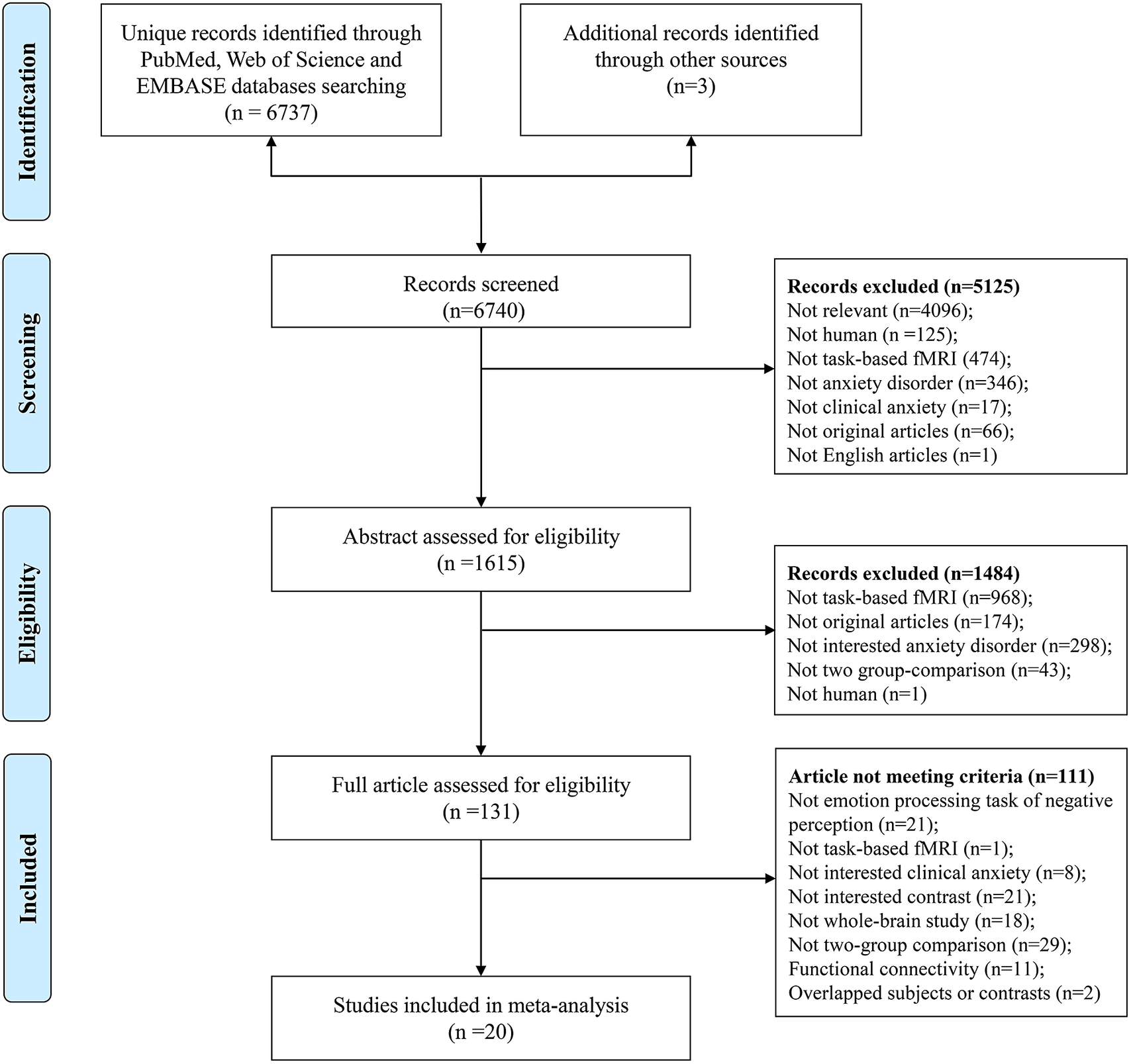
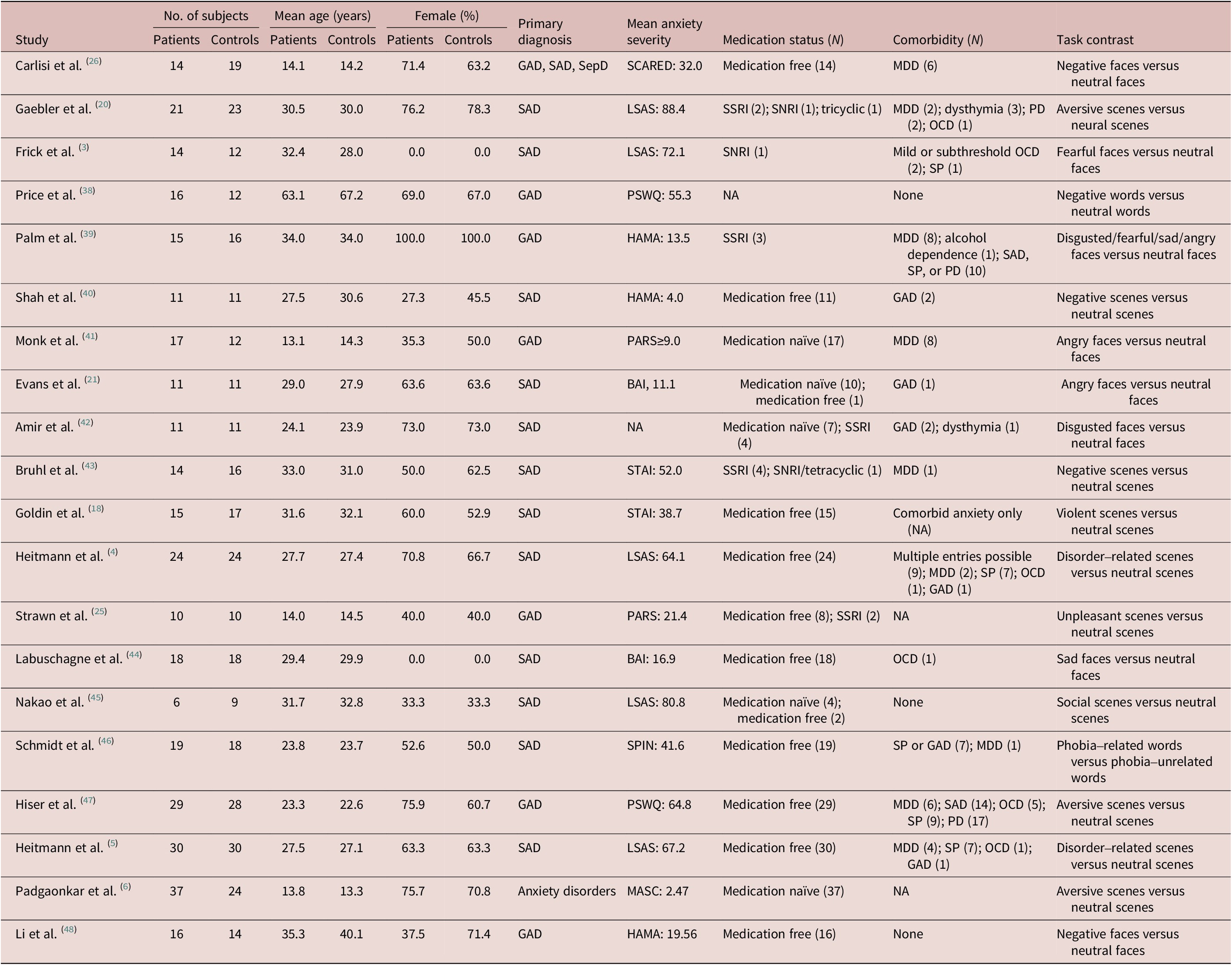
Our search generated 6740 unique records (6737 from the databases and 3 from reference lists of previous publications). Twenty studies employing negative versus neutral condition contrasts and using the same type of stimuli in negative and neutral conditions were included in the meta-analysis (Figure 1). The 20 included studies evaluated 348 individuals with anxiety disorders and 335 controls and primarily evaluated adults (
 $ \kappa =15,75\% $
). Twelve studies focused on SAD; 6 evaluated patients with GAD; and the other 2 evaluated mixed samples of patients with SAD, GAD, and separation anxiety disorder. Of note, 10 studies used negative/neutral scenes as stimuli, 8 used negative/neutral faces as stimuli, and the remaining 2 used negative/neutral words as stimuli. Demographic and clinical characteristics of participants are summarized in Table 1.
Preferred reporting items for systematic reviews and meta-analyses flowchart of literature search and selection criteria.

Summary of the Characteristics of Studies Included in the Meta-Analysis

Abbreviations: BAI, Beck Anxiety Inventory; GAD, generalized anxiety disorder; HAMA, Hamilton Anxiety Rating Scale; LSAS, Liebowitz Social Anxiety Scale; MASC, Multidimensional Anxiety Scale for Children; MDD, major depressive disorder; OCD, obsessive–compulsive disorder; PARS, Pediatric Anxiety Rating Scale; PD, panic disorder; PSWQ, Penn State Worry Questionnaire; SAD, social anxiety disorder; SCARED, Self-Report for Childhood Anxiety Related Disorders; SepD, separation anxiety disorder; SNRI, selective noradrenalin reuptake inhibitor; SPIN, Social Phobia Inventory; SP, specific phobia; SSRI, selective serotonin reuptake inhibitor; STAI, State-Trait Anxiety Inventory.
Generalized activation alterations during negative emotion processing across the scene- and face-stimulus tasks in patients with anxiety disorders
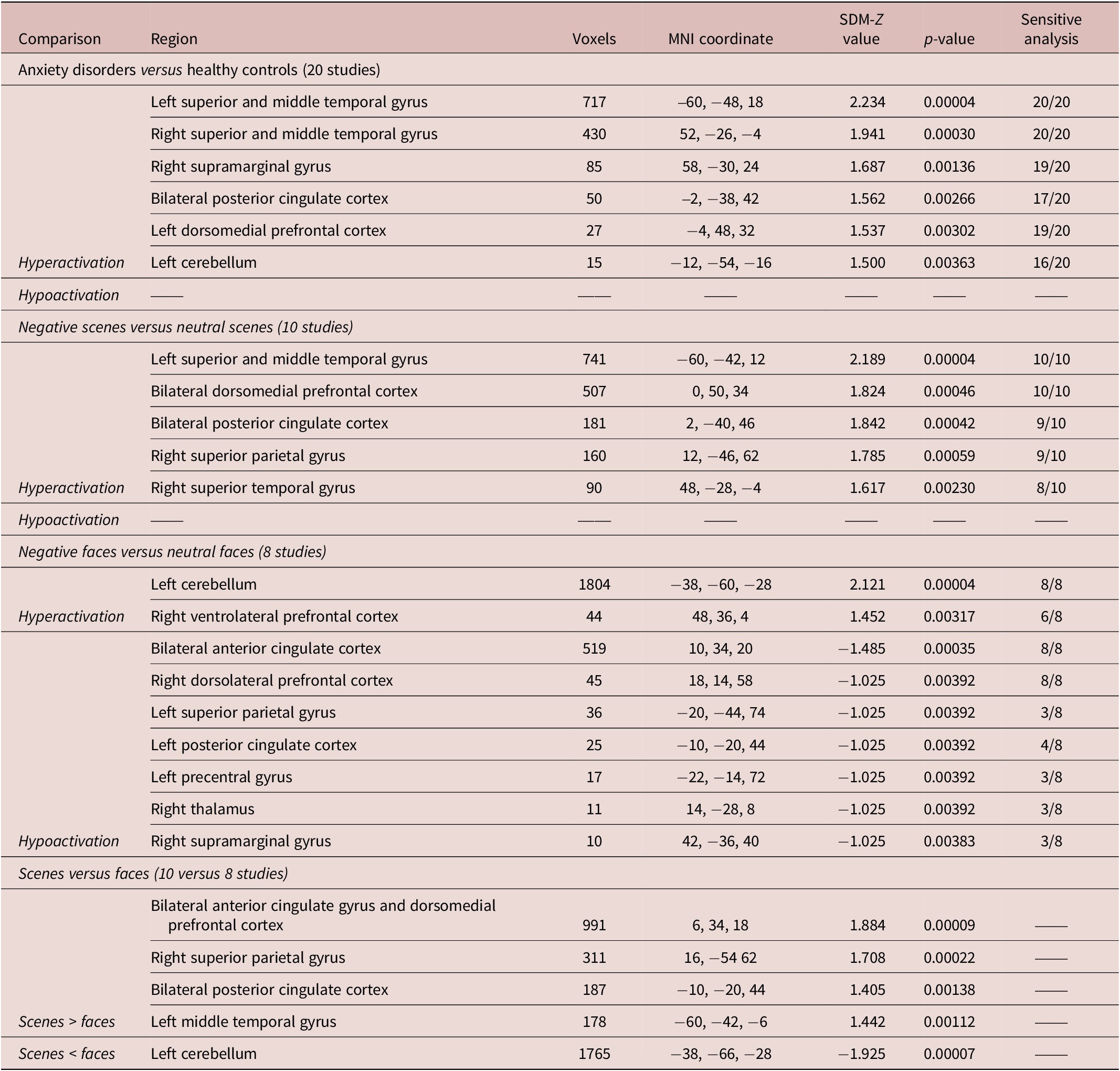
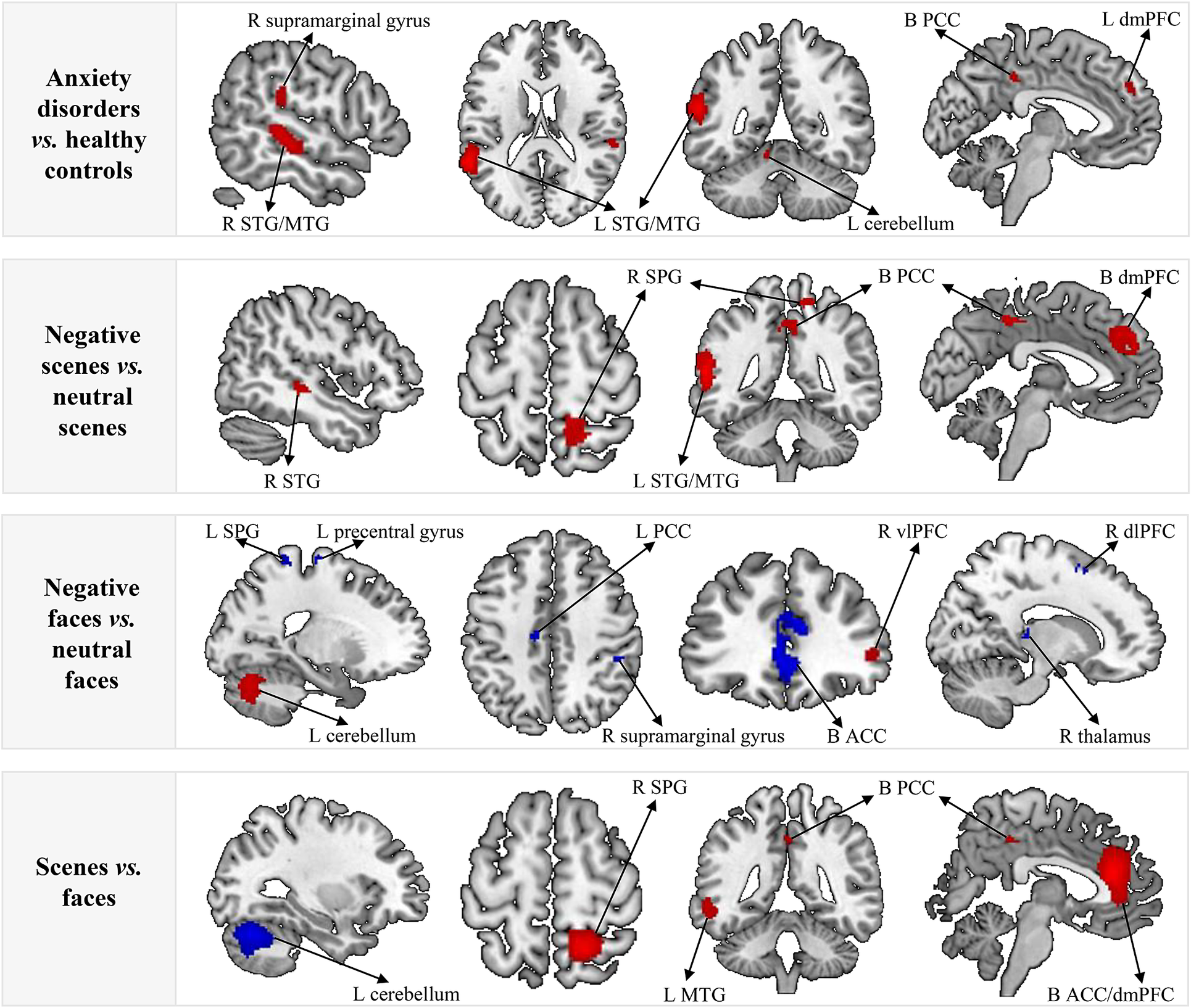
Taking studies with contrasts of negative scenes versus neutral scenes and negative faces versus neutral faces together, compared with controls, patients with anxiety disorders had significantly generalized greater activation during negative emotion processing in the bilateral STG extending to the middle temporal gyrus (MTG), right supramarginal gyrus, bilateral posterior cingulate cortex (PCC), left dmPFC, and left cerebellum (Table 2 and Figure 2). No regions with decreased activation during negative emotion processing were observed in patients with anxiety disorders compared to controls. The subgroup analyses of patients with SAD and adults with anxiety disorders revealed similar results (Supplementary Table S1 and Supplementary Figure S1). When using SDM-PSI for validation, no significant clusters survived correction for multiple comparisons.
Brain Activations during Negative Emotion Processing in Patients with Anxiety Disorders Compared to Controls

Abbreviations: MNI, Montreal Neurological Institute; SDM, seed-based d mapping.
Activations during negative emotion processing in patients with anxiety disorders compared to controls. The red clusters indicate increased activation (hyperactivation); the blue clusters indicate decreased activation (hypoactivation). Abbreviations: ACC, anterior cingulate cortex; B, bilateral; dlPFC, dorsolateral prefrontal cortex; dmPFC, dorsomedial prefrontal cortex; L, left; MTG, middle temporal gyrus; PCC, posterior cingulate cortex; R, right; SPG, superior parietal gyrus; STG, superior temporal gyrus; vlPFC, ventrolateral prefrontal cortex.

Differed activation alterations during negative emotion processing between scene-stimulus tasks and face-stimulus tasks in patients with anxiety disorders
In studies comparing negative scenes to neutral scenes, patients with anxiety disorders had significantly increased activation in the left STG extending to the MTG, bilateral dmPFC, bilateral PCC, right superior parietal gyrus (SPG), and right STG relative to controls. In studies comparing negative faces to neutral faces, activation was increased in the left cerebellum and right ventrolateral PFC and decreased in the bilateral ACC, right dorsolateral PFC, left SPG, left PCC, left precentral gyrus, right thalamus, and right supramarginal gyrus (Table 2 and Figure 2).
In the comparison of face-stimulus studies and scene-stimulus studies, the latter produced greater activation in bilateral ACC extending to dmPFC, right SPG, bilateral PCC, and left MTG, as well as decreased activation in the left cerebellum in patients with anxiety disorders (Table 2 and Figure 2).
Meta-regression
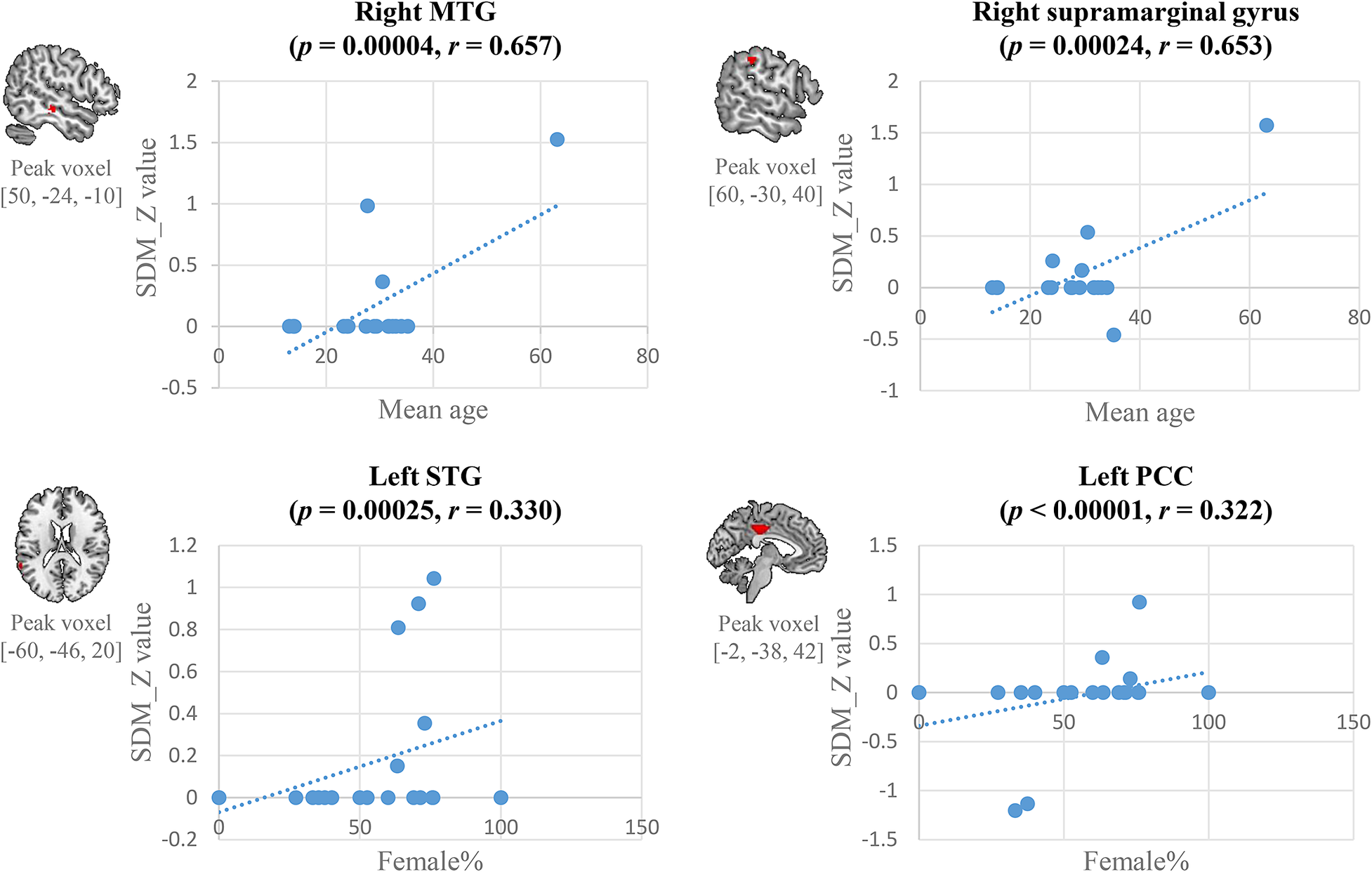
In meta-regression analyses, older patients with anxiety disorders had greater activation in the right MTG (r = 0.657, p < 0.001) and supramarginal gyrus (r = 0.653, p < 0.001). Additionally, studies with more female patients had greater activation in left STG (r = 0.330, p < 0.001) and PCC (r = 0.322, p < 0.001) (Figure 3).
The results of meta-regression analysis. Abbreviations: MTG, middle temporal gyrus; PCC, posterior cingulate cortex; STG, superior temporal gyrus.

Jackknife sensitivity, heterogeneity, and publication bias analysis
For the results of the main analysis, sensitivity analyses revealed that the clusters in bilateral STG extending to MTG were highly replicable, with all 20 combinations of studies preserved. The right supramarginal gyrus and left dmPFC remained significant in all but one combination, while the remaining clusters in bilateral PCC and cerebellum remained significant in all but 3 or 4 combinations. Most results in subgroup analyses were robust enough to survive more than 80% of iterations under jackknife sensitivity analyses (Supplementary Tables S2–S6).
Only bilateral PCC and the left cerebellum among significant clusters identified in our primary analysis showed considerable heterogeneity between studies. Egger’s tests were nonsignificant, and funnel plots (Supplementary Figure S2) suggested a low likelihood of publication bias.
Discussion
Our meta-analysis is unique in that it focuses on task-based fMRI studies employing the same stimulus type in both negative and neutral emotion stimuli and direct contrasts of negative and neutral stimuli. This approach minimizes confounding effects arising from experimental paradigms, a significant limitation of prior meta-analyses. It reveals distinct patterns of brain activation during negative emotion processing in patients with anxiety disorders. Herein, we found that, compared with controls, patients with anxiety disorders had increased activation in the STG, MTG, supramarginal gyrus, PCC, dmPFC, and cerebellum. These findings were robust in subgroup analyses, including studies with patients with a primary diagnosis of SAD only, adults with anxiety disorders only, and negative and neutral scenes as stimuli only. Our meta-analysis excluded studies with unmatched contrast and stimulus types, such as using resting-state fMRI or positive emotion as the comparator for stimuli with a negative emotional valence, thus improving the utility of study findings for understanding the alterations in brain processing of negative emotion in patients with anxiety disorders.
The dmPFC, PCC, STG, and MTG belong to the default mode network (DMN),Reference Koster, Crombez, Verschuere, Van Damme and Wiersema49 which subserves self-referential processing (eg, “Is this relevant to me?” or “Can I handle this?”) predominantly negative self-referential processing (eg, “I am ugly” and “you are boring to me”).Reference Ford, Grasso, Elhai, Courtois, Ford, Grasso, Elhai and Courtois50, Reference Moran, Heatherton and Kelley51 Accumulating evidence indicates that self-referential processes involve increased DMN activation, especially during exposure to negative stimuli in patients with anxiety disorders.Reference Blair, Geraci and Otero52, Reference Yoon, Seo, Kim and Choo53 DMN hyperactivation in patients with anxiety disorders suggests accentuated self-referential thinking in the presence of negative stimuli, which might relate to negative interpretation biases (ie, interpreting negative, neutral, and other emotionally ambiguous stimuli more negative)—a hallmark of these disorders.Reference Chen, Short and Kemps54 SPG and supramarginal gyrus, important frontal–parietal network (FPN) regions, are associated with emotion and cognitive regulation. Increased FPN activation may suggest that patients with anxiety disorders needed more regulation and involvement to handle the negative stimuli compared with healthy controls. In addition to the increased DMN and FPN activation, we found here, prior studies reveal that patients with anxiety disorders have decreased functional connectivity between DMN and FPN during restReference Xu, Van Dam and Feng55 and emotion processing.Reference Strawn, Bitter and Weber25 Increased FPN activation might be a compensatory alteration of the impaired FPN–DMN connectivity that facilitates regulation of a patient’s response to emotional stimuli.
In this meta-analysis, negative scenes and faces evoked distinct brain networks, consistent with findings from healthy individuals.Reference Sabatinelli, Fortune and Li24 In contrasts of negative scenes and negative faces, we observed greater robustness of case–control differences during processing negative scenes compared to negative faces. Negative scenes evoked greater activation in ACC, dmPFC, PCC, SPG, and MTG (ie, DMN and FPN) but lower activation in the cerebellum relative to the negative faces. There are several potential reasons for this difference that may relate to psychological aspects of anxiety disorders. First, negative scenes may induce greater emotional arousal than negative facesReference Britton, Taylor, Sudheimer and Liberzon23 and thus produce greater self-referential processing and increased DMN activation. Second, the increased complexity of negative scenes may require greater perception and regulation demands on the FPN,Reference Adhikari, Lerner and Finkelstein56–Reference Pico-Perez, Radua, Steward, Menchon and Soriano-Mas59 which may contribute to the greater activation we observed in SPG when processing negative scenes relative to negative faces. Third, when viewing negative scene stimuli, patients with anxiety disorders may allocate more top-down attentional resources for threat scanning,Reference Adhikari, Lerner and Finkelstein56, Reference Hariri, Tessitore, Mattay, Fera and Weinberger58 which might interpret why negative scenes induce greater activation in the FPN. The negative face-related cerebellar hyperactivation is consistent with the study by Reisch et al. in which cerebellar activation was observed when individuals viewed negative faces but not when they viewed negative scenes or words.Reference Reisch, Wegrzyn, Woermann, Bien and Kissler60 This direct condition comparison is in line with the evolving consensus that implicates the cerebellum in perceiving, encoding, and discriminating facial expressions,Reference Fusar-Poli, Placentino and Carletti16, Reference Adamaszek, D’Agata and Kirkby61–Reference Turner, Paradiso and Marvel63 especially negative emotions.Reference Adamaszek, D’Agata and Ferrucci64, Reference Northoff and Bermpohl65 Moreover, the face-specific increased activation in the cerebellum as well as the opposite activation pattern in PCC in the face- and scene-specific subgroup analyses also explained the heterogeneity we found in these two regions, indicating different neural responses of emotion processing between scenes and faces although they both belonged to the visual modality. Taken together, our results suggested that the type of stimuli influences the robustness of alterations in brain responses to negative emotions in patients with anxiety disorders. While it is difficult to compare the intensity and specific aspects of emotion elicited by the facial and scene stimuli used in prior studies, our findings suggest that scene stimuli may be more useful for studying alterations in brain systems supporting emotional responses in patients with anxiety disorders, which could be important in selecting stimuli for genetic association studies or studies of treatment response.
The meta-regression analyses revealed that the mean age and proportion of female participants were positively correlated with the degree of brain activation. Specifically, studies with older patients had greater activation in the right MTG and supramarginal gyrus during negative emotion processing. In contrast, studies with more women had more activation in the left STG and PCC. This age effect on brain activation in response to negative emotions, together with a prior study, showed that older patients with GAD had stronger resting-state functional connectivity within DMN,Reference Andreescu, Sheu, Tudorascu, Walker and Aizenstein66 which raises the possibility that anxiety disorders might exacerbate the effect of age on brain functional activation and connectivity especially within the DMN. The relationship between STG activation and the proportion of female patients replicates an earlier study of healthy adults suggesting that unpleasant stimuli induce greater STG activation in women compared to men,Reference Hofer, Siedentopf and Ischebeck67 which could relate to the clinical observations of more severe or persistent symptoms in women.Reference Hallers-Haalboom, Maas, Kunst and Bekker68
Compared to previous meta-analysesReference Bruhl, Delsignore, Komossa and Weidt10–Reference Yu, Ruan and Zhang15 that allowed for significant contrasts and stimulus heterogeneity, we did not observe differences in brain activation while processing negative emotion in the amygdala, insula, fusiform gyrus, or occipital cortex. The fusiform gyrus contributes to face perception,Reference Haxby, Hoffman and Gobbini69–Reference Puce, Allison, Asgari, Gore and McCarthy71 and its activation in previous meta-analyses could thus in part be related to the inclusion of negative faces to resting-state conditions or geometric figures rather than neutral faces. Studies comparing complex negative scenes with simple perceptual conditions or rest may produce greater occipital activation related to visual perception. Unexpectedly, our analysis did not show increased activation in the amygdala and insula, the key regions of the limbic network that are associated with emotional response, in anxiety disorders. Instead, our findings suggest that alterations in neocortical areas that provide self-referential and regulatory control of these emotional response regions may be more impaired than regions that are more primary to emotional responses. Several factors may account for the lack of group differences in amygdala and insula activation. First, the initial emotional responses to task stimuli may be robust in both patients and controls, which could limit the ability to detect differences in these deeper limbic regions. Consistent with this speculation, Dixon et al. found that patients with SAD and healthy controls had similar brain activation during emotion processing and did not observe group differences.Reference Dixon, Moodie and Goldin72 Parametric studies regulating the degree of emotionality of task stimuli might help address these issues. Second, the small sample sizes, inadequate correction for multiple comparisons, and region of interest analyses focused on the amygdala in previous studies may artificially boost the amygdala’s significance of this structure’s role in the pathoetiology of anxiety disorders. Consistent with this possibility, a recent meta-analysis of brain activation during emotion processing and mega-analysis examining structural alterations in anxiety disorders at the whole-brain level failed to confirm a significant difference in the amygdala.Reference Weber-Goericke and Muehlhan14, Reference Harrewijn, Cardinale and Groenewold73 Last, emotion processing is associated with distributed and interconnected brain regions, so it might be the functional connectivity between regions of emotional response—amygdala and insula—and regions of emotion regulation but not the activation in regions of emotional response per se that is impaired in anxiety disorders.
While this is the first meta-analysis of anxiety disorders to focus on studies with homogeneous task-control comparisons, there are several limitations. First, like all voxel-wise meta-analytic approaches, we used coordinates reported in original studies rather than raw t-statistic maps or individual image data, which limits the precision of activation effects and group differences.Reference Radua, van den Heuvel, Surguladze and Mataix-Cols31, Reference Radua and Mataix-Cols32 Meta-analysis with subject-level behavior and imaging data during emotion processing may provide more granular results for this field. Second, most of the included studies recruited patients with anxiety disorders and comorbid conditions (eg, major depressive disorder). While this increases the real-world applicability, it may decrease the specificity of findings. The influence of comorbidity on brain activation during negative emotion processing is unclear.Reference Beesdo, Lau and Guyer74 Third, most patients in the included studies were currently or previously treated with medication, which may alter activation profiles in response to emotion tasks. Studies of untreated patients are needed to define such possible effects. Fourth, although the leave-one-out jackknife sensitivity analysis was performed to test the robustness and replicability of findings from meta-analyses, it might fail to overcome bias from a small set of highly influential studies since it only removed one study each time and repeated the analysis. Last, unfortunately, our findings did not survive the Family Wise Error–Threshold-Free Cluster Enhancement (FWE-TFCE) correction of SDM-PSI, and we believed two factors might have caused this discrepancy. First, AES-SDM has been reported to be more voxel-based sensitive than SDM-PSI when only peak coordinates are available. Second, SDM-PSI is developed with a deep focus on the control of FWE correction, which may be associated with too conservative p values.Reference Albajes-Eizagirre, Solanes, Vieta and Radua33 Future meta-analysis, including more high-quality studies and having higher statistical power, is needed for validation.
Conclusion
We examined brain activation to negative emotions in patients with anxiety disorders using task-based fMRI studies contrasting responses to negative and neutral stimuli that employed similar, affectively neutral control stimuli. Patients with anxiety disorders (compared to healthy individuals) had increased activation in core regions of DMN and FPN in response to negative relative to neutral emotions. Further, the processing of affective scenes (compared to faces) produced differential and greater regional activation. Our findings underscore the importance of self-reference- and cognitive regulation-related functional disturbances during negative emotion processing in anxiety disorders. Taken together, these results suggest that more robust effects can be observed by using emotional scenes compared to emotional faces, and a lack of consistent activation in emotional response-related limbic regions, including amygdala and insula. Finally, our findings underscore the need to develop a better understanding of neurofunctional biomarkers using more standardized paradigms and the challenge when comparing results between studies that have used differing approaches.Reference Baumel, Lu and Huang75
Supplementary material
The supplementary material for this article can be found at http://doi.org/10.1017/S1092852924000166.
Author contribution
Data curation: R.W., L.L.; Formal analysis: R.W., H.X.; Methodology: R.W., H.Q.; Resources: R.W., J.H., L.L.; Software: R.W., Y.G.; Writing – original draft: R.W., L.L.; Supervision: J.H., L.L., J.R.S.; Conceptualization: L.L., J.A.S.; Funding acquisition: L.L., Q.G.; Writing – review & editing: L.L., J.R.S., J.A.S., Q.G.; Investigation: H.X.; Validation: H.X.; Project administration: Q.G.
Financial support
This work was supported by the National Natural Science Foundation of China (LL, grant number 82202116), the Natural Science Foundation of Sichuan Province (LL, grant number 24NSFSC7937), the National Key R&D Program of China (QG, grant number 2022YFC2009900), and the National Institute of Child Health and Development (JRS, grant number R01 HD098757).
Disclosure
Dr. Sweeney consulted with VeraSci. Dr. Strawn has received research support from AbbVie, PCORI, and the National Institutes of Health. He provided consultation to Otsuka, Intracellular Therapeutics, and Cerevel. He received royalties from Springer Publishing, Cambridge University Press, and UpToDate, as well as material support from Myriad. He has also received honoraria from the American Academy of Pediatrics, the American Academy of Child & Adolescent Psychiatry, and Medscape. The remaining authors have nothing to disclose.