Introduction
Oxyuris equi is a common inhabitant of the lower colon, or occasionally the large colon, of horses. Urquhart et al. (Reference Urquhart, Armour, Duncan, Dunn and Jennings1998) and Bowman et al. (Reference Bowman, Lynn, Eberhard and Alacaraz2006) considered that most pathogenic effects of this parasite in the intestine are caused by the feeding habits of third- and fourth-stage infective larvae, resulting in small erosions in the mucosa. However, a more important effect is the irritation caused by adult females during oviposition. Bowman et al. (Reference Bowman, Lynn, Eberhard and Alacaraz2006) also stated that pregnant female parasites, instead of eliminating their eggs in the faecal bulk, migrate to the lower part of the colon and rectum and out of the anus to adhere egg masses (8000–60,000 eggs) in the anal skin and the perianal region, causing anal itching.
The commercial raising of wild animals in countries with large regional differences has been recognized as an important protein source for the subsistence of poor populations. There are, however, some important barriers to the commercial production of these animals, particularly gastrointestinal nematodiosis (Bonuti et al., Reference Bonuti, Nascimento, Mapeli and Arantes2002). Austroxyuris finlaysoni, of the family Oxyuridae, is a gastrointestinal nematode parasite of Petauroides volans (Petauridae, Marsupiala). Marsupials are primitive mammals found only in the Americas and Australia. Gomes et al. (Reference Gomes, Cruz and Vicente2003) reported that some marsupials, for instance the white-eared opossum (Didelphis albiventris), have great medical–veterinary importance as a reservoir for gastrointestinal nematode parasites and a definitive host of potentially zoonotic intracellular parasites with a wide distribution in neo-tropical regions. Studies on marsupial endoparasites are scarce and generally aimed at helminth systematics (Hugot & Bougnoux, Reference Hugot and Bougnoux1988).
Alternative treatments may help to reduce the continued use of the same class of antihelminths, as well as doses above the recommended dose. Biological control carried out with natural nematode antagonists, such as nematophagous fungi, is among these alternatives. These fungi are biologically very important in the environment, playing a role in recycling carbon, nitrogen and other elements derived from nematode degradation (Araújo et al., Reference Araújo, Mota and Campos2004).
These organisms comprise various types of fungi characterized as predators, endoparasites and opportunists; their action is concentrated in the faecal environment and aimed against free-living parasite larvae. Predatory fungi and egg parasites are certainly the most studied groups and show the greatest biological control potential (Nordbring-Hertz et al., Reference Nordbring-Hertz, Jansson and Tunlid2002). In the predatory group, Duddingtonia flagrans and Monacrosporium thaumasium are promising species in the control of gastrointestinal nematode parasites of domestic animals. Pochonia chlamydosporia, in the ovicidal group, stands out as an important biological control agent of parasite eggs (Braga et al., Reference Braga, Araújo, Campos, Carvalho, Silva and Tavela2008; Araújo et al., Reference Araújo, Araújo, Braga, Carvalho, Silva and Campos2009.
This study evaluated the in vitro effects of the nematophagous fungi D. flagrans, M. thaumasium and P. chlamydosporia on eggs of O. equi and A. finlaysoni in two experimental assays (A and B) at intervals of 5, 10 and 15 days of interaction.
Material and methods
Four nematophagous fungal isolates, AC001 from D. flagrans, NF34a from M. thaumasium and VC1 and VC4 from P. chlamydosporia, were kept in test tubes containing 2% corn-meal-agar (2% CMA), at 4°C in the dark for 10 days. The isolates were stored previously at the Laboratory of Parasitology in the Department of Veterinary Medicine, Federal University of Viçosa, Minas Gerais, Brazil.
Culture discs, 4 mm in diameter, were extracted from the fungal cultures and transferred to 9.0-cm diameter Petri dishes containing 20 ml of 2% potato-dextrose-agar (2% PDA), at 26°C, in the dark for 10 days. After fungal growth, new 4-mm culture discs were transferred to 9.0-cm diameter Petri dishes containing 20 ml of 2% water-agar (2% WA) and kept at 26°C in the dark for 10 days.
Eggs of O. equi were recovered by dissection of adult female specimens from faeces of an infected horse and identified according to Urquhart et al. (Reference Urquhart, Armour, Duncan, Dunn and Jennings1998). Eggs of A. finlaysoni were recovered by dissection of adult specimens obtained from the autopsy of a white-eared opossum (D. albiventris) that had died of natural causes. Adult parasites were identified according to Hugot & Bougnoux (Reference Hugot and Bougnoux1988).
The work was divided into two experimental tests carried out at different stages over 15 days. The action of fungal isolates D. flagrans, M. thaumasium and P. chlamydosporia against O. equi and A. finlaysoni eggs at 5, 10 and 15 days was studied according to Araújo et al. (Reference Araújo, Braga, Araújo, Silva and Tavela2008), in Petri dishes (9-cm diameter) containing only 2% WA medium, with ten repetitions per group. Each Petri dish contained 1000 eggs with one fungal isolate. The control group contained 1000 eggs without fungi. At 5, 10 and 15 days post-inoculation, 100 eggs were removed from each plate containing the fungal isolates and from the control (without fungi) as described by Araújo et al. (Reference Araújo, Santos and Ferraz1995). The eggs were placed on glass slides with a drop of 1% Amam blue and evaluated under light microscopy (40 × objective lens) according to Lysek et al. (Reference Lysek, Fassatiová, Pineda and Hernández1982), as follows. Type 1: physiological and biochemical effect with no morphological damage to the eggshell and with hyphal adherence; type 2: lytic effect with morphological changes in eggshells and embryos, without hyphal penetration; and type 3: lytic effect with morphological change in eggshells and embryos, with hyphal penetration and internal egg colonization.
Data were examined by the nonparametric Friedman test at the 1% probability level (Ayres et al., Reference Ayres, Ayres, Ayres and Santos2003).
Results and discussion
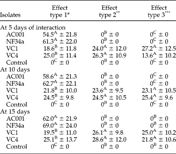
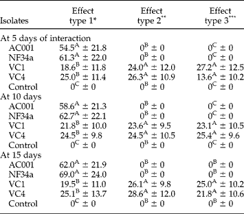
Table 1 shows the percentage results of the fungal effect on O. equi eggs at 5, 10 and 15 interaction days. The fungal isolate D. flagrans AC001 showed 54.5%, 58.6% and 62.0% of type 1 effect on O. equi eggs at 5, 10 and 15 days, respectively. Type 2 and 3 effects were not detected for this isolate in the studied periods; AC001 hyphae were only found adhered to O. equi eggshells, characterizing a type 1 effect. Isolate M. thaumasium (NF34a) had percentage results similar to AC001; 61.3%, 62.7% and 69.0% of only type 1 effect at 5, 10 and 15 days, respectively.
Percentages and standard deviations of the ovicidal activity of nematophagous fungi Duddingtonia flagrans (AC001), Monacrosporium thaumasium (NF34a) and Pochonia chlamydosporia (VC1 and VC4), and the control group without fungi, on eggs of Oxyuris equi at 5, 10 and 15 days of interaction.
Percentages followed by same letter in the same column are not significantly different (P>0.01) by Friedman test.
* Physiological, biochemical effect without morphological damage to eggshell, with hyphae adhered to the shell.
** Lytic effect with morphological alteration of embryo and eggshell, without hyphal penetration through the eggshell.
*** Lytic effect with morphological alteration of embryo and eggshell, besides hyphal penetration and internal colonization.
No significant difference was found (P>0.01) for type 1 effect when comparing the effect of isolates AC001 and NF34a on O. equi eggs. Isolates VC1 and VC4 of P. chlamydosporia showed type 3 effect on O. equi eggs in the three studied intervals. In this experiment, VC1 showed percentage results for this effect of 27.2%, 23.1% and 25.0% at 5, 10 and 15 days, respectively. Similarly, VC4 showed percentages of 13.6%, 25.4% and 21.8% for type 3 effect in the same intervals.
VC1 also showed results for type 1 and 2 effects, with percentages of 18.6%, 21.8% and 19.5% at 5, 10 and 15 days for type 1 effect, respectively; and 24.0%; 23.6% and 26.1% for type 2 effect. VC4 presented percentage results for type 1 effect of 25.0%, 24.5% and 25.1% and for type 2 effect of 26.3%, 24.5% and 28.6% at 5, 10 and 15 days, respectively. No differences (P>0.01) were found between VC1 and VC4 for type 1, 2 and 3 effects on O. equi eggs during the interaction days.
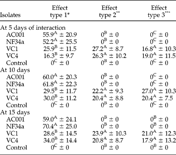
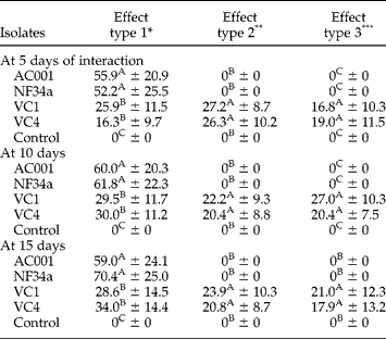
Table 2 shows the percentage results for the effect of different fungi on A. finlaysoni eggs at 5, 10 and 15 interaction days. Duddingtonia flagrans AC001 showed percentage results of 55.9%, 60.0% and 59.0% for type 1 effect on A. finlaysoni eggs at 5, 10 and 15 days, respectively, but no type 2 and 3 effects were recorded. Therefore, D. flagrans was not classified as ovicidal. M. thaumasium (NF34a) showed results similar to AC001; only a type 1 effect with percentage results of 52.2%, 61.8% and 70.4% at 5, 10 and 15 days, respectively; also showing no ovicidal activity.
Percentages and standard deviations of the ovicidal activity of nematophagous fungi Duddingtonia flagrans (AC001), Monacrosporium thaumasium (NF34a) and Pochonia chlamydosporia (VC1 and VC4), and the control group without fungi, on eggs of Austroxyuris finlaysoni at 5, 10 and 15 interaction days.
Percentages followed by same letter in the same column are not significantly different (P>0.01) by Friedman test.
* Physiological, biochemical effect without morphological damage to eggshell, with hyphae adhered to the shell.
** Lytic effect with morphological alteration of embryo and eggshell, without hyphal penetration through the eggshell.
*** Lytic effect with morphological alteration of embryo and eggshell, besides hyphal penetration and internal colonization.
Comparison of AC001 and NF34a for type 1 effect on A. finlaysoni eggs showed no significant difference (P>0.01); both had the same action.
The isolate VC1 showed type 1, 2 and 3 effects at 5, 10 and 15 interaction days, respectively, on A. finlaysoni eggs. The fungus had type 3 effects of 16.8%, 27.0% and 21.0% at 5, 10 and 15 days, respectively; type 1 effects of 25.9%, 29.5% and 28.6%; and type 2 effects of 27.2%, 22.2% and 23.9%. Isolate VC4 also showed type 3 effects of 19.0%, 20.4% and 17.9%; type 1 effects of 16.3%, 30.0% and 34.0%; and type 2 effects of 26.3%, 20.4% and 20.8% at 5, 10 and 15 days, respectively.
No differences (P>0.01) were found between VC1 and VC4 for type 1, 2 and 3 effects on A. finlaysoni eggs, demonstrating that there was no difference between their forms of interaction with A. finlaysoni eggs.
This work confirmed the ovicidal activity of P. chlamydosporia isolates (VC1 and VC4) on O. equi and A. finlaysoni eggs, as well as the capacity of interaction of predatory fungal isolates AC001 and NF34a with parasite eggs.
Biological control using nematophagous fungi, which is very important in the environment, is a promising strategy in the control of gastrointestinal nematode parasites in wild and domestic animals (Larsen, Reference Larsen1999; Sanyal et al., Reference Sanyal, Sarkar, Patel, Mandal and Pal2008). Nematophagous fungi, especially the genera Duddingtonia and Monacrosporium, have predatory capacity against L3 of gastrointestinal nematode parasites (Araújo et al., Reference Araújo, Mota and Campos2004, Reference Araújo, Braga, Araújo, Silva and Tavela2008). Ovicidal nematophagous fungi have been tested successfully as biological control agents of gastrointestinal nematode parasite eggs, mainly the genus Pochonia (Braga et al., Reference Braga, Araújo, Campos, Carvalho, Silva, Tavela and Maciel2007; Araújo et al., Reference Araújo, Braga, Araújo, Silva and Tavela2008). Although some studies have shown the action of the nematophagous fungi Duddingtonia, Monacrosporium and Pochonia in the biological control of gastrointestinal nematodiosis of domestic animals (Araújo et al., Reference Araújo, Braga, Araújo, Silva and Tavela2008), there are no reports demonstrating their action on eggs of O. equi and gastrointestinal nematodes of wild animals, such as A. finlaysoni.
The appressorium is the first mechanical structure formed by ovicidal fungi, allowing adhesion to the host surface and providing mechanical strength for egg penetration (Lysek et al., Reference Lysek, Fassatiová, Pineda and Hernández1982). According to Stirling & West (Reference Stirling and West1991), the direct effect of parasitism of these fungi on embryo development is an enzymatic action on the eggshell, increasing its permeability and favouring the passage of toxins. As a biological control agent, a fungus has to survive passage through the gastrointestinal tract of animals, such as that shown by the chlamydospore-producing species D. flagrans and P. chlamydosporia (Braga et al., Reference Braga, Araújo, Campos, Carvalho, Silva and Tavela2008).
Morgan-Jones et al. (Reference Morgan-Jones, White and Rodriguez-Kábana1983) suggested that the ideal procedure would be to feed animals with eggs, after prior contact with the fungus for 15 days, to verify whether or not eggs and embryos become unviable.
As suggested by Araújo et al. (Reference Araújo, Santos and Ferraz1995), the experimental tests were carried out with longer intervals of interaction between fungi and parasite eggs to observe a possible type 3 effect. After 15 days, nevertheless, hyphae and conidia of isolates AC001 and NF34a were found colonizing only the egg surface, characterizing a type 1 effect. Isolates VC1 and VC4 produced hyphae adhering to the surface and colonizing inside the eggs, and, later, egg rupture was also observed, characterizing a type 3 effect.
According to Lysek et al. (Reference Lysek, Fassatiová, Pineda and Hernández1982) the ovicidal condition of a fungus is characterized by the type 3 effect during egg infection; and, as pointed by Gams & Zare (Reference Gams and Zare2001), mechanical and enzymatic actions are involved in the process.
Differences in the interaction of P. chlamydosporia with eggs of some helminth genera have been attributed to peculiarities of these eggs. For instance, eggs of Fasciola hepatica and Schistosoma mansoni are large, with diameters between 150 μm and 190 μm, and very resistant (Burger & Stoye, Reference Burger, Stoye and Kelly1978). This could reduce the ovicidal activity of P. chlamydosporia (Braga et al., Reference Braga, Araújo, Campos, Carvalho, Silva and Tavela2008). Eggs of Ascaris lumbricoides and A. suum have a thick chitin–protein capsule (Silva & Massara, Reference Silva, Massara and Neves2005; Bowman et al., Reference Bowman, Lynn, Eberhard and Alacaraz2006). This is important, since P. chlamydosporia has enzymatic and mechanical action on eggs during the interaction, and eggs without a gross capsule would undergo less damage when in contact with the fungus.
The penetration mechanism of ovicidal fungi on parasitized eggs is not yet fully understood. Still, several authors agree that enzymatic activity is a key component in the attack and penetration of eggs by fungi (Lysek & Sterba, Reference Lysek and Sterba1991).
Results of the experiment showed that P. chlamydosporia (VC1 and VC4) negatively influenced eggs of O. equi and A. finlaysoni, and is considered to be a potential candidate for the biological control of nematodes.