Introduction
Weaning is a critical period in a piglet’s life and is accompanied by nutritional, social and environmental stress. At weaning, the diet is changed from a highly digestible and liquid diet (the milk) to a solid, more complex and less-digestible diet, and post-weaning gut disorders cause important economic losses in the pig industry. A transient anorexia as usually observed after weaning leads to gut dysfunction, increased sensitivity to enteric infections and diarrhoea. The most consistent patho-physiological changes effect the anatomy and function of the small intestine (Pluske et al., Reference Pluske, Hampson and Williams1997), and these include a 20–30% reduction in mucosal weight associated with villous atrophy (Lallès et al., Reference Lallès, Boudry, Favier, Le Floc’h, Luron, Montagne, Oswald, Pié, Piel and Sève2004). Further recent studies have shown that intestinal barrier function is compromised, resulting in increased secretion of electrolytes and water and increased permeability to potentially toxic substances (reviews by Vente-Spreeuwenberg and Beynen, Reference Vente-Spreeuwenberg and Beynen2003; Lallès et al., Reference Lallès, Boudry, Favier, Le Floc’h, Luron, Montagne, Oswald, Pié, Piel and Sève2004). In addition to alterations in gut physiology, weaning causes disturbances in the homeostasis of the gut microbiota and delays the anatomical and functional development of the mucosal immune system (review by Lallès et al., Reference Lallès, Bosi, Smidt and Stokes2007).
In-feed antibiotic growth promoters (AGP) have been long considered as acting essentially on the gut microbiota by decreasing the potentially harmful effects of pathogenic bacteria (review by Dibner and Richards, Reference Dibner and Richards2005). Such AGP were more effective in promoting growth in young animals such as in piglets after weaning, and when hygiene conditions are poor (review by Anderson et al., Reference Anderson, McKracken, Amirov, Sympson, Mackie, Verstegen and Gaskins1999). Complementary mechanisms of action centred on the gut microbiota have been described together with the consequences of supplemental AGP on improvements of gut homoeostasis, feed component digestibility, feed efficiency and growth performance. However, AGP may also act directly on the host and one recent hypothesis would suggest that AGP would display non-antibiotic anti-inflammatory properties (Niewold, Reference Niewold2007). This in turn would explain a lower protein catabolism related to a reduced level of inflammatory cytokines and, therefore, a better growth performance.
The use of in-feed AGP has been banned in the EU since 1 January 2006 and many attempts at alternative approaches have been applied to replace them. However, the success rate is low due to the complexity of the aetiology and pathogenesis of gastrointestinal tract (GIT) disorders on the one hand, and to the frequent lack of knowledge on the mechanisms of action and effective doses of alternative substances on the other hand. This has led to intensive research over Europe to increase our understanding on the mechanisms of action of known substances, used alone or in combination, and for exploring the possibilities offered by new substances. Such potential bioactive feed additives include nutrients (e.g. particular amino acids (AA)), mineral and organic acids (OA), minerals, growth factors, prebiotics and probiotics, and finally phytogenic compounds. All these substances have in common their potential for protecting the gut, albeit through very different ways, including mucosal growth, intestinal barrier function strengthening, anti-oxidant and anti-inflammatory capacities, and finally anti-bacterial properties.
The aim of this review is to gather recent findings on a limited number of categories of AGP alternatives, including OA (and sodium butyrate (SB)), specific AA (Gln, Trp, Arg, Cys, Thr), animal proteins (e.g. spray-dried plasma (SDP) and bovine colostrum) and plant extracts with known anti-bacterial activities in vitro. These substances have been studied more extensively within the frame of two EU projects (Healthypigut, 2000; Feed for Pig Health, 2003) and some of these substances offer a real alternative to AGP for protecting the gut and maintaining animal performance, whilst others display interesting protective properties in vitro or ex vivo but there are limited data in vivo and/or evidence of their protective efficacy.
Organic acids
The effects and modes of action of OA have been reviewed recently (Partanen, Reference Partanen2001; Roselli et al., Reference Roselli, Finamore, Britti, Bosi, Oswald and Mengheri2005; Bannink et al., Reference Bannink, Dijkstra, Koopmans and Mroz2006; Mroz et al., Reference Mroz, Koopmans, Bannink, Partanen, Krasucki, Øverland and Radcliffe2006). Some highlights from these reviews involve the in vivo trials with acidified diets in weaned piglets. From formic, fumaric and citric acids and potassium diformate, sufficient data could be gathered for meta-analysis (Partanen, Reference Partanen2001). From this analysis it was concluded that formic acid and potassium diformate were most potent in increasing feed consumption and average daily gain (ADG). Increased feed intake is thought to reflect improved health after weaning and is crucial for high growth. Another highlight from these reviews includes the bactericidal potency of OA. In a porcine intestinal organ culture model, it was established that coliform bacteria were most effectively killed by benzoic > fumaric > lactic > butyric > formic > propionic acid. With respect to Salmonella typhimurium the order was benzoic > sorbic > lactic > propionic > formic > acetic acid. Overall, benzoic acid is most effective, lactic acid is intermediate and both formic and propionic acids are least effective at killing both coliforms and S. typhimurium.
The stomach contents play an important role in limiting the entry of bacteria into the intestine. The inclusion of a combination of 1% lactic acid and 1% formic acid in the diet reduced gastric pH and concentration of lactic acid bacteria and enterobacteria (Hansen et al., Reference Hansen, Riis, Bresson, Højbjerg and Jensen2007). Lactic acid bacteria were also reduced by 0.5% benzoic acid (Guggenbuhl et al., Reference Guggenbuhl, Séon, Piñón Quintana and Simões Nunes2007) and by the addition of 200 mEq/kg of a 1 : 1 formic : fumaric acid mixture, but not by a 1 : 1 formic : lactic acid mixture (Franco et al., Reference Franco, Fondevila, Lobera and Castrillo2005). Benzoic acid at 0.5% or 1.0% reduced the number of total aerobic, total anaerobic, lactic acid forming and Gram-negative bacteria in the stomach (Kluge et al., Reference Kluge, Broz and Eder2006). In another study benzoic acid at 0.5% improved growth and feed efficiency, but had no effect on reducing the number of culturable Lactobacilli, Enterococci, Escherichia coli and Clostridium perfringens from the ileum and caecum as measured with traditional culture methods (Torrallardona et al., Reference Torrallardona, Badiola and Broz2007a). However, these authors did observe a marked increase in the biodiversity of the ileal microbiota, which suggests a more stable microbial ecosystem (Zoetendal et al., Reference Zoetendal, Collier, Koike, Mackie and Gaskins2004). Potassium diformate has also been shown to reduce Salmonella shedding in Salmonella-challenged piglets (Papenbrock et al., Reference Papenbrock, Stemme, Amtsberg, Verspohl and Kamphues2005). Finally, inclusion of an OA mixture with a lipid matrix was found to increase the concentration of OA in the distal GIT and reduced the number of coliforms in the caudal jejunum and in the caecum (Piva et al., Reference Piva, Pizzamiglio, Morlacchini, Tedeschi and Piva2007).
Very little attention has been paid to the impact of OA on the stomach. Gastric mucosa absorbs and transports volatile fatty acids (VFA) at substantial rates and OA are rapidly recovered from the blood after a single feed (Hanzlik et al., Reference Hanzlik, Fowler and Eells2005). Concentrations of OA decline rapidly between the stomach and the duodenum (e.g. formic acid; Canibe et al., Reference Canibe, Hojberg, Hojsgaard and Jensen2005), suggesting that pig proximal GIT is an important site of OA absorption. VFA are transported by the monocarboxylate transporters (MCT). The transporter MCT1 is most widely distributed in the GIT and has a specific role in lactate and butyrate transport. The presence of MCT1 has been detected by immuno-staining on the surface of adult mouse gastric surface epithelium (Nakai et al., Reference Nakai, Chen and Nowak2006). The distribution of MCT1 was studied in the small intestine and colon of pigs (Ritzhaupt et al., Reference Ritzhaupt, Ellis, Hosie and Shirazi-Beechey1998a and Reference Ritzhaupt, Wood, Ellis, Hosie and Shirazi-Beechey1998b; Sepponen et al., Reference Sepponen, Ruusunen, Pakkanen and Poso2007), and its presence was enhanced by the luminal production of butyrate after bacteria fermentation. The addition of different OA to the diet can differently affect the morphology and function of the stomach of weaned pigs. In the fundic mucosa, Ca-formate reduced the number of HCl-secreting parietal cells and H+/K+ ATPase gene expression (Bosi et al., Reference Bosi, Mazzoni, De Filippi, Casini, Trevisi, Petrosino and Lalatta-Costerbosa2006). Conversely, it increased numbers of cells secreting somatostatin, which is suppressive to HCl secretion. Fat-protected formic acid was also tested in piglet feeding, to explore the effect of a delayed release of this OA on gut barrier function. Interestingly, this product did not cause the gastric changes observed with free OA, confirming its direct impact on oxyntic mucosa (Bosi et al., Reference Bosi, Mazzoni, De Filippi, Casini, Trevisi, Petrosino and Lalatta-Costerbosa2006).
The effect of OA supplementation on gastric emptying and on the rate of digesta passage has not been studied in pigs. In humans, oral intake of propionate reduced the gastric emptying rate (Darwiche et al., Reference Darwiche, Ostman, Liljeberg, Kallinen, Bjorgell, Bjorck and Almer2001), and this could explain the increased weight of gastric content and dry matter (DM) percentage after supplementation with formic acid to early-weaned pigs (Manzanilla et al., Reference Manzanilla, Perez, Martin, Kamel, Baucells and Gasa2004). An increased stasis of feed in the stomach can permit a deeper action of acid secretion and this can be seen particularly favourable in a dirty environment. Conversely, it can also reduce feed intake. Addition of OA can have negative effect (Eisemann and van Heugten, Reference Eisemann and van Heugten2007), no effect (Partanen, Reference Partanen2001; Ettle et al., Reference Ettle, Mentschel and Roth2004) or favourable effect (Partanen, Reference Partanen2001) on feed intake in piglets. This could be related to the type or dose of OA used, the differences in taste and smell, or from differences in local sensing of OAs. More research attention should be placed on the role of stomach in the action of OA in the GIT of young pigs.
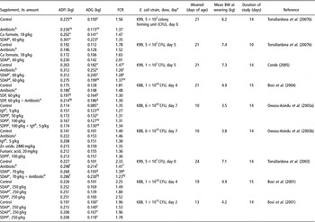
The dietary effect of OA has also been studied in weaning pigs orally infected with pathogenic Escherichia coli K88 (Table 1). Dietary supplementation with free Ca-formate, but not with fat-protected formate, improved post-challenge growth, feed intake, gain to feed ratio and reduced the faecal score, days of diarrhoea and faecal excretion of total (but not K88-specific) E. coli, and increased villous height in the small intestine (Bosi et al., Reference Bosi, Sarli, Casini, De Filippi, Trevisi, Mazzoni and Merialdi2007). The effects were seen independent of the individual piglet phenotypes for intestinal adhesion of E. coli K88. After a challenge with E. coli K99, Ca-formate improved neither growth performance nor total E. coli count in ileum, but jejunal villous height was reduced (Torrallardona et al., Reference Torrallardona, Conde, Badiola and Polo2007b). Earlier work with pathogenic challenges indicated that 2% fumaric acid tended to improve average daily gain (ADG) over the first 2 weeks post-weaning and to reduce scours, faecal shedding of E. coli K88 and mortality (Owusu-Asiedu et al., Reference Owusu-Asiedu, Nyachoti and Marquardt2003b). In a study with Lawsonia intracellularis challenge 2.4% lactic acid, but not 1.8% fumaric acid was able to reduce intestinal lesions induced by this pathogen (Boesen et al., Reference Boesen, Jensen, Schmidt, Jensen, Jensen and Møller2004).
Average daily feed intake (ADFI), weight gain (average daily gain (ADG)) and feed conversion ratio (FCR) obtained in different studies on weaning piglets experimentally challenged with different Escherichia coli strains when spray-dried plasma (SDP) or other substances were fed to the animals

aDay post-weaning at which the experimental infection was performed.
bColistin 300 mg/kg diet.
cIgG-standardised and pelleted spray-dried plasma from animal (bovine and porcine) origin.
dNon-standardised spray-dried plasma from animal (bovine and porcine) origin in powder form.
eColistin 250 mg/kg diet and amoxycycline 500 mg/kg diet.
fStatistically significant effect of antibiotic addition over control.
gStatistically significant effect of SDAP addition over control.
hEgg yolk antibodies from hens immunised against E. coli K88 and E. coli F18.
iSpray-dried plasma from porcine origin.
jCarbadox 55 mg/kg diet.
x,yDifferent superscripts mark significant difference between values in a column within one study.
The case of sodium butyrate
As a result of their well-known beneficial effects on colonic mucosa, butyrate and butyrate precursors have been the focus of special attention (review by Hamer et al., Reference Hamer, Jonkers, Venema, Vanhoutvin, Troost and Brummer2008). However, little is known of the effect of oral butyrate on the small intestine.
SB is more frequently used in pig feeding due to its less-strong odour than the acid. It is presumed that the effect inside the gut is not different, because SB dissociates. In experiments conducted by Manzanilla et al. (Reference Manzanilla, Nofrarias, Anguita, Castillo, Perez, Martin-Orue, Kamel and Gasa2006) and Castillo et al. (Reference Castillo, Martin-Orue, Roca, Manzanilla, Badiola, Perez and Gasa2006), SB was included in the starter diet at 3 g/kg and fed for 14 days to piglets weaned at 18–22 days of age. SB supplementation improved feed gain ratio in week 2 post-weaning and over the 14-day post-weaning period. SB was detected only in the stomach, suggesting quick gastric and (or) duodenal absorption or catabolism (Gálfi and Bakori, Reference Gálfi and Bakori1990; Manzanilla et al., Reference Manzanilla, Nofrarias, Anguita, Castillo, Perez, Martin-Orue, Kamel and Gasa2006). SB increased gastric DM percentage (Manzanilla et al., Reference Manzanilla, Nofrarias, Anguita, Castillo, Perez, Martin-Orue, Kamel and Gasa2006). SB supplementation also induced large changes in both the biodiversity of the microbial ecosystem and species composition of the bacterial community in the jejunum (Castillo et al., Reference Castillo, Martin-Orue, Roca, Manzanilla, Badiola, Perez and Gasa2006). Total microbial activity in digesta was lower in the caecum and distal colon when SB was present in the diet. Manzanilla et al. (Reference Manzanilla, Nofrarias, Anguita, Castillo, Perez, Martin-Orue, Kamel and Gasa2006) suggested that SB supplementation may have contributed to stabilise the gastrointestinal ecosystem while depressing amilolytic bacteria, thus improving the health status of the pigs and the efficiency of the use of nutrients for growth.
A range of doses of SB (0, 1, 2 and 4 g/kg) was tested for 6 weeks in another study of piglets weaned at 28 days (Biagi et al., Reference Biagi, Piva, Moschini, Vezzali and Roth2007). SB had effects on neither growth performance (despite a numerical improvement with the highest dose) nor intestinal mucosa morphology. The only reported changes were increased caecal pH and isobutyric acid concentration with a tendency for increased ammonia concentration. This suggested that oral SB stimulated protein catabolism in the caecum of pigs. However, the underlying mechanisms are unresolved thus far. The authors considered the bacterial hypothesis as unlikely because the counts of clostridia, enterobacteria and lactic acid bacteria along the intestines were unaffected by SB (Biagi et al., Reference Biagi, Piva, Moschini, Vezzali and Roth2007).
The period of oral SB administration has also recently been investigated by another group (Le Gall et al., Reference Le Gall, Sève, Sahar, Leborgne, Lallès and Guilloteau2007). SB (0.3 g/kg of milk or feed intake) was provided to piglets either during the suckling period (day 4 to day 28) or after weaning (day 28 to day 39–40) or both before and after weaning (day 4 to day 39–40). SB stimulated growth rate before weaning and over the entire period of observation, whilst feed intake after weaning was enhanced when SB was fed during the suckling period. Supplementing SB before and (or) after weaning increased the number of HCl-secreting parietal cells per gland observed at 7-day post-weaning (Mazzoni et al., Reference Mazzoni, Le Gall, De Filippi, Minieri, Trevisi, Wolinski, Lalatta-Costerbosa, Lallès, Guilloteau and Bosi2008). This effect was seen notwithstanding the parallel increase of somatostatin-secreting cells. The weights of the whole tissue and mucosa of the small intestine were reduced with SB feeding, with minor changes in villous-crypt architecture and digestive enzyme-specific activities (Le Gall et al., Reference Le Gall, Sève, Sahar, Leborgne, Lallès and Guilloteau2007). These changes were very similar to previous findings in a study with plasma protein (Jiang et al., Reference Jiang, Chang, Stoll, Fan, Arthington, Weaver, Campbell and Burrin2000b) that showed suppression of both the inflammatory response and AA catabolism in the intestine. Whether SB alters intestinal inflammation and AA metabolism is presently unknown.
The protective effects of butyrate are well documented (review by Hamer et al., Reference Hamer, Jonkers, Venema, Vanhoutvin, Troost and Brummer2008) and an alternative approach to delivering butyrate to the gut has been by providing the animal with butyric acid precursors, including tributyrin and lactitol (Piva et al., Reference Piva, Prandini, Fiorentini, Morlacchini, Galvano and Luchansky2002) or gluconic acid (Tsukahara et al., Reference Tsukahara, Koyama, Okada and Ushida2002; Biagi et al., Reference Biagi, Piva, Moschini, Vezzali and Roth2006a and Reference Biagi, Vezzali, Piva and Roth2006b). Supplementing the piglets with tributyrin or lactitol reduced mortality and this protective effect was enhanced when both were fed together (Piva et al., Reference Piva, Prandini, Fiorentini, Morlacchini, Galvano and Luchansky2002). The combined supplementation increased growth performance and jejunal villous height while reducing histamine levels in jejunal and caecal tissues and caecal crypt depth. Tributyrin alone had adverse effects on growth performance. It did not influence jejunal villus height but it reduced drastically caecal crypt depth. Lactitol alone stimulated jejunal villous height and the level of lactic acid in the caecum while reducing caecal crypt depth. Gluconic acid tended to improve growth rate without changes in intestinal morphology (Biagi et al., Reference Biagi, Piva, Moschini, Vezzali and Roth2006a) and to support longer ileal crypts and shorter caecal crypts (Biagi et al., Reference Biagi, Vezzali, Piva and Roth2006b).
Collectively, the results obtained with an oral supplementation of SB or with butyrogenic precursors indicate that the outcome may depend on various factors including dose, supplementation duration, site of observation, age of pigs at initiation of butyrate supplementation and probably other uncontrolled factors. As an example, an interaction between butyrate infused in the caecum of pigs and dietary inulin was recently demonstrated (Kien et al., Reference Kien, Blauwiekel, Bunn, Jetton, Frankel and Holst2007). Butyrate alone stimulated epithelial cell proliferation along the intestines but dietary inulin tended to block this effect. The precise modes of action of butyrate in the small intestine are not yet fully understood.
Specific amino acids
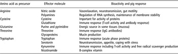
Apart from their role as building blocks for peptides and proteins, several essential and non-essential AA are thought to have therapeutic effects on the GIT and on the whole organism (review by Kim et al., Reference Kim, Mateo, Yin and Wu2007). Specific AA can promote health by improving (gut) tissue anabolism, by reducing the impact of stress and by modulating local immunology (Table 2). This could be beneficial when growth is inhibited by infection, inflammation and stress (Gruys et al., Reference Gruys, Toussaint, Landman, Tivapasi, Chamanza and Van Veen1998). Biologically active AA can be added to the diet in case of a deficiency during disease and experimental results suggest that the profile of AA required for the immune system differs substantially from that for growth (Reeds et al., Reference Reeds, Fjeld and Jahoor1994). In addition, surplus administration of certain dietary AA has been shown to be necessary for inducing significant bioactivity for therapeutic purposes (Massey et al., Reference Massey, Blakeslee and Pitkow1998). From the literature, five AA, namely glutamine, tryptophan, arginine, cysteine and threonine, showed consistent bioactivity with respect to physiology, immunology, endocrinology and metabolism.
Summary of the effector molecules and bioactivities of five bioactive amino acids (adapted from D’Mello, Reference D’Mello2003)

Glutamine (Wu et al., Reference Wu, Knabe, Yan and Flynn1995) and glutamate (Reeds et al., Reference Reeds, Burrin, Stoll and Jahoor2000) are important fuels for intestinal epithelial cells. Notwithstanding the stresses of weaning, these AA were able to maintain the high turnover of the gut mucosa, as indicated by longer villi in pigs supplemented with glutamine (Ayonrinde et al., Reference Ayonrinde, Williams, McCauley and Mullan1995; Liu et al., Reference Liu, Peng, Xiong, Zhou and Cheng2002; Lee et al., Reference Lee, Cheng, Wu, Sato, Shinzato, Cheng and Yen2003; Domeneghini et al., Reference Domeneghini, Di Giancamillo, Savoini, Paratte, Bontempo and Dell’Orto2004 and Reference Domeneghini, Di Giancamillo, Bosi and Arrighi2006) or glutamate (Liu et al., Reference Liu, Peng, Xiong, Zhou and Cheng2002). For glutamine, this was determined by a decrease in the apoptotic to mitotic mucosal cell ratio (Domeneghini et al., Reference Domeneghini, Di Giancamillo, Savoini, Paratte, Bontempo and Dell’Orto2004 and Reference Domeneghini, Di Giancamillo, Bosi and Arrighi2006). The positive influence of glutamine on intestinal architecture and growth performance in healthy swine persists in swine at risk of colibacillosis, as shown in E. coli K88-challenged piglets (Yi et al., Reference Yi, Carroll, Allee, Gaines, Kendall, Usry, Toride and Izuru2005). Interestingly, the small intestine utilises glutamine to synthesise citrulline and after rotavirus infection, blood plasma citrulline values were reduced, while glutamine increased (Rhoads et al., Reference Rhoads, Corl, Harrell, Niu, Gatlin, Phillips, Blikslager, Moeser, Wu and Odle2007). Thus utilisation of glutamine in the intestine was reduced for the compromised number of villous cells. Systemic glutamine may also influence the gut since its parenteral administration decreased intestinal permeability and endotoxin levels in burns patients (Zhou et al., Reference Zhou, Jiang, Sun, He and Hong2004).
Glutamine is rapidly utilised by cells of the immune system in culture and is required to support optimal lymphocyte proliferation and production of cytokines by lymphocytes and macrophages. Glutamine may stimulate the immune system in mucosal tissue (Calder and Yaqoob, Reference Calder and Yaqoob1999). For example, in infected, early-weaned pigs, supplemental dietary glutamine (+40 g/kg diet) restored lymphocyte function and intramuscular glutamine concentrations (Yoo et al., Reference Yoo, Field and McBurney1997). Locally, supplemental dietary glutamine (+5 g/kg diet) increased ileal mucosa densities of macrophages and intra-epithelial lymphocytes (Domeneghini et al., Reference Domeneghini, Di Giancamillo, Savoini, Paratte, Bontempo and Dell’Orto2004). Enteral glutamine supplementation (+45 g/kg diet) of rats with gut-derived sepsis enhanced peritoneal macrophage phagocytic activity (Yeh et al., Reference Yeh, Lai, Shang, Lin and Chen2004), preserved CD4+ T cells and maintained the pool of circulating T lymphocytes. In piglets, glutamine supplementation (+40 g/kg diet) of piglets stimulated cell proliferation to mitogens and Th-1-type cytokine response of T lymphocytes present in mesenteric lymph node cells (Johnson et al., Reference Johnson, Ball, Baracos and Field2006). Whilst it could be argued that such responses can be beneficial in the face of mucosal infection, it is not clear whether the immuno-stimulation by glutamine is direct, or indirect via the extra energy provided to the immune system (Bannink et al., Reference Bannink, Dijkstra, Koopmans and Mroz2006). Finally, glutamine supplementation reduced blood cortisol on the first day post weaning in pigs (Zhou et al., Reference Zhou, Peng, Liu and Fang2006).
Tryptophan is the most potent AA for stimulating cholecystokinin secretion and subsequent pancreatic enzyme production (Massey et al., Reference Massey, Blakeslee and Pitkow1998). Of the total body serotonin, 80% is located within the gut and dietary tryptophan may locally boost the serotoninergic system in the GIT and as such enhance peristaltic activity by smooth muscle and stimulate intestinal secretion by enterocytes (Spiller, Reference Spiller2001). The tryptophan-driven effect on gut serotonin may be (partly) responsible for the observed increases in digestion and/or absorption. In addition, supplemental dietary tryptophan (+5 g/kg diet) may increase gut functionality indirectly through its inhibitory effect on the peripheral sympathetic nervous system (Koopmans et al., Reference Koopmans, Ruis, Dekker, van Diepen, Korte and Mroz2005). Supplemental dietary tryptophan (+5 g/kg diet) has been shown to reduce villous atrophy in weaning piglets (Koopmans et al., Reference Koopmans, Guzik, Van der Meulen, Dekker, Kogut, Kerr and Southern2006), to control the inflammatory response and to preserve growth in weaned pigs submitted to immune stress. Adequate dietary tryptophan (20% tryptophan to lysine ratio) reduced plasma haptoglobin concentrations, while its catabolism increased under indoleamine-2,3-dioxygenase activity during chronic lung inflammation in pigs (Le Floc’h et al., Reference Le Floc’h, Melchior and Sève2004; Melchior et al., Reference Melchior, Mézière, Sève and Le Floc’h2004 and Reference Melchior, Mézière, Sève and Le Floc’h2005). This highlights that during inflammatory stress in the gut, the tryptophan requirement to maintain growth could be greater than during ‘health’. Indeed, supplementing a standard weaning diet with 100 mg/kg l-tryptophan improved daily gain and also stimulated feed intake in piglets susceptible to intestinal adhesion of E. coli K88 during the first 4 days post challenge with this pathogen strain (Trevisi et al., Reference Trevisi, Melchior, Mazzoni, Casini, De Filippi, Minieri, Lalatta-Costerbosa and Bosi2008). Tryptophan, via its conversion to serotonin, has been shown to reduce cortisol and noradrenaline concentrations in blood. In addition, surplus dietary tryptophan (+5 g/kg diet) enhanced the recovery from the ‘social stress’ of mixing piglets at weanling (Koopmans et al., Reference Koopmans, Ruis, Dekker, van Diepen, Korte and Mroz2005 and Reference Koopmans, Guzik, Van der Meulen, Dekker, Kogut, Kerr and Southern2006). The protective effect of tryptophan against stress may spare arginine and glutamine catabolism in enterocytes of post-weaning pigs since cortisol stimulates the degradation of arginine and glutamine (Flynn and Wu, Reference Flynn and Wu1997). Inhibition of the adreno-cortical and sympatho-adrenal axis activity by tryptophan and arginine during periods of stress (e.g. post weaning) may contribute to improved gut integrity, digestion and tissue anabolism.
Arginine has been used as a therapeutic agent for the treatment of necrotic enterocolitis, an inflammation of the gut induced by enterotoxins. Arginine-mediated production of nitric oxide (NO) causes smooth muscle relaxation, protects the gut from blood-borne toxins and tissue-destructive mediators (Di Lorenzo et al., Reference Di Lorenzo, Bass and Krantis1995; Di Lorenzo and Krantis, Reference Di Lorenzo and Krantis2002), and enhances wound healing (Shi et al., Reference Shi, Efron, Most, Tantry and Barbul2000). Supplementation of diets with ornithine, a metabolite of arginine, increased villous height and crypt depth in the jejunum and ileum of starved rats (Cynober, Reference Cynober1994). Supplemental dietary arginine (or glutamine) has also been reported to reduce bacterial translocation and to decrease atrophy of intestinal villi in rats with obstructive jaundice (Zulfikaroglu et al., Reference Zulfikaroglu, Zulfikaroglu, Ozmen, Ozalp, Berkem, Erdogan, Besler, Koc and Korkmaz2003). In weaned pigs challenged with lipopolysaccharide (LPS), arginine supplementation (at 5 and 10 g/kg feed) in weaned pigs partly protected from changes in villous–crypt architecture, in crypt cell proliferation and villous cell apoptosis induced by LPS (Liu et al., Reference Liu, Huang, Hou, Zhu, Zhao, Ding, Yin, Yi, Shi and Fan2008). Arginine supplementation to septic pigs restored the intestinal motility pattern and improved microcirculation and protein anabolism (Luiking et al., Reference Luiking, Poeze, Ramsay and Deutz2005). Arginine (264 mg of N/kg rat per day) improves histone and acute-phase protein synthesis during Gram-negative sepsis (Léon et al., Reference Léon, Redmond, Stein, Shou, Schluter, Kelly, Lanza-Jacoby and Daly1991), stimulates the bactericidal actions of macrophages (Massey et al., Reference Massey, Blakeslee and Pitkow1998) and restores depressed immunity (Cynober, Reference Cynober1994). Enteral arginine supplementation enhanced macrophage phagocytic activity and reduced total bacterial counts in the peritoneum of septic rats (Wang et al., Reference Wang, Shang, Lai and Yeh2003; Yeh et al., Reference Yeh, Lai, Shang, Lin and Chen2004).
Mucus is a semi-solid gel, which creates an important barrier for microorganisms and their toxins in the lumen of the gut. Cysteine and threonine are important building blocks for mucin; consequently, disease-induced stimulation in mucus secretion leads to extra requirement for cysteine and threonine (Lobley and Lapierre, Reference Lobley and Lapierre2003; Bannink et al., Reference Bannink, Dijkstra, Koopmans and Mroz2006). Similarly, diets that stimulate mucin production decrease pig threonine retention (Myrie et al., Reference Myrie, Bertolo, Sauer and Ball2003), indicating that in this case the AA requirement should be adjusted. However, an excess of threonine has a negative effect on intestinal protein synthesis (Wang et al., Reference Wang, Qiao, Yin, Yue, Wang and Wu2007), and this may be explained by its possible competitive inhibition on the absorption of other indispensable AA. Conversely, a threonine-deficient diet (6.5 v. 9.3 g/kg feed) increased intestinal para-cellular permeability and reduced villous height, villous height to crypt depth ratio and amino-peptidase N activity in the ileum of young piglets (Hamard et al., Reference Hamard, Mazurais, Boudry, Luron, Sève and Le Floc’h2007a and Reference Hamard, Sève and Le Floc’h2007b). Whilst these data highlight the functional role of threonine at the intestinal level, no studies have specifically assessed threonine requirements in piglets stressed with enterobacteria or reared in dirty environment. Therefore, in practice, the recommended values should be over the NRC (1998) requirement, but not increased up to 150% of this value (Wang et al., Reference Wang, Qiao, Yin, Yue, Wang and Wu2007). Finally, cysteine provided at 25% above the NRC for pigs decreased jejunal mass, possibly due to its mucolytic properties (Harte et al., Reference Harte, Shoveller, Bertolo and Ball2003).
Spray-dried animal plasma
SDP is an abattoir by-product obtained from animal blood after exclusion of cells, concentration and spray drying. Three types of SDP products, from porcine (SDPP), bovine (SDBP) and unknown or mixed animal origin (SDAP) are available commercially. Following the temporary ban in Europe as a consequence of the bovine spongiform encephalopathy crisis, feeding SDPP is now allowed for pigs. Hygienic collection and processing of the blood are essential, and irradiation or formaldehyde treatment of SDP improved growth only in poor-quality SDP containing a high number of bacteria (DeRouchey et al., Reference DeRouchey, Tokach, Nelssen, Goodband, Dritz, Woodworth, James, Webster and Hastad2004). The possibility that plasma may provide a source for the transmission of infectious diseases has also been considered (van Dijk et al., Reference Van Dijk, Everts, Nabuurs, Margry and Beynen2001), and spray drying process has been shown to be effective in inactivating pseudorabies and porcine respiratory and reproductive syndrome viruses (Polo et al., Reference Polo, Quigley, Russell, Campbell, Pujols and Lukert2005).
It is now well established that both porcine and bovine SDP incorporated into weaning diets stimulate growth performance and feed intake (Coffey and Cromwell, Reference Coffey and Cromwell2001; van Dijk et al., Reference Van Dijk, Everts, Nabuurs, Margry and Beynen2001), and that SDPP was more effective than SDBP (Pierce et al., Reference Pierce, Cromwell, Lindemann, Russell and Weaver2005). This observation could suggest some degree of specificity in the effect of IgG in SDPP against porcine pathogens. Pierce et al. (Reference Pierce, Cromwell, Lindemann, Russell and Weaver2005) showed that it was the immunoglobulin-rich fraction that was responsible for the beneficial effects of SDP. These effects are greater in piglets weaned at 22 days as compared to 32 days (Torrallardona et al., Reference Torrallardona, Conde, Esteve-Garcia and Brufau2002) and higher under poorer health conditions. This supports the view that the protective effect of SDP may be via the immune system or directly against pathogens (Coffey and Cromwell, Reference Coffey and Cromwell1995; Bergstrom et al., Reference Bergstrom, Nelssen, Tokach, Goodband, Dritz, Owen and Nessmith1997). Indeed, SDP lowered the incidence of diarrhoea in pigs challenged with pathogens (Owusu-Asiedu et al., Reference Owusu-Asiedu, Nyachoti, Baidoo, Marquardt and Yang2003a and Reference Owusu-Asiedu, Nyachoti and Marquardt2003b; Conde, Reference Conde2005) and improved body condition (Van Dijk et al., Reference Van Dijk, Enthoven, Van den Hoven, Van Laarhoven, Niewold, Nabuurs and Beynen2002) and performance (Table 1).
It is widely accepted that the beneficial effects of plasma are mediated by its immunoglobulin fraction and their inhibitory activities against pathogens and enterotoxins. Therefore, differences in IgG contents (both quantitative and qualitative) may influence the efficacy of different SDP sources. SDP with guaranteed high levels of immunoglobulins were shown to be superior to conventional plasma (Bosi et al., Reference Bosi, Han, Jung, Heo, Perini, Castellazzi, Casini, Creston and Gremokolini2001; Conde, Reference Conde2005). Furthermore, Bosi et al. (Reference Bosi, Han, Jung, Heo, Perini, Castellazzi, Casini, Creston and Gremokolini2001 and Reference Bosi, Casini, Finamore, Cremokolini, Merialdi, Trevisi, Nobili and Mengheri2004) observed that challenge of piglets with E. coli K88 resulted in a lower concentration of specific IgA anti-K88 in plasma and saliva if they were fed a plasma source rich in IgG, suggesting a protective effect against the adhesion of E. coli K88 to the enterocytes.
A lower basal activation of the immune system was observed in piglets fed SDP than in piglets without SDP (Touchette et al., Reference Touchette, Carroll, Allee, Matteri, Dyer, Beausang and Zannelli2002). This is in line with the reduced production of pro-inflammatory cytokines (tumour necrosis factor-α, interleukin-8 and interferon-γ) in the jejunum of SDP-fed piglets challenged with enterotoxigenic E. coli (ETEC) K88 (Bosi et al., Reference Bosi, Casini, Finamore, Cremokolini, Merialdi, Trevisi, Nobili and Mengheri2004). Porcine plasma was found to have levels of specific ETEC antibodies higher than SDAP of mixed origin (Owusu-Asiedu et al., Reference Owusu-Asiedu, Baidoo, Nyachoti and Marquardt2002), and when tested in E. coli F18-challenged piglets, the source with more E. coli F18 antibodies (SDPP) showed numerically higher growth and feed intake. In contrast, other studies with piglets fed a plasma source without specific immunoglobulins for E. coli F18 have shown that SDP impedes E. coli F18 binding to the enterocytes by receptor competition by a non-specific protection mechanism (Nollet et al., Reference Nollet, Deprez, Van Driessche and Muylle1999). Finally, hyper-immune SDPP obtained from pigs vaccinated against specific pathogens resulted in little production advantages over conventional SDPP in piglets challenged with the same pathogen (Conde, Reference Conde2005; Niewold et al., Reference Niewold, Van Dijk, Geenen, Roodink, Margry and Van Der Meulen2007). This could suggest that non-specific mechanisms may play a role or that pre-existing antibodies may already be present in conventional SDPP as a consequence of ‘natural’ challenge.
Studies evaluating SDP in the presence or absence of antimicrobial medication have shown no (Coffey and Cromwell, Reference Coffey and Cromwell1995; Torrallardona et al., Reference Torrallardona, Conde, Esteve-Garcia and Brufau2002; Bosi et al., Reference Bosi, Casini, Finamore, Cremokolini, Merialdi, Trevisi, Nobili and Mengheri2004) or little (Torrallardona et al., Reference Torrallardona, Conde, Badiola, Polo and Brufau2003; Bikker et al., Reference Bikker, van Dijk, Dirkzwager, Fledderus, Ubbink-Blanksma and Beynen2004) interactions between SDP and antimicrobials. These observations would suggest that SDP and antimicrobials might have essentially different modes of action and additive effects. This may be due to differences in antimicrobial efficacy against different pathogens. For example, SDP may not contain IgG against a particular pathogen that is sensitive to the antibiotic tested whereas SDP may be effective against pathogens that are resistant to the antibiotics. In addition, it must be considered that besides IgG, SDP contains growth factors, cytokines and other biologically active compounds that may also contribute to its positive effects on performance. Finally, SDP may reduce both the influx of LPS from the gut and LPS plasma levels, as shown with bovine colostrum in patients with Gram-negative bacterial infections (Struff and Sprotte, Reference Struff and Sprotte2008).
In most studies in which SDP was compared directly to antibiotics, OA or other sources of immunoglobulins, SDP resulted in superior or equivalent performance (Coffey and Cromwell, Reference Coffey and Cromwell1995; Owusu-Asiedu et al., Reference Owusu-Asiedu, Baidoo, Nyachoti and Marquardt2002; Torrallardona et al., Reference Torrallardona, Conde, Esteve-Garcia and Brufau2002, Reference Torrallardona, Conde, Badiola, Polo and Brufau2003 and Reference Torrallardona, Conde, Badiola and Polo2007b; Owusu-Asiedu et al., Reference Owusu-Asiedu, Nyachoti, Baidoo, Marquardt and Yang2003a and Reference Owusu-Asiedu, Nyachoti and Marquardt2003b; Bikker et al., Reference Bikker, van Dijk, Dirkzwager, Fledderus, Ubbink-Blanksma and Beynen2004; Bosi et al., Reference Bosi, Casini, Finamore, Cremokolini, Merialdi, Trevisi, Nobili and Mengheri2004; Conde, Reference Conde2005; Pierce et al., Reference Pierce, Cromwell, Lindemann, Russell and Weaver2005; Nofrarias et al., Reference Nofrarias, Manzanilla, Pujols, Gibert, Majo, Segales and Gasa2006).
Early studies argued that the effect of SDP is mediated by an increase in feed intake due to the improved palatability of the diets. This was supported by a double choice feed preference study in which piglets preferred a diet containing SDP to a diet containing dried skimmed milk (Ermer et al., Reference Ermer, Miller and Lewis1994). However, the higher feed intake can also be related to the improved health and body weight of the piglets. Piglets pair-fed a feed with or without plasma protein have shown that its effects are independent of intake, suggesting a specific biological effect (Jiang et al., Reference Jiang, Chang, Stoll, Fan, Arthington, Weaver, Campbell and Burrin2000b). Protein utilisation was improved by SDP in piglets and this could be explained by a lower protein catabolism by the microbiota (Jiang et al., Reference Jiang, Chang, Stoll, Ellis, Shypailo, Weaver, Campbell and Burrin2000a and Reference Jiang, Chang, Stoll, Fan, Arthington, Weaver, Campbell and Burrin2000b). SDP reduced intestinal mass and the cellularity of the lamina propia, suggestive of an antimicrobial activity, but data directly addressing the effects of SDPP on gut bacteria are scarce. In one study, it was shown to increase the number of lactobacilli in the ileal and caecal contents of piglets (Torrallardona et al., Reference Torrallardona, Conde, Badiola, Polo and Brufau2003), but this could not be confirmed in others (Conde, Reference Conde2005; Torrallardona et al., Reference Torrallardona, Conde, Badiola and Polo2007b). Finally, SDP may (Owusu-Asiedu et al., Reference Owusu-Asiedu, Nyachoti, Baidoo, Marquardt and Yang2003a and Reference Owusu-Asiedu, Nyachoti and Marquardt2003b) or may not (Jiang et al., Reference Jiang, Chang, Stoll, Fan, Arthington, Weaver, Campbell and Burrin2000b; Owusu-Asiedu et al., Reference Owusu-Asiedu, Baidoo, Nyachoti and Marquardt2002; Torrallardona et al., Reference Torrallardona, Conde, Badiola, Polo and Brufau2003; Nofrarias et al., Reference Nofrarias, Manzanilla, Pujols, Gibert, Majo, Segales and Gasa2006; Torrallardona et al., Reference Torrallardona, Conde, Badiola and Polo2007b) increase villous height in the jejunum of pigs.
It can be concluded that SDP is a protein source of high interest for pre-starter piglet diets. Besides its clear positive effects on growth, feed intake and feed efficiency, there is enough evidence to support that SDP (mainly its IgG fraction) prevents the binding of pathogens to the gut wall and reduces the incidence of post-weaning diarrhoea. Therefore, SDP can be put forward as an alternative to in-feed antimicrobials for weanling piglets.
Bovine colostrum
Bovine colostrum is a by-product of milk production, and earlier studies on its inclusion in weaner diets led to increased growth performance and reduced days to slaughter (Pluske et al., Reference Pluske, Pearson, Morel, King, Skilton and Skilton1999). Such an effect on growth was associated with a drastic enhancement of feed intake post-weaning when bovine colostrum was provided at 40 g/kg starter feed to pigs (Le Huërou-Luron et al., Reference Le Huërou-Luron, Huguet, Callarec, Leroux and Le Dividich2004; Boudry et al., Reference Boudry, Dehoux, Wavreille, Portetelle, Théwis and Bulgen2008). Interestingly, the positive effect of colostrum lasted for 5 weeks after the end of the supplementation (Le Huërou-Luron et al., Reference Le Huërou-Luron, Huguet, Callarec, Leroux and Le Dividich2004). In another study, when the pigs were pair-fed the colostrum-supplemented and the control diets, the only significant effects of colostrum supplementation were a decreased gastric pH at 1 and 2 weeks post-weaning and an increased duodenal lactobacilli to coliform ratio caused by numerically lower coliform counts (Huguet et al., Reference Huguet, Sève, Le Dividich and Le Huërou-Luron2006). This would suggest that colostrum supplementation might predominantly act by stimulating appetite post weaning. Intestinal explants incubated in media with or without colostrum revealed that it may stimulate the expression of genes involved in epithelial cell migration along the crypt–villous axis and genes bearing anti-apoptotic properties (Huguet et al., Reference Huguet, Le Normand, Fauquant, Kaeffer and Le Huërou-Luron2007). In contrast with these results, Boudry et al. (Reference Boudry, Buldgen, Portetelle, Collard, Théwis and Dehoux2007a and Reference Boudry, Buldgen, Porterelle, Gianello, Thewis and Dehoux2007b), studying the effect of the dose of bovine colostrum fed to pigs (0, 1 or 5 g/pig per day) for 21 days after weaning, did not observe any effect on feed intake and growth rate. The effects of bovine colostrum on immunity were unclear in these latter studies.
Plant extracts with anti-bacterial activities
Herbs have been in use in human nutrition for thousands of years due to renowned antiseptic qualities (Cowan, Reference Cowan1999). Numerous plants, their extracts or other natural substances possess anti-bacterial activity. Research has focused on natural components with antimicrobial activity as this was thought to be one of the modes of actions of AGP. Here we focus on essential oils from oregano, thyme, clove and cinnamon as increasing interest in feed science is given to these components.
Essential oils – occurrence and composition
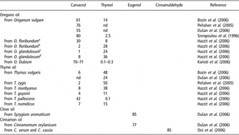
Volatile or essential oils, distilled from non-woody parts of herbs, contain principally terpenoids and minor constituents including various aliphatic hydrocarbons, acids, alcohols, aldehydes and other compounds (Dorman, Reference Dorman1999; Bozin et al., Reference Bozin, Mimica-Dukic, Simin and Anackov2006). The composition of the oils is highly variable within plants due to different environmental and climatic conditions (Table 3). The distillation method also influences the final composition of essential oils (Yang et al., Reference Yang, Kayan, Bozer, Pate, Baker and Gizir2007). Oregano oil (Origanum vulgare) and thyme oil (Thymus vulgaris) contain mainly carvacrol and thymol (Table 3) (Peñalver et al., Reference Peñalver, Huerta, Borge, Astorga, Romero and Perea2005; Bozin et al., Reference Bozin, Mimica-Dukic, Simin and Anackov2006). Eugenol dominates in oils from clove (Syzygium aromaticum) and cinnamon (Cinnamomum zeylanicum) (Dušan et al., Reference Dušan, Marian, Katarina and Dobroslava2006). Cinnamaldehyde is the main component of C. verum or C. cassia (Ooi et al., Reference Ooi, Li, Kam, Wang, Wong and Ooi2006). Essential oils themselves make 1.5% to 4.5% of the plant (Bozin et al., Reference Bozin, Mimica-Dukic, Simin and Anackov2006; Hazzit et al., Reference Hazzit, Baaliouamer, Faleiro and Miguel2006; Yang et al., Reference Yang, Kayan, Bozer, Pate, Baker and Gizir2007).
Main components of selected essential oils (%)

nd: no data in reference.
aCollected in Chrea National Park.
bCollected in Hammam Melouane.
cCollected in Souhane.
dCollected in Ighzer mokrane.
In vitro activity of essential oils
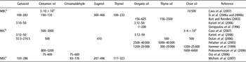
The minimal inhibitory concentration (MIC) of carvacrol and essential oils from O. dubium against different bacterial strains (Table 4) was determined to be the same for pure component and the extract (Karioti et al., Reference Karioti, Vrahimi-Hadjilouca, Droushiotis, Rancic, Hadjipavlou-Litina and Skaltsa2006). The MIC of essential oils from O. vulgare and T. zygis against several Enterobacteriaceae strains isolated from poultry and pigs with clinical disease outcome or carrier state were shown to vary widely, the highest MIC being observed for E. coli strains (Peñalver et al., Reference Peñalver, Huerta, Borge, Astorga, Romero and Perea2005). Hammer et al. (Reference Hammer, Carson and Riley1999) obtained MIC for O. vulgare essential oil against several Gram-positive and Gram-negative bacteria and Candida albicans. Pseudomonas aeruginosa was the least sensitive bacterium, having the highest MIC. The authors recorded similar values for essential oils from S. aromaticum and T. vulgaris. The MIC for clove and thyme essential oils against S. aureus and E. coli were half the minimal bactericidal concentration (Hammer et al., Reference Hammer, Carson and Riley1999).
Antimicrobial activity of selected essential oils in vitro

aMBC (μg/ml): minimal bactericidal concentration.
bMIC (μg/ml): minimal inhibitory concentration.
cMEC (μg/ml): minimal effect concentration.
The reader is asked to look in references for details.
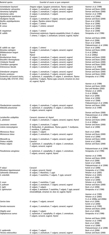
Essential oils extracted from O. vulgare and T. vulgaris (Table 3) showed activity against Gram-positive and Gram-negative bacteria (Table 5) and their strains multi-resistant to antibiotics (Bozin et al., Reference Bozin, Mimica-Dukic, Simin and Anackov2006). Generally, Gram-positive bacteria seem to be more sensitive to essential oils than Gram-negative bacteria. Dorman and Deans (Reference Dorman and Deans2000) determined equal anti-bacterial activity of volatile oils distilled from O. vulgare ssp. hirtum and S. aromaticum (clove) against several Gram-positive and Gram-negative bacteria, whereas essential oils from thyme showed stronger activity against Gram-positive organisms. The widest spectrum of activity was found for T. vulgaris, followed by oregano and clove essential oils. The pure components showed reduced activity against a wide spectrum of bacteria: thymol > carvacrol > eugenol. Hazzit et al. (Reference Hazzit, Baaliouamer, Faleiro and Miguel2006) observed little inhibition of Listeria monocytogenes growth caused by essential oils from different oregano and thyme species. By contrast, oregano and thyme extracts were found to be inhibitory against several Bacillus sp. (Özcan et al., Reference Özcan, Sagdic and Ozkan2006). Cinnamon and clove oils have shown highly variable MIC across bacteria (Prabuseenivasan et al., Reference Prabuseenivasan, Jayakumar and Ignacimuthu2006, Table 4). Clove oil was less active than cinnamon oil.
Bacterial species inhibited in in vitro studies when plant essential oils or pure oil components were added to growth medium

Oregano oil containing 74% carvacrol showed high anti-bacterial and antifungal activity and considerable reduction in bacterial growth even at very low concentrations (Sivropoulou et al., Reference Sivropoulou, Papanikolaou, Nikolaou, Kokkini, Lanaras and Arsenakis1996; Adam et al., Reference Adam, Sivropoulou, Kokkini, Lanaras and Arsenakis1998). Essential oils from C. verum and C. cassia and pure cinnamaldehyde were inhibitory for bacteria (Table 5), yeasts (C. albicans, C. glabrata, C. tropicalis and C. krusei), for moulds and dermatophytes (Ooi et al., Reference Ooi, Li, Kam, Wang, Wong and Ooi2006). Also Matan et al. (Reference Matan, Rimkeeree, Mawson, Chompreeda, Haruthaithanasan and Parker2006) found that the antifungal activity of cinnamon and clove oils volatile against spoilage fungi, yeasts and bacteria. Park et al. (Reference Park, Gwak, Yang, Choi, Jo, Chang, Jeung and Choi2007) found eugenol to be the active component of clove oil against several dermatophytes.
Extrapolation of in vitro results to in vivo
Simulation of gastric environment (artificial substrate containing several different sugars, casein, soybean oil, vitamins, minerals, etc., in a buffer solution of pH 3) to test anaerobic bacteria inhibition by essential oils revealed relatively high minimal effect concentration (MEC) for carvacrol, thymol, eugenol and trans-cinnamaldehyde (Michiels et al., Reference Michiels, Missotten, Fremaut, De Smet and Dierick2007). Trans-cinnamaldehyde displayed a strong inhibitory activity in the small intestinal environment (containing the same substrate as for the gastric environment with bile salts in a buffer of pH 6.5 inoculated with fresh small intestine supernatant of a weaned piglet). A synergistic action of carvacrol and thymol against total anaerobic bacteria was observed at ratios >1, strongly suggesting a different, dose-dependent mode of action of the essential oil components against E. coli and other coliform bacteria. Si et al. (Reference Si, Gong, Tsao, Zhou, Yu, Poppe, Johnson and Du2006b) noticed strong anti-bacterial activity of carvacrol, thymol, cinnamon oil and eugenol in vitro (Table 4). When added to pig caecal digesta, all the tested oils reduced the growth of the indigenous E. coli, exogenous E. coli O157:H7 when added and coliforms present in the digesta, with no effect on lactobacilli (Si et al., Reference Si, Gong, Tsao, Zhou, Yu, Poppe, Johnson and Du2006b). This study clearly showed no detrimental effect of pig digesta on the anti-bacterial essential oil activity in vitro.
It is difficult to relate the minimum effective dose in vitro to the dietary dose required in animals. In an in vitro study of the inhibition of S. enterica serovar Typhimurium, it was shown that the dose required for thymol was 700-fold greater than that required with the antibiotic gentamycin (Merialdi, personal communication), which was also able to inhibit Salmonella translocation to pig mesenteric lymph nodes (Modesto et al., Reference Modesto, Stefanini, D’Aimmo, Mazzoni, Trevisi, Tittarelli, Bosi and Biavati2007). Based on the concentration of gentamycin required to protect weaning pigs, the amount of thymol required to be added to the diet has been estimated to be 4%.
The antimicrobial activity of essential oils in vitro is not always reflected in tests in vivo. For example, Si et al. (Reference Si, Gong, Chanas, Cui, Yu, Caballero and Friendship2006a and Reference Si, Gong, Tsao, Zhou, Yu, Poppe, Johnson and Du2006b) showed that Salmonella shedding in challenged pigs was not reduced as the inhibitory effect of essential oils disappeared when they were mixed with the diet. This was probably because of binding of the phenolic and other components to fats and other hydrophobic materials of the diet. Emulsifiers like fenugreek or xanthan gum have no effect on essential oil activity and they are required for stabilising oils during storage (Si et al., Reference Si, Gong, Chanas, Cui, Yu, Caballero and Friendship2006a). However, whether emulsified components keep their activity when added to feed is not known. A possible way to overcome these problems could be micro-encapsulation of the oils before adding to the diet (Meunier et al., Reference Meunier, Cardot, Manzanilla, Wysshaar and Alric2007) and first studies considering this route have already been performed (Janczyk et al., unpublished).
Feeding studies on weaning piglets
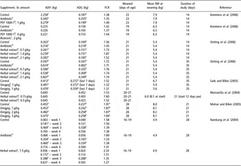
The published results of in vivo studies, whilst highly variable, suggest that relatively high concentrations of essential oils are required for displaying beneficial effects in young pigs (Table 6).
Average daily feed intake (ADFI), weight gain (average daily gain (ADG)) and feed conversion ratio (FCR) obtained in different studies on weaning piglets when herbs or herbal extracts were fed to the animals

aCarbadox 50 mg/kg diet.
bPEP-1000-1 – essential oil mix containing anis oil, citrus oil, oregano oil and natural flavours.
cBiotronic – organic and inorganic acids mix containing phosphoric and lactic acids.
dZn-bacitracin, olaquindox, colistin, 50 mg/kg diet each.
eHerbal extract in this study contained 20% of essential oils: 3.3% clove oil, 3.3% eugenol, 3.3% oregano oil, 3.3% carvacrol and 6.7% thyme oil.
fOregpig – 500 g oregano oil, 33.4 g thymol, dried oregano flowers and leaves ad 1000 g. Concentration of carvacrol – 60 g/kg, thymol – 55 g/kg.
gHerbal extract used in this study: carvacrol 50 g/kg, cinnamaldehyde 30 g/kg and capsicum oleoresin 20 g/kg.
hLincomycin 110 mg/kg diet.
iHerbal extract used in this study contained extracts of cinnamon, thyme and oregano on clay carrier.
x,y,zDifferent superscripts mark significant difference between values in a column within one study.
Kommera et al. (Reference Kommera, Mateo, Neher and Kim2006) observed no effect on feed intake, growth performance or feed efficiency when a mix of essential oils was added to the diet (1 to 4 g/kg) of weaning pigs, without or with a combination of OA. Supplementation of piglet diet with 2.1 g/kg of herbal extract resulted in an improved growth performance similar to an antibiotic control group, with intermediate feed intake and feed efficiency (Oetting et al., Reference Oetting, Utiyama, Giani, Ruiz and Miyada2006). The only variables influenced by the highest dose tested were the apparent digestibility of DM and the weight of the small intestine, which were higher with herbal extract supplementation. Improved growth performance and feed efficiency were recorded in weaned piglets fed a product containing oregano extract at a dose of 1 g/kg (Sads and Bilkei, Reference Sads and Bilkei2003). By contrast, no effect could be observed when 0.15 or 0.3 g/kg of herbal mix was fed to early-weaned piglets (Manzanilla et al., Reference Manzanilla, Perez, Martin, Kamel, Baucells and Gasa2004), except for a decreased gastric emptying rate (Manzanilla et al., Reference Manzanilla, Perez, Martin, Kamel, Baucells and Gasa2004) and a reduction in the lactobacilli to enterobacteria ratio in caecum at a dose of 0.3 g/kg (Castillo et al., Reference Castillo, Martin-Orue, Roca, Manzanilla, Badiola, Perez and Gasa2006).
Molnar and Bilkei (Reference Molnar and Bilkei2005) conducted a study in a pig herd in Hungary where post-weaning colibacillosis had been present for years. Interestingly, the mortality rate decreased from 3.7% in the control to 2% in the group supplemented with 1 g/kg of a product containing oregano extract and to 1% in the groups supplemented with 2 and 3 g/kg (P < 0.05). An increase in growth performance was observed with high doses (2 or 3 g/kg, P < 0.05). A reduction in feed conversion was observed already at the level of 1 g/kg of this product (P < 0.05). Contrasting results were obtained by Namkung et al. (Reference Namkung, Li, Gong, Yu, Cottrill and de Lange2004) who recorded a reduction in growth performance and feed intake in the piglets supplemented with 7.5 g/kg of herbal extract (P < 0.05) without effects on gut anatomy or immunity. However, coliform counts were reduced in the ileum and faeces (P < 0.05), but not in the colon.
Based on the published literature it is clear that the effective concentration of essential oils in feed still needs to be established. A recent study performed was therefore aimed towards this issue. The effect of thymol addition to a starter diet (10 g/kg), determined from in vitro test and toxicology information, was tested in pigs with or without S. typhimurium challenge (Trevisi et al., Reference Trevisi, Merialdi, Mazzoni, Casini, Tittarelli, De Filippi, Minieri, Lalatta-Costerbosa and Bosi2007). The final body weight after 29 days of trial was not affected by thymol but feed intake was reduced. Thymol protected against a rise in body temperature 1-day post challenge, but it did not reduce faecal shedding of Salmonella. Thymol increased the density of acid-secreting cells in gastric oxintic glands. Bacterial diversity in the jejunum was also affected. Actinobacillus minor was present in almost all the pigs fed thymol, Citrobacter freundii being absent (Janczyk et al., Reference Janczyk, Trevisi, Souffrant and Bosi2008).
In terms of palatability, Jugl-Chizzola et al. (Reference Jugl-Chizzola, Ungerhofer, Gabler, Hagmuller, Chizzola, Zitterl-Eglseer and Franz2006) observed that piglets had a preference for diets with low inclusion rates of herbs in diets but they could not show differences between thyme and oregano.
Finally, the bioavailability and pharmacokinetics of essential oils have received little attention in pigs. Studies in humans suggest a rapid absorption of these molecules (e.g. thymol, Kohlert et al., Reference Kohlert, Schindler, Marz, Abel, Brinkhaus, Derendorf, Grafe and Veit2002). This could reduce the action of essential oils on commensal and exogenous microflora in vivo. On the other hand, different essential oils may be sensitive to oxidation and thus they may undergo degradation (Pérez-Alonso et al., Reference Pérez-Alonso, Cruz-Olivares, Barrera-Pichardo, Rodríguez-Huezo, Báez-González and Vernon-Carter2008). Protection of essential oils, e.g. by micro-encapsulation, may be a valuable strategy in animal feeding (Meunier et al., Reference Meunier, Cardot, Gauthier, Beyssac and Alric2006 and Reference Meunier, Cardot, Manzanilla, Wysshaar and Alric2007), and more studies are clearly required in this area.
Conclusions and perspectives
Relating to the recent ban on in-feed antibiotics, many efforts have been made in the past years for optimising further diet composition in terms of protein, AA and energy. Additional substances including OA and compounds of plant origin with known antimicrobial properties have also been evaluated. It has become clear that diet supplementation with plasma protein is probably the best way for preventing post-weaning gut disorders, provided it is of high hygienic quality. Such a success lies mostly in its richness in immunoglobulins that act at two complementary levels: protecting the intestinal mucosa from luminal aggression and stimulating the immune system for defence against pathogens. Interestingly, the mode of action of plasma protein appears to be different from, and additive to, that of in-feed antibiotics. Positive results have been obtained with particular AA (Gln, Trp, Arg, Cys, Thr) probably because their requirements are increased during periods of general and immune stress, especially in younger animals and under poor hygienic conditions. Many OA have also proven successful, acting on gut ecology and preventing the outgrowth of pathogenic bacteria. Variable results have been obtained with SB administered orally. However, when it displays positive effects in vivo, its mechanisms of action are still unclear, thus calling for additional work. Investigations in vitro clearly show antimicrobial properties of essential oils. They are supported by one or two major molecules or by more subtle synergistic interactions within the extract mixture. However, major discrepancies do exist between in vitro and in vivo results, the latter being variable and rather inconsistent. It appears that the diet itself may neutralise the antimicrobial activity of essential oils in vivo. Finally, the bioavailability and pharmacokinetics of these essential oils in pigs are mostly unknown. This will undoubtedly constitute an area for future investigation.
In terms of cost effectiveness, this review in some way confirms the practices that are now more sprayed among the experts of piglet feed formulation: to include consistently more than one OA and to raise some AA when the gut environment is more challenged.
Acknowledgements
The authors wish to thank Professor C. R. Stokes (University of Bristol, UK) for correcting this text for English. The European Union is greatly acknowledged by the authors of the present review for supporting financially research in gut health and nutrition in piglets around weaning (Healthypigut project no. QLK5-CT2000-00522 and Feed for Pig Health project no. FP6-FOOD-1-506144).