Childhood maltreatment is a severe stressor that jeopardizes normative development and can compromise adaptive functioning (Cicchetti & Toth, Reference Cicchetti and Toth2005). In 2013 alone, there were an estimated 679,000 cases of child maltreatment in the United States (US Department of Health and Human Services, Administration for Children and Families, Administration on Children, Youth and Families, Children's Bureau, 2015). Unfortunately, this number likely underrepresents the true magnitude of the problem, because it does not account for the vast number of unreported cases. Childhood maltreatment disrupts development across many domains, and individuals who experience childhood maltreatment are at higher risk for the development of psychopathology across the lifespan (Cicchetti & Toth, Reference Cicchetti and Toth1995; Kim & Cicchetti, Reference Kim and Cicchetti2010).
Despite increased understanding of early adversity, and the importance of supporting young children, many social service agencies struggle to provide effective services for children facing maltreatment (Jaffee & Christian, Reference Jaffee and Christian2014). This disconnect may be due in part to the fact that there is still much to be learned about the precise mechanisms by which childhood maltreatment alters developmental trajectories, including the neurobiological impact of maltreatment. The neuroimaging literature on childhood maltreatment has yet to fully evaluate the development of brain networks critical in supporting social, cognitive, and emotion regulation behaviors that are known to be vulnerable to adversity.
Emotion Processing and Maltreatment
Behaviorally, maltreated children experience a host of socioemotional disruptions (Aber & Cicchetti, Reference Aber and Cicchetti1984; Pechtel & Pizzagalli, Reference Pechtel and Pizzagalli2011). These differences can lead to a cascade of effects that confer vulnerability for affective and behavioral disorders (Masten & Cicchetti, Reference Masten and Cicchetti2010). Individuals who experience childhood maltreatment are at risk for a wide range of psychopathology, including depression, anxiety, bipolar disorder, suicidality, conduct disorder, and substance abuse (Brodsky et al., Reference Brodsky, Mann, Stanley, Tin, Oquendo and Birmaher2008; Cannon, Bonomi, Anderson, Rivara, & Thompson, Reference Cannon, Bonomi, Anderson, Rivara and Thompson2010; Edalati & Krank, Reference Edalati and Krank2015; Garno, Goldberg, Ramirez, & Ritzler, Reference Garno, Goldberg, Ramirez and Ritzler2005; Jaffee et al., Reference Jaffee, Caspi, Moffitt, Dodge, Rutter and Taylor2005; Kaufman, Reference Kaufman1991; Springer, Sheridan, Kuo, & Carnes, Reference Springer, Sheridan, Kuo and Carnes2007; Widom, DuMont, & Czaja, Reference Widom, DuMont and Czaja2007).
Converging evidence suggests that maltreatment leads to an altered processing of emotions and social stimuli (da Silva Ferreira, Crippa, & de Lima Osorio, Reference da Silva Ferreira, Crippa and de Lima Osorio2014). Overall, maltreated children tend to be less accurate in emotion recognition (Camras, Grow, & Ribordy, Reference Camras, Grow and Ribordy1983; Camras et al., Reference Camras, Ribordy, Hill, Martino, Spaccarelli and Stefani1988, Reference Camras, Ribordy, Hill, Martino, Sachs and Spaccarelli1990; During & McMahon, Reference During and McMahon1991; Pollak, Cicchetti, Hornung, & Reed, Reference Pollak, Cicchetti, Hornung and Reed2000). Differences also have been observed among maltreatment subtypes, with neglected children displaying difficulty discriminating between emotions, and physically abused children showing a response bias for angry facial expressions, requiring less perceptual information to recognize angry expressions (Pollak et al., Reference Pollak, Cicchetti, Hornung and Reed2000; Pollak & Kistler, Reference Pollak and Kistler2002; Pollak, Messner, Kistler, & Cohn, Reference Pollak, Messner, Kistler and Cohn2009; Pollak & Sinha, Reference Pollak and Sinha2002). Physically abused children show selective attention to threatening stimuli (Pollak & Tolley-Schell, Reference Pollak and Tolley-Schell2003), although severe physical abuse has also been associated with an attentional bias away from threat (Pine et al., Reference Pine, Mogg, Bradley, Montgomery, Monk and McClure2005). Evidence from event-related potentials also suggests that children with a history of maltreatment process positive and negative facial expressions differently than do nonmaltreated children (Cicchetti & Curtis, Reference Cicchetti and Curtis2005; Curtis & Cicchetti, Reference Curtis and Cicchetti2011, Reference Curtis and Cicchetti2013; Pollak, Cicchetti, Klorman, & Brumaghim, Reference Pollak, Cicchetti, Klorman and Brumaghim1997). Although most of the maltreatment research on emotion processing has been conducted with children, recent data suggest similar emotion recognition deficits in adults who experienced maltreatment in childhood (Young & Widom, Reference Young and Widom2014).
Altered emotion processing following maltreatment points to potential neural differences in emotion-related regions of the brain. Of particular interest are the amygdala and the hippocampus, subcortical limbic structures involved in emotion and memory (LeDoux, Reference LeDoux2000), and prefrontal regulatory regions, including the orbital frontal cortex (OFC) and medial prefrontal cortex (mPFC; Etkin, Egner, & Kalisch, Reference Etkin, Egner and Kalisch2011). Structural and functional connectivity of the amygdala and mPFC is related to efficient emotion regulation, fear conditioning, and extinction (Kim et al., Reference Kim, Gee, Loucks, Davis and Whalen2011). Although the dorsolateral PFC (dlPFC) and ventrolateral PFC (vlPFC) do not have direct structural connections with the amygdala, research suggests that these regions are also indirectly involved in regulation of the amygdala (Delgado, Nearing, LeDoux, & Phelps, Reference Delgado, Nearing, LeDoux and Phelps2008; Ochsner & Gross, Reference Ochsner and Gross2005; Wager, Davidson, Hughes, Lindquist, & Ochsner, Reference Wager, Davidson, Hughes, Lindquist and Ochsner2008). The importance of these limbic and frontolimbic circuits for emotion processing and regulation suggests they may be prime targets for long-term, downstream consequences of childhood maltreatment.
Structural Brain Differences in Maltreatment
Childhood maltreatment is associated with a range of differences in brain structure. Globally, children with a history of maltreatment and posttraumatic stress disorder (PTSD) have shown reduced cerebral volumes (De Bellis et al., Reference De Bellis, Keshavan, Shifflett, Iyengar, Beers and Hall2002) and lower overall gray matter volumes (De Brito et al., Reference De Brito, Viding, Sebastian, Kelly, Mechelli and Maris2013). Data from diffusion tensor imaging studies suggest that childhood maltreatment is associated with reduced integrity of white matter tracts throughout the brain (Choi, Jeong, Polcari, Rohan, & Teicher, Reference Choi, Jeong, Polcari, Rohan and Teicher2012; Choi, Jeong, Rohan, Polcari, & Teicher, Reference Choi, Jeong, Rohan, Polcari and Teicher2009; Hanson et al., Reference Hanson, Adluru, Chung, Alexander, Davidson and Pollak2013; Huang, Gundapuneedi, & Rao, Reference Huang, Gundapuneedi and Rao2012; Jackowski et al., Reference Jackowski, Douglas-Palumberi, Jackowski, Win, Schultz and Staib2008). Similarly, a number of studies have reported reduced prefrontal volumes or cortical thickness following maltreatment (Andersen et al., Reference Andersen, Tomada, Vincow, Valente, Polcari and Teicher2008; De Bellis et al., Reference De Bellis, Keshavan, Shifflett, Iyengar, Beers and Hall2002; Edmiston et al., Reference Edmiston, Wang, Mazure, Guiney, Sinha and Mayes2011), particularly in emotion regulatory regions such as the OFC (Dannlowski et al., Reference Dannlowski, Stuhrmann, Beutelmann, Zwanzger, Lenzen and Grotegerd2012; De Brito et al., Reference De Brito, Viding, Sebastian, Kelly, Mechelli and Maris2013; Edmiston et al., Reference Edmiston, Wang, Mazure, Guiney, Sinha and Mayes2011; Hanson et al., Reference Hanson, Chung, Avants, Shirtcliff, Gee and Davidson2010; Kelly et al., Reference Kelly, Viding, Wallace, Schaer, De Brito and Robustelli2013; Lim, Radua, & Rubia, Reference Lim, Radua and Rubia2014), anterior cingulate cortex (Dannlowski et al., Reference Dannlowski, Stuhrmann, Beutelmann, Zwanzger, Lenzen and Grotegerd2012; Kelly et al., Reference Kelly, Viding, Wallace, Schaer, De Brito and Robustelli2013) and mPFC (Van Harmelen et al., Reference Van Harmelen, van Tol, Van Der Wee, Veltman, Aleman and Spinhoven2010).
Structural differences in the hippocampus and amygdala as a result of maltreatment have been of great interest, given the relevance of these structures to mood disorders (Price & Drevets, Reference Price and Drevets2010) and the animal literature suggesting that these regions are vulnerable to stress (McEwen, Reference McEwen1999; Moriceau, Roth, Okotoghaide, & Sullivan, Reference Moriceau, Roth, Okotoghaide and Sullivan2004; Poeggel et al., Reference Poeggel, Helmeke, Abraham, Schwabe, Friedrich and Braun2003; Vyas, Mitra, Shankaranarayana Rao, & Chattarji, Reference Vyas, Mitra, Shankaranarayana Rao and Chattarji2002). A meta-analysis of maltreatment-related PTSD found smaller hippocampal volumes in adults with a history of childhood maltreatment, but not in children who had been maltreated (Woon & Hedges, Reference Woon and Hedges2008). Recent work in populations without PTSD has supported this finding in adults, with consistently smaller hippocampal volumes observed in maltreated groups (Chaney et al., Reference Chaney, Carballedo, Amico, Fagan, Skokauskas and Meaney2014; Dannlowski et al., Reference Dannlowski, Stuhrmann, Beutelmann, Zwanzger, Lenzen and Grotegerd2012; Riem, Alink, Out, van Ijzendoorn, & Bakermans-Kranenburg, Reference Riem, Alink, Out, van Ijzendoorn and Bakermans-Kranenburg2015; Samplin, Ikuta, Malhotra, Szeszko, & DeRosse, Reference Samplin, Ikuta, Malhotra, Szeszko and DeRosse2013; Teicher, Anderson, & Polcari, Reference Teicher, Anderson and Polcari2012). Volumetric findings from the amygdala have been more mixed, with some studies of maltreated adolescents showing decreased amygdala volumes (Edmiston et al., Reference Edmiston, Wang, Mazure, Guiney, Sinha and Mayes2011) and others showing increased volumes (Mehta et al., Reference Mehta, Golembo, Nosarti, Colvert, Mota and Williams2009; Pechtel, Lyons-Ruth, Anderson, & Teicher, Reference Pechtel, Lyons-Ruth, Anderson and Teicher2014); however, at least two studies of adults have reported no difference in amygdala volumes between childhood maltreatment and control groups (Andersen et al., Reference Andersen, Tomada, Vincow, Valente, Polcari and Teicher2008; Bremner et al., Reference Bremner, Randall, Vermetten, Staib, Bronen and Mazure1997).
Functional Brain Differences in Maltreatment
Despite the growing evidence of structural alterations associated with childhood maltreatment, there are still relatively few studies that probe functional differences in brain activity following maltreatment. The existing functional magnetic resonance imaging (fMRI) studies on maltreatment lend support to a hypothesis of altered limbic circuitry. A number of recent studies point to an association between maltreatment and increased amygdala reactivity (Dannlowski et al., Reference Dannlowski, Stuhrmann, Beutelmann, Zwanzger, Lenzen and Grotegerd2012, Reference Dannlowski, Kugel, Huber, Stuhrmann, Redlich and Grotegerd2013; De Bellis & Hooper, Reference De Bellis and Hooper2012; Grant, Cannistraci, Hollon, Gore, & Shelton, Reference Grant, Cannistraci, Hollon, Gore and Shelton2011; McCrory et al., Reference McCrory, De Brito, Sebastian, Mechelli, Bird and Kelly2011, Reference McCrory, De Brito, Kelly, Bird, Sebastian and Mechelli2013; Van Harmelen et al., Reference Van Harmelen, van Tol, Demenescu, van der Wee, Veltman and Aleman2013; White et al., Reference White, Bogdan, Fisher, Muñoz, Williamson and Hariri2012). Differences have also been found in reward processing regions such as the basal ganglia (Dillon et al., Reference Dillon, Holmes, Birk, Brooks, Lyons-Ruth and Pizzagalli2009) and in frontal regulatory regions, including the mPFC (Fonzo et al., Reference Fonzo, Flagan, Sullivan, Allard, Grimes and Simmons2013; Lim et al., Reference Lim, Hart, Mehta, Simmons, Mirza and Rubia2015; Mueller et al., Reference Mueller, Maheu, Dozier, Peloso, Mandell and Leibenluft2010; Van Harmelen et al., Reference Van Harmelen, Hauber, Moor, Spinhoven, Boon and Crone2014).
Although it is necessary to understand functional differences in specific brain regions, it is perhaps equally important to investigate interactions between regions. Measures of functional connectivity can provide a better understanding of how communication among networks of regions may be influenced by early life stress. Data from resting-state fMRI studies have identified altered emotion-processing networks in individuals who experienced childhood maltreatment (Cisler et al., Reference Cisler, James, Tripathi, Mletzko, Heim and Hu2013; Herringa et al., Reference Herringa, Birn, Ruttle, Burghy, Stodola and Davidson2013; van der Werff et al., Reference van der Werff, Pannekoek, Veer, van Tol, Aleman and Veltman2013). However, there is little consensus about the direction of effects in these networks, perhaps due to large heterogeneity in sample characteristics. In a study with adolescents, Herringa et al. (Reference Herringa, Birn, Ruttle, Burghy, Stodola and Davidson2013) found that childhood maltreatment predicted reduced connectivity between the amygdala and vmPFC, but greater amygdala–dlPFC connectivity. In an adult sample, childhood emotional maltreatment was related to decreased connectivity between the amygdala and the hippocampus, insula, and frontal structures such as the OFC (van der Werff et al., Reference van der Werff, Pannekoek, Veer, van Tol, Aleman and Veltman2013). However, in a pilot study of drug users who had been maltreated as children, maltreatment was associated with increased connectivity between the amygdala and the hippocampus, hippocampal gyrus, and OFC (Dean, Kohno, Hellemann, & London, Reference Dean, Kohno, Hellemann and London2014). Overall, these studies examined participants of different ages, with different types of maltreatment that occurred at different points during development. The high degree of heterogeneity within the maltreated population makes differences in resting-state connectivity measures challenging to interpret.
While data on maltreatment and resting-state functional connectivity are scarce, even less is known about the coordination of neural networks while performing a task. One study observed maltreatment-related differences in limbic circuitry during a face-viewing task in women with PTSD subsequent to partner violence (Fonzo et al., Reference Fonzo, Flagan, Sullivan, Allard, Grimes and Simmons2013). Specifically, results from this study suggested a positive relationship between a continuous measure of maltreatment (Child Trauma Questionnaire) and amygdala–prefrontal connectivity, but a negative association between Child Trauma Questionnaire scores and amygdala–insula connectivity. Similar maltreatment-related differences in connectivity have also been observed in cognitive control regions during an inhibitory control task (Elton et al., Reference Elton, Tripathi, Mletzko, Young, Cisler and James2014). Identifying and characterizing how networks of brain regions differ for individuals with a history of childhood maltreatment is as important to our understanding of the effects of early maltreatment as is the identification of regions that differ in their level of activation alone.
Current Study
In the current study, we sought to further examine the association between childhood maltreatment and task-related functional connectivity of frontolimbic circuits. The vast majority of neuroimaging research with maltreated samples relies on self-reported measures of maltreatment. However, a reliance on self-report alone may not capture the extent of maltreatment and may include substantial measurement error (Hardt & Rutter, Reference Hardt and Rutter2004). Our sample consisted of participants who were studied longitudinally as children, providing prospective data on maltreatment histories and early experiences. In addition, some prior studies have used healthy comparison groups that are not well matched on socioeconomic status (SES) or other demographic variables associated with maltreatment risk. In our study, the maltreated and comparison participants were drawn from similarly low-income, high-risk environments. This design helps minimize the contribution of other forms of early adversity and comes closer to isolating the unique relationship between childhood maltreatment and brain development.
The current study used an fMRI task known to elicit limbic activation to examine differences in functional connectivity related to childhood maltreatment. Because mounting data suggest an association between maltreatment and amygdala activation in response to emotionally valenced stimuli, we predicted a hyperactive amygdala response to emotional stimuli in maltreated participants relative to SES-matched comparison participants. There is a paucity of data regarding maltreatment and functional connectivity of limbic circuits during emotion processing. Consequently, there is not enough evidence to predict the direction of effects, or the specific nature of amygdala connectivity. However, given the importance of frontal regions in emotion regulation, as well as documented maltreatment-related behavioral deficits in this domain (Kim & Cicchetti, Reference Kim and Cicchetti2010; Shields & Cicchetti, Reference Shields and Cicchetti1997), we hypothesized differences in amygdala–prefrontal connectivity between the maltreated and comparison groups.
Methods
Participants
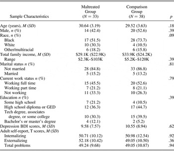
Eighty-seven adults (M = 30.1 years, SD = 3.6, range = 23–37 years) were tested. Participants were part of an existing longitudinal sample, first recruited to a research summer camp for low-income, high-risk children when they were 6–12 years of age, and subsequently assessed at two time points in adolescence (Rogosch, Oshri, & Cicchetti, Reference Rogosch, Oshri and Cicchetti2010). Forty-four participants had a history of childhood maltreatment as documented by Department of Human Services records; 43 adults had no history of maltreatment as documented by a lack of Department of Human Services records (see Table 1 for sample characteristics). In addition to examining records, maltreatment was ruled out using the Maternal Maltreatment Classification Interview (Cicchetti, Toth, & Manly, Reference Cicchetti, Toth and Manly2003). The Maltreatment Classification System (Barnett, Manly, & Cicchetti, Reference Barnett, Manly, Cicchetti, Cicchetti and Toth1993) was used to classify the type and developmental timing of each report of substantiated maltreatment. Maltreatment experiences included emotional maltreatment, physical neglect, physical abuse, and sexual abuse. The majority (70%) of participants in the maltreatment group experienced more than one type of maltreatment.
Demographics and sample characteristics for the maltreated and comparison groups

Note: BDI, Beck Depression Inventory.
Exclusion criteria included current or past history of neurological disorders or trauma; known intellectual impairment; uncorrected visual or auditory impairments; or MRI contraindications, including claustrophobia, metal in the body, or extreme obesity. Sixteen individuals participated but were excluded from the final sample of 71 for the following reasons: task accuracy > 2 SD below the mean on control trials (<75.7% correct; 4 maltreated, 1 comparison); serious mental illness (2 maltreated; 1 schizophrenia, 1 bipolar disorder); structural brain anomalies (1 maltreated, 1 comparison); or excessive motion during the fMRI task (a minimum of three blocks of each stimulus type was required for inclusion, with no more than 5 out of 15 repetition times exceeding motion criteria within each block [see fMRI Analysis]; 4 maltreated, 3 comparison). An additional 53 longitudinal participants were unable to participate for the following reasons: size/obesity (19), metal in body (6), incarceration (11), refusal or scheduling conflicts (14), pregnancy (1), or deceased (2). All participants provided informed consent in compliance with the University of Rochester's institutional review board.
Self-report measures
Participants completed several questionnaires, including the Adult Self-Report (Achenbach & Rescorla, Reference Achenbach and Rescorla2003), the Beck Depression Inventory—II (BDI-II; Beck, Steer, & Brown, Reference Beck, Steer and Brown1996), and a demographics questionnaire. The Adult Self-Report includes normed scales for adaptive functioning, empirically based syndromes, substance use, internalizing, externalizing, and total problems. The BDI-II is a 21-item self-report instrument designed to assess severity of depression. Total scores for the BDI-II indicate ranges of symptom severity. The groups were not significantly different in income levels, education, internalizing, externalizing, or depression ratings (see Table 1).
Behavioral fMRI paradigm
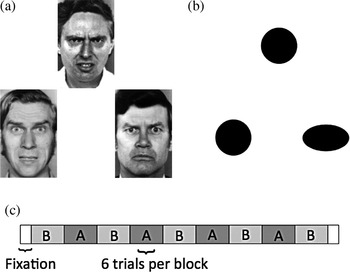
In the scanner, participants completed an emotion-matching task (Hariri, Bookheimer, & Mazziotta, Reference Hariri, Bookheimer and Mazziotta2000) that has been found to robustly activate the amygdala. The task was a block design with alternating blocks of emotion-matching trials or shape-matching trials (Figure 1). Each trial contained three images, one on the top half of the screen and two in the lower half. Participants were asked to match one of the two images on the bottom with the image on the top. For emotion-matching trials, images were angry or fearful faces posed by three different actors of the same sex. Participants were instructed to match based on the emotion of the faces. The emotion to be matched (anger or fear) was balanced across trials. Face stimuli came from a standardized set of emotional faces (Ekman & Friesen, Reference Ekman and Friesen1976). For shape-matching trials, images were black shapes (circles, or horizontal or vertical ellipses) and participants were instructed to match based on shape. The task consisted of a total of nine blocks, each containing six trials, with alternating blocks of shape and emotion matching (five and four blocks, respectively). Stimuli were presented for 4500 ms, with a 500-ms interstimulus interval.
Emotion matching task. (a) Example emotion-matching trial with angry and fear faces (Ekman & Friesen, Reference Ekman and Friesen1976). (b) Example shape-matching trial. (c) Task design included alternating emotion-matching and shape-matching blocks. Each block had six trials (5000 ms per trial).

MRI acquisition
Structural and functional MRI data were acquired on a Siemens 3-Tesla Trio scanner using a 32-channel head coil. High-resolution, T1 weighted, three-dimensional images were acquired for each participant using an MPRAGE sequence (echo time [TE] = 3.44 ms, repetition time [TR] = 2530 ms, field of view = 256 mm, matrix = 256 × 256, slice thickness = 1 mm, flip angle = 7°, 192 sagittal slices). Functional data were acquired using an echo-planar imaging sequence (TE = 30 ms, TR = 2000 ms, field of view = 224 mm, matrix = 64 × 64, slice thickness = 3.5 mm with a 29% gap, flip angle = 90°, 30 interleaved oblique axial slices). To correct geometric distortion in the functional data, a fieldmap volume was collected immediately prior to the functional data acquisition using the same positioning prescription (TE1 = 5.19 ms, TE2 = 7.65 ms, TR = 400 ms, field of view = 224 mm, matrix = 64 × 64, slice thickness = 3.5 mm with a 29% gap, flip angle = 60°, 30 interleaved oblique axial slices).
fMRI analysis
MRI data were analyzed using FSL (FMRIB Software Library, v. 4.1.9; http://fsl.fmrib.ox.ac.uk/fsl; Jenkinson, Beckmann, Behrens, Woolrich, & Smith, Reference Jenkinson, Beckmann, Behrens, Woolrich and Smith2012). Preprocessing involved motion correction with MCFLIRT (Motion Correction FMRIB's Linear Image Registration Tool), skull stripping using the Brain Extraction Tool, slice timing correction, geometric unwarping based on a fieldmap volume, spatial smoothing using a 6-mm FWHM Gaussian kernel, and high-pass temporal filtering with a filter cut off of 60 s based on task design. Volumes were assessed for exclusion (censoring) based on the following parameters: (a) motion exceeding one voxel of overall displacement from the first volume in the series or (b) motion exceeding one-half voxel from one volume to the next. Volumes immediately preceding and following those that met the second criterion were also excluded. Motion displacement was quantified as the root mean square across the six motion correction parameters. A separate confound predictor was included in a given participant's initial regression analysis for each excluded volume. Finally, each participant's functional images were registered to the corresponding high-resolution anatomical image (6 df), which was in turn registered to MNI standard space (Montreal Neurological Institute's MNI 152 T1 2-mm template; 12 df).
Individual data were entered into a general linear model (GLM) using a predictor for emotion-matching blocks, with shape matching as the baseline. Additional predictors of noninterest included fixation (one predictor coding for short fixation periods at the beginning and end of the task), motion (3 rotation and 3 linear translation), and motion censoring predictors. To compare group differences in activation, higher level analyses were conducted using a random-effects GLM. This analysis was run once with maltreatment status as the sole predictor, and then a second time adding sex, depression symptoms, and task accuracy (emotion trials) as covariates. Group differences were tested at a voxel-wise significance threshold of Z = 2.575 (p < .005) with a cluster threshold of 10 contiguous raw voxels (552 mm3; Forman et al., Reference Forman, Cohen, Fitzgerald, Eddy, Mintun and Noll1995; Lieberman & Cunningham, Reference Lieberman and Cunningham2009).
Psychophysiological interaction (PPI) analysis
PPI analyses were conducted to examine task-based functional connectivity (Friston et al., Reference Friston, Buechel, Fink, Morris, Rolls and Dolan1997; O'Reilly, Woolrich, Behrens, Smith, & Johansen-Berg, Reference O'Reilly, Woolrich, Behrens, Smith and Johansen-Berg2012). Whole-brain analyses were run using the bilateral amygdala as the seed region. This analysis identified regions where activation was temporally correlated (either negatively or positively) with activation in the bilateral amygdala during the emotion-matching component of the task relative to the shape-matching baseline. We created our bilateral amygdala mask based on the Harvard–Oxford subcortical anatomical atlas included with FSL (http://www.HarvardOxford-sub-maxprob-thr0-2mm.nii.gz), and dilated the mask by 3 mm to ensure adequate coverage across individual variations in anatomy. We obtained the bilateral amygdala signal for each participant by back-projecting this mask into each participant's original functional data space and extracting the mean physiological time series from within the mask. In addition to the predictors included in the initial task-related GLM, the individual-level PPI GLM included the bilateral amygdala physiological time series and the interaction of the emotion-matching predictor and the physiological time series. The interaction predictor was used to identify regions that covaried in a task-dependent manner with amygdala activity. Higher level analyses to test group differences in functional connectivity were conducted using a random-effects GLM. Again, this model was run with and without covariates (sex, depression symptoms, and task accuracy), with a voxelwise significance threshold of p < .005 and a cluster threshold of 10 contiguous raw voxels (552 mm3).
Results
Behavioral results
Accuracy and response time were examined using separate 2 × 2 mixed model analyses of variance with condition (shape matching, emotion matching) as a within-subjects factor and group (maltreated, comparison) as a between-subjects factor. Trials without a response, due to either failure to respond or a late response outside of the allotted time window, were coded as inaccurate. For accuracy, there was a main effect of condition, F (1, 69) = 153.73, p < .001, and a main effect of group, F (1, 69) = 6.44, p < .05. Participants were more accurate in shape matching (M = 96.5%, SD = 5.1%) than in emotion matching (M = 63.7%, SD = 22.3%). The maltreated group had lower accuracy than the comparison group for both shape (M mal = 95.2%, SD = 6.5%; M comp = 97.6%, SD = 2.7%) and emotion matching (M mal = 57.8%, SD = 20.4%; M comp = 68.8%, SD = 22.9%). There were no interaction effects. For response time, there was a main effect of condition, F (1, 68) = 540.99, p < .001, with participants showing faster response times for shape-matching trials (M = 1372 ms, SD = 374 ms) than for emotion-matching trials (M = 2583 ms, SD = 424 ms). There were no significant group or interaction effects for response time.
fMRI results
Main effect of task
An analysis of the basic task effect (emotion matching greater than shape matching) across all subjects revealed significant activation in multiple brain regions, including the bilateral amygdala, medial frontal gyrus, middle frontal gyrus, thalamus, basal ganglia, and occipital lobe. These regions are consistent with previous studies using this emotion-matching task (e.g., Fakra, Salgado-Pineda, Delaveau, Hariri, & Blin, Reference Fakra, Salgado-Pineda, Delaveau, Hariri and Blin2008; Hariri et al., Reference Hariri, Bookheimer and Mazziotta2000).
Group differences in task activation
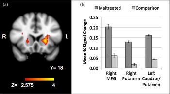
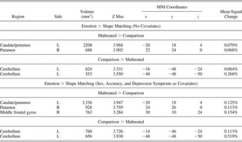
An analysis of group differences in task activation revealed two regions where the maltreated group had greater activation than the comparison group for the contrast of emotion matching greater than shape matching. These regions included the bilateral putamen and left caudate (Table 2, Figure 2). In addition, the comparison group had greater activation than the maltreated group in two regions in the left cerebellum. After controlling for sex, depression symptoms (as measured by the BDI-II), and accuracy on emotion trials, group differences in these regions remained significant. The inclusion of these covariates also revealed a region in the right middle frontal gyrus where the maltreated group had greater activation than comparison participants. There were no significant effects of group on amygdala activation.
(Color online) Group differences (maltreated > comparison) in task activation for the contrast of emotion matching > shape matching. (a) Brain image shows significant clusters in right putamen and left putamen/caudate (medial frontal gyrus not pictured). (b) The results from significant clusters are represented in graphical form (M±SE across voxels in cluster).

Group differences in task activation for the comparison of emotion greater than shape matching

Note: The results are reported with and without covariates (sex, accuracy, and depression symptoms).
Functional connectivity: PPI analysis
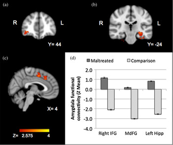
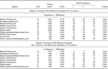
In the PPI analyses, we examined general patterns of limbic connectivity across all subjects. There were no regions that exhibited positive connectivity with the amygdala. However, many regions were negatively correlated (anticorrelated) with amygdala activation. These regions included the medial prefrontal gyrus, anterior cingulate cortex, middle frontal gyrus, caudate, insula, posterior cingulate, parahippocampal gyrus, and thalamus. A group contrast revealed regions showing group differences in negative amygdala connectivity. Relative to the comparison group, the maltreated group had altered connectivity between the bilateral amygdala and left hippocampus, bilateral parahippocampal gyrus, right inferior frontal gyrus (orbital), right inferior frontal gyrus (lateral), medial frontal gyrus, left frontal pole, right middle frontal gyrus, and right inferior parietal lobule/angular gyrus (Table 3, Figure 3). In each of these regions, the comparison group showed a negative correlation with the amygdala, while the maltreated group showed a positive correlation. All regions remained significant after controlling for sex, depression symptoms, and task accuracy, with the exception of the cluster in right middle frontal gyrus, which fell below the cluster threshold of 552 mm3 (to 512 mm3).
(Color online) Group differences in functional connectivity with bilateral amygdala. Representative significant clusters are depicted in parts a–c. (a) Group differences in inferior frontal gyrus. (b) Group differences in left hippocampus. (c) Group differences in medial frontal gyrus. (d) Representative significant contrasts of amygdala connectivity are depicted in graphical form (M±SE across voxels in cluster).

Group differences in functional connectivity as revealed through the PPI analysis

Note: The results are reported with and without covariates (sex, accuracy, depression symptoms).
Discussion
In the present study, we investigated differences in brain function and connectivity associated with childhood maltreatment. We employed an emotion-processing fMRI task in a sample of adults who experienced childhood maltreatment, and in a comparison group of nonmaltreated adults with similar SES backgrounds, to target the neural circuitry underlying behavioral alterations associated with early maltreatment. In the emotion-matching task, we identified regions in the basal ganglia and dlPFC that showed greater activation for maltreated participants than for the comparison group. The PPI analysis revealed that, although the full sample had predominantly negative connectivity between the frontal and limbic regions and the amygdala during the emotional component of the task, maltreated participants had positive connectivity in a number of frontal and subcortical regions. The primary results remained significant even after controlling for sex, accuracy, and depression. These findings suggest that lasting alterations to frontolimbic circuitry occur as a consequence childhood maltreatment.
Participants with a history of maltreatment were significantly less accurate behaviorally during the task than were participants in the comparison group. This result is congruent with previous studies of emotion identification deficits in maltreated children (e.g., Camras et al., Reference Camras, Grow and Ribordy1983). However, accuracy differences between groups were not specific to emotion matching, suggesting that the entire task was more challenging for the maltreated group. Five additional participants were excluded for extremely poor performance on the shape trials. Four of these individuals were from the maltreated group. Overall, these group differences may reflect more general effects of maltreatment on cognitive and motor function rather than specific effects on emotion processing.
Group differences were observed in task activation, with maltreated participants showing increased activity in the basal ganglia (specifically caudate and putamen). After controlling for task accuracy, sex, and depression, this increased activity in the basal ganglia persisted, and a similar group difference in the middle frontal gyrus emerged. Analogous frontostriatal effects have been found in relation to early life stress, with increased activation in the caudate, putamen, and frontal regulatory regions during a cognitive control task (Mueller et al., Reference Mueller, Maheu, Dozier, Peloso, Mandell and Leibenluft2010). Greater activation of the basal ganglia is also supported by Dannlowski et al. (Reference Dannlowski, Kugel, Huber, Stuhrmann, Redlich and Grotegerd2013), who found that maltreatment was positively associated with putamen activation in response to sad faces. However, the direction of effects in the basal ganglia may be context dependent because maltreated participants have also shown reduced basal ganglia activation during a reward-processing task (Dillon et al., Reference Dillon, Holmes, Birk, Brooks, Lyons-Ruth and Pizzagalli2009). Increased recruitment of the basal ganglia, which, in addition to reward processing, plays a central role in performance and response control (Casey et al., Reference Casey, Davidson, Hara, Thomas, Martinez and Galvan2004; O'Doherty et al., Reference O'Doherty, Dayan, Schultz, Deichmann, Friston and Dolan2004), may be indicative of the challenging nature of the task for maltreated participants. Greater activation of the PFC in the maltreated group also supports this hypothesis. The dlPFC is involved in attending to difficult task demands (e.g., MacDonald, Cohen, Stenger, & Carter, Reference MacDonald, Cohen, Stenger and Carter2000) and in the conscious downregulation of emotional responses using cognitive reappraisal (Eippert et al., Reference Eippert, Veit, Weiskopf, Erb, Birbaumer and Anders2007). It is reasonable to expect that cognitively and emotionally challenging tasks would require greater recruitment of these regions. Certainly, the difficulty of the task for the maltreated group is reflected behaviorally in lower accuracy. However, maltreatment accounted for differences in neural activation even after controlling for behavior, suggesting that regardless of performance during emotion matching, the maltreated group experienced the task differently than the comparison group. Taken together, the results from the PFC and basal ganglia suggest that the cognitive regulatory demands of the task may have differed across groups.
We did not find the predicted relationship between maltreatment and amygdala activation that has been observed in other studies (e.g., Dannlowski et al., Reference Dannlowski, Kugel, Huber, Stuhrmann, Redlich and Grotegerd2013). The use of a comparably high-risk comparison group may have made it difficult to detect subtle differences in amygdala activation. We know from behavioral data that alterations in emotion processing differ by maltreatment subtype (e.g., Pollak et al., Reference Pollak, Cicchetti, Hornung and Reed2000); however, we did not have sufficient power to divide groups into maltreatment subtypes, and many participants experienced more than one subtype. Therefore, by pooling across participants, we may have been unable to capture the heterogeneity of maltreatment experience. Comparison to the existing literature also proves challenging because of wide variation among samples and methods. Some previously reported results have come from children or adolescents (e.g., De Bellis & Hooper, Reference De Bellis and Hooper2012; McCrory et al., Reference McCrory, De Brito, Sebastian, Mechelli, Bird and Kelly2011, Reference McCrory, De Brito, Kelly, Bird, Sebastian and Mechelli2013), some from patient groups (e.g., Grant et al., Reference Grant, Cannistraci, Hollon, Gore and Shelton2011), and some from samples that did not include control subjects matched on demographic variables such as SES. In our sample, both groups came from high-risk backgrounds with multiple potential sources of early adversity, aside from maltreatment. We know that diverse forms of early life stress, including low SES, have been associated with structural alterations to the amygdala (e.g., Hanson et al., Reference Hanson, Nacewicz, Sutterer, Cayo, Schaefer and Rudolph2015). Therefore, it is possible that other risk factors contributed to amygdala alterations in both groups, making it challenging to detect differences unique to maltreatment.
The PPI analysis with all participants revealed a number of frontal and subcortical regions that showed negative correlations with amygdala activity. Negative amygdala–prefrontal connectivity parallels previous work (Gee et al., Reference Gee, Gabard-Durnam, Flannery, Goff, Humphreys and Telzer2013; Hare et al., Reference Hare, Tottenham, Galvan, Voss, Glover and Casey2008; Hariri et al., Reference Hariri, Bookheimer and Mazziotta2000; Kim, Somerville, Johnstone, Alexander, & Whalen, Reference Kim, Somerville, Johnstone, Alexander and Whalen2003), and it has been theorized that this inverse relationship reflects frontal, top-down regulation of amygdala reactivity (Hariri, Mattay, Tessitore, Fera, & Weinberger, Reference Hariri, Mattay, Tessitore, Fera and Weinberger2003; Kim, Loucks, et al., Reference Kim, Gee, Loucks, Davis and Whalen2011). Gee et al. (Reference Gee, Gabard-Durnam, Flannery, Goff, Humphreys and Telzer2013) reported a developmental shift from positive amygdala–mPFC connectivity in childhood to negative connectivity in adulthood. The group connectivity from our adults matches this research, suggesting that when the mPFC was more active, amygdala activity decreased. Although our data cannot speak to the causal directionality of this relationship, the findings are consistent with the hypothesis of top-down frontal regulation.
A number of brain regions showed negative amygdala connectivity for the comparison group, but exhibited positive connectivity for the maltreated group. We did not observe this effect in the vmPFC, which has been examined frequently in this context due to structural connections to the amygdala (Kim, Loucks, et al., Reference Kim, Loucks, Palmer, Brown, Solomon and Marchante2011). We did, however, observe connectivity differences in a number of other frontal regions. Specifically, the maltreated group had positive connectivity between the amygdala and the OFC, vlPFC, dmPFC, and dlPFC. The vlPFC (or inferior frontal gyrus) has been associated with cognitive control and response inhibition (Aron, Robbins, & Poldrack, Reference Aron, Robbins and Poldrack2014; Hampshire, Chamberlain, Monti, Duncan, & Owen, Reference Hampshire, Chamberlain, Monti, Duncan and Owen2010; Levy & Wagner, Reference Levy and Wagner2011) and has been found to activate in response to emotional distractors (Yamasaki, LaBar, & McCarthy, Reference Yamasaki, LaBar and McCarthy2002). Increasing evidence suggests that the OFC, dmPFC, and dlPFC contribute to emotion processing and regulation, particularly through cognitive mechanisms such as reappraisal and attentional control (Banks, Eddy, Angstadt, Nathan, & Phan, Reference Banks, Eddy, Angstadt, Nathan and Phan2007; Etkin et al., Reference Etkin, Egner and Kalisch2011; Ochsner & Gross, Reference Ochsner and Gross2005). These findings suggest that when the amygdala was activated in response to faces, the maltreated group upregulated activation of cognitive control regions. While we are the first to identify this relationship during an emotion-processing task, similar amygdala associations to the OFC and dlPFC have been reported in maltreated samples during resting-state fMRI (Dean et al., Reference Dean, Kohno, Hellemann and London2014; Herringa et al., Reference Herringa, Birn, Ruttle, Burghy, Stodola and Davidson2013).
There are also data to suggest that the observed amygdala–prefrontal connectivity could reflect an elevated neural response to stress or threat. In healthy adults, greater amygdala–OFC and amygdala–dlPFC connectivity have been associated with threat-induced anxiety (Gold, Morey, & McCarthy, Reference Gold, Morey and McCarthy2015). In addition, previous data from both resting-state and task-based functional connectivity have indicted that amygdala–dmPFC connectivity is positively correlated with anxiety (Kim, Gee, Loucks, Davis, & Whalen, Reference Kim, Gee, Loucks, Davis and Whalen2011; Robinson, Charney, Overstreet, Vytal, & Grillon, Reference Robinson, Charney, Overstreet, Vytal and Grillon2012) and is associated with a higher inflammatory response to stress (Muscatell et al., Reference Muscatell, Dedovic, Slavich, Jarcho, Breen and Bower2015). Increased connectivity in the maltreated group could signify higher vigilance to the threatening aspect of the angry and fearful faces used in the task. The task may have comprised a fundamentally different experience for maltreated versus comparison participants, being more emotionally disturbing or stressful.
Finally, the PPI analysis revealed group differences in amygdala–hippocampal and parahippocampal connectivity, again with positive connectivity observed for maltreated participants and negative connectivity for the comparison group. This finding is novel, because functional connectivity between these regions has not been examined previously within the context of the emotion-matching task. The normative state of amygdala–hippocampal connectivity during emotion processing is not well characterized at this time. However, data from resting-state connectivity analyses has described a positive relationship between the amygdala and the hippocampus/parahippocampal gyrus (Roy et al., Reference Roy, Shehzad, Margulies, Kelly, Uddin and Gotimer2009). Associations between childhood maltreatment and resting-state connectivity of the amygdala and hippocampus have been mixed, with results showing both decreased (van der Werff et al., Reference van der Werff, Pannekoek, Veer, van Tol, Aleman and Veltman2013) and increased (Dean et al., Reference Dean, Kohno, Hellemann and London2014) connectivity. The manner of interaction between the two regions may be dependent on the nature of the cognitive function elicited by a task. For instance, connectivity between the amygdala and hippocampus has been shown to increase during emotional memory retrieval compared to the retrieval of memories without emotional valence (Smith, Stephan, Rugg, & Dolan, Reference Smith, Stephan, Rugg and Dolan2006). Although this interpretation is speculative, the strong amygdala–hippocampal connectivity observed in our results could indicate that maltreated participants are linking threatening stimuli to reactivation of negative memory traces. Pollak, Cicchetti, and Klorman (Reference Pollak, Cicchetti and Klorman1998) have argued that the association between affective stimuli and traumatic memories alters the subsequent interpretation of emotions following maltreatment. Our data support this hypothesis, showing long-term alterations in the interaction between the hippocampus and amygdala during emotion processing.
Strengths and limitations
The current study makes several important contributions over and above the existing literature. First, unlike many studies of maltreatment, this project includes a control group matched on socioeconomic and risk status. Second, both maltreated and comparison groups were assessed prospectively and did not rely on retrospective self-report. Third, the current study provides further understanding of altered brain networks as a function of childhood maltreatment using functional connectivity analyses. Such analyses complement more traditional measures of task-based functional brain activation. Studies including multiple levels of analysis are necessary to understand the developmental pathways associated with complex risk factors such as childhood maltreatment.
The current study also has several limitations. Although the participants in the study have been followed since childhood, MRI assessments were limited to one time point in adulthood. Consequently, we have no way to map the trajectory of brain alterations associated with early experience. We cannot rule out the possibility of preexisting differences in brain structure and function that preceded maltreatment, including prenatal risks such as exposure to drugs or alcohol in utero. Maltreatment is known to co-occur with many other types of risk. Our design mitigates this confound by recruiting all participants from high-risk, low-SES backgrounds; however, we cannot be certain that we accounted for all relevant factors. As mentioned previously, there was wide heterogeneity of maltreatment experience within the maltreated sample, including type of maltreatment, duration and severity of maltreatment, and age of onset. Given the size of our sample and the high degree of comorbidity in maltreatment subtypes, we were unable to examine the unique influence of different subtypes of maltreatment. A larger sample would be desirable to examine subtypes of maltreatment. However, our sample size was consistent with or exceeded previous imaging studies of maltreatment. Because a number of the longitudinal participants were unable to participate in our study, we did not get the full range of possible cases. Participants who were potentially more successful (e.g., having either moved away or refused because of employment conflicts) or less successful (e.g., incarcerated) were screened out, as well as a number of cases with health-related exclusions (e.g., obesity or metal in body). Consequently, our sample may represent the midrange of outcomes following maltreatment. Finally, in relation to the PPI analysis, it is worth repeating that we did not have measures of directionality between regions. Therefore, although we speculate about a top-down relationship between frontal regions and amygdala, the evidence was correlational, and no causation can be inferred.
Conclusions and future directions
Our investigation highlights a number of avenues for future research. In particular, we provide further motivation for examining functional neural networks, in addition to the investigation of regional differences in activity. At present, there is very little literature on the effect of maltreatment on the functional relationship among brain regions. Frontolimbic circuitry is likely to be just one of many neural systems affected by early trauma. More research is necessary to understand outcomes associated with individual differences in experience. Timing, chronicity, and type of maltreatment are all critical factors that likely shape neural development and behavioral outcomes (e.g., Cowell, Cicchetti, Rogosch, & Toth, Reference Cowell, Cicchetti, Rogosch and Toth2015). Not all children who are maltreated go on to experience negative outcomes; many demonstrate remarkable resilience in the face of early adversity (Cicchetti, Reference Cicchetti2013). Consequently, it is also imperative to investigate individual differences in relation to adaptive functioning.
The current study highlights altered emotion circuitry and identifies a network of regions vulnerable to long-term effects of maltreatment. Brain and behavioral data suggest that our sample of maltreated individuals may have experienced the task as more cognitively taxing and emotionally stressful than did the nonmaltreated group. The disrupted neural circuitry observed may help explain the differential response to emotionally salient stimuli and may contribute to the heightened risk for psychopathology in individuals with a history of childhood maltreatment. Our study provides added evidence of the lasting neurobiological impact of child maltreatment, but there is clearly much more to be learned about the complex processes involved in the brain's response to extreme stress.