Introduction
Surgical treatment of cholesteatoma aims to create a dry, safe ear and to avoid recurrence. In past years, the ‘gold standard’ for cholesteatoma management has been the canal wall down technique. This procedure allows excellent visualisation of areas where cholesteatoma may be hidden, and enables complete removal of disease. The advantages and disadvantages of this technique have been extensively discussed.Reference Berenholz, Rizer, Burkey, Schuring and Lippy1–Reference Tos and Lau4
After complete eradication of disease, the secondary goal of cholesteatoma surgery is to restore hearing. With the boundaries of biomedical technology ever expanding, ear surgeons now have a wide spectrum of prostheses available, usually obviating the need to use the patients' own tissues (e.g. ossicles, cartilage or cortical bone). These tissues (especially ossicles) may harbour cholesteatoma, which may lead to recurrence. In addition, the sculpting of the replacement ossicle depends on the manual dexterity of the surgeon, as well as the amount and shape of the ossicle left intact, leading to non-standard outcomes.
In this study, we analysed patients undergoing canal wall down mastoidectomy for cholesteatoma, together with ossicular reconstruction with a titanium prosthesis, in order to identify factors associated with favourable and unfavourable anatomical and functional results.
Patients and methods
We reviewed the clinical records of patients diagnosed with cholesteatoma in a tertiary academic hospital between 2005 and 2010. Ninety-seven patients who had been operated upon in a single-stage procedure were included in this study. These patients ranged in age from eight to 71 years (mean, 41 years). The post-operative observation period ranged from six to 60 months (mean, 38 months). We excluded from the study any patients who had undergone staged reconstruction due to mucosal pathology.
All patients were operated upon via a postauricular approach, under general anaesthesia. Following intra-operative evaluation of the cholesteatoma extent, the decision was made to proceed with canal wall down mastoidectomy, in all patients. The major criteria for removing the posterior canal wall were: the presence of extensive disease developing in an inaccessible setting (especially posterior mesotympanic cholesteatoma (sinus or tensa cholesteatomas)); extensive posterior epitympanic cholesteatoma; and the presence of a hypopneumatised mastoid.
The presence of stapes superstructure was the major factor determining the method of ossicular reconstruction. The incus (or its remnants) was removed in every case. The malleus head was removed and the malleus handle preserved whenever possible. A Kurz titanium ossicular prosthesis was used to reconstruct the ossicular chain. Most cases were reconstructed with a 3.75 mm total ossicular replacement prosthesis (TORP) (Ariel, Kurz, Dusslingen, Germany) or a 1.75 mm partial ossicular replacement prosthesis (PORP) (Bell, Kurz, Dusslingen, Germany). Two patients required a 4 mm TORP and one patient a 3.5 mm TORP. In the PORP group, three patients required a 2 mm prosthesis and one patient a 2.5 mm prosthesis. The facial ridge was lowered, leaving 1 to 2 mm of bone over the nerve to avoid exposing it. A conchal cartilage island graft, along with its perichondrium, was interposed between the prosthesis and the fascial graft. After ossicular chain reconstruction, a meatoplasty was performed.
Patients were followed every month during the first three post-operative months, and then every three months for the first year. Early audiological evaluation was performed at three months and at one year. After the first year, patients were followed up yearly. Audiological evaluation was based on audiograms performed within one month pre-operatively, and the most recent post-operative audiogram. Mean pre- and post-operative air conduction and bone conduction hearing thresholds were calculated for 500, 1000, 2000 and 3000 Hz.
The Austin classification was used to define each patient's ossicular status. Hearing results were compared between the various classification groups.
Hearing results were reported in concordance with the Academy of Otolaryngology–Head and Neck Surgery Committee on Hearing and Equilibrium guidelines for the evaluation of results for the treatment of conductive hearing loss. Successful hearing was defined as closure of the air–bone gap to within 10 dB, and a pure tone average of 30 dB or less.
Statistical analysis was performed using the Statistical Package for the Social Sciences version 13.0 for Windows software program. The paired sample t-test or Wilcoxon signed rank test were used to compare continuous variables. The chi-square test was used to compare categorical data.
Results
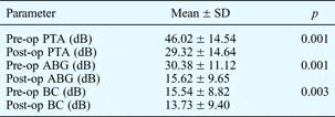
The patients' overall hearing results are shown in Table I.
Overall hearing results

SD = standard deviation; pre-op = pre-operative; PTA = pure tone average; post-op = post-operative; ABG = air–bone gap; BC = bone conduction
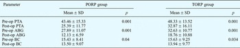
The patients' mean pure tone average ± standard deviation (SD) improved from 46.02 ± 14.54 dB pre-operatively to 29.32 ± 14.64 dB post-operatively (i.e. both TORP and PORP groups combined). The mean air–bone gap ± SD improved from 30.38 ± 11.12 dB pre-operatively to 15.62 ± 9.65 dB post-operatively, for both groups combined. Table II compares results for pre- and post-operative pure tone average and air–bone gap, for the separate PORP and TORP groups.
Post-operative hearing for PORP and TORP groups

PORP = partial ossicular replacement prosthesis; TORP = total ossicular replacement prosthesis; SD = standard deviation; pre-op = pre-operative; PTA = pure tone average; post-op = post-operative; ABG = air–bone gap; BC = bone conduction
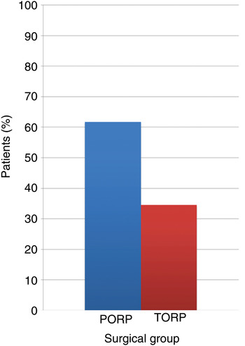
Success (i.e. closure of the air–bone gap to within 10 dB) was achieved in 61.7 per cent of the PORP group and 34.6 per cent of the TORP group. Figure 1 shows these success rates. The mean air conduction hearing threshold in the PORP group was 43.8 dB pre-operatively and 25.5 dB post-operatively; in the TORP group, it was 48.3 dB pre-operatively and 32.8 dB post-operatively.
Mean percentage of patients with a successful post-operative hearing outcome (i.e. closure of the air–bone gap to within 10 dB) in the total ossicular replacement prosthesis (TORP) and partial ossicular replacement prosthesis (PORP) groups.

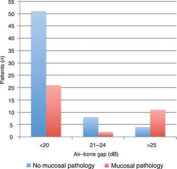
The post-operative air–bone gap was significantly influenced by the status of the middle-ear mucosa (p < 0.05). Mucosal pathology (fibrotic or oedematous) was observed in 34 of the 97 cases. The overall results are summarised in Figure 2.
Patients' post-operative air–bone gap, by presence or absence of middle-ear mucosal pathology.

For practical purposes, patients' cholesteatomas were subdivided into two groups – epitympanic or mesotympanic – according to their localisation. Twenty-eight (28.9 per cent) cholesteatoma cases were epitympanic and 69 (71.1 per cent) were mesotympanic. Cholesteatoma localisation had no statistically significant effect on post-operative air–bone gap (Table III).
Post-operative hearing by cholesteatoma location

p = 0.724, epitympanic vs mesotympanic groups (chi-square test). ABG = air–bone gap; pts = patients
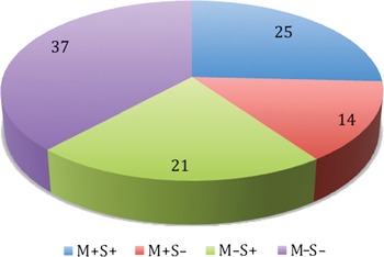
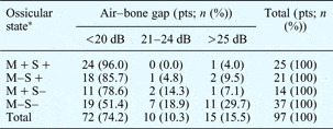
Figure 3 shows the patients' Austin classification of ossicular chain state, while Table IV presents data for Austin ossicular chain classification and hearing results.
Patients' Austin ossicular chain classification. M + = malleus handle present; S + = stapes superstructure present; M– = malleus handle absent; S– = stapes superstructure absent

Post-operative hearing by ossicular chain state*

p<0.005, M + S+ vs M–S– groups (chi-square). *Austin classification. Pts = patients; M + =malleus handle present; S + =stapes superstructure present; M– = malleus handle absent; S– = stapes superstructure absent
Operative findings were similar in the TORP and PORP groups. The facial nerve was dehiscent in 13 patients (seven TORP patients and six PORP patients). A high jugular bulb was observed in three patients (one TORP patient and two PORP patients). There were four cases of lateral semicircular canal dehiscence, in four TORP patients. Concomitant tympanosclerosis was seen in three patients (one TORP patient and two PORP patients). Six patients suffered a recurrence of cholesteatoma (four TORP patients and two PORP patients). Graft failure occurred in four patients (one TORP patient and three PORP patients).
Discussion
In order to obtain the best possible treatment results, the acquired cholesteatoma cases in our series were subdivided into pars tensa and attic cholesteatomas, according to the system of Tos and Lau.Reference Tos and Lau4 Tensa retraction cholesteatoma is defined as arising from a retraction or perforation of the whole pars tensa cholesteatoma, whereas attic cholesteatoma is defined as arising from a retraction or perforation of the posterosuperior part of the pars tensa. In our series, the recurrence rate was lowest (6.6 per cent) for attic cholesteatomas and highest (13.3 per cent) for pars tensa cholesteatomas. Hearing results were best for attic cholesteatomas and poorest for pars tensa cholesteatomas.
We believe that cholesteatoma surgery should be individualised, and that both canal wall up and canal wall down methods have their place. During surgical management of attic cholesteatomas, one of the most important factors is preservation of the middle-ear cavity, in whole or part, in order to improve the functional result. Thus, one may predict better results for patients with epitympanic cholesteatoma, because of the greater possibility of middle-ear cavity preservation. However, in our study the cholesteatoma location (i.e. epitympanic versus mesotympanic) did not affect patients' hearing outcomes in terms of post-operative air–bone gap. This result may have arisen because all the epitympanic cholesteatomas in this series were advanced, necessitating removal of the canal wall.
A number of studies have attempted to determine the prognostic significance of peri-operative inflamed mucosa on ossiculoplasty outcomes.Reference Felek, Islam, Celik, Demirci, Samim and Kose5–Reference Vos, Gersdorff and Gerard7 Some authors, studying series consisting of both primary and staged procedures, have found an association between inflammation and worse hearing outcome, although the strongest prognostic indicators tend to be ‘staging-independent’ (in particular, ossicular chain status and canal wall preservation). In our study, the presence of mucosal pathology had a statistically significant effect on functional results, when both PORP and TORP groups were considered together. However, when the PORP group was considered alone, the presence of mucosal pathology did not have a statistically significant effect on the post-operative air–bone gap.
When considering the debate regarding surgical staging, it is important to bear in mind the practical issues raised by Tos and Lau in 1989.Reference Tos and Lau4 It remains true that two operations are more costly than one; a second operation places an additional financial burden upon the patient and the healthcare system. Furthermore, it is difficult to predict what proportion of cases scheduled for a second stage will actually undergo that surgery. Many patients will be satisfied with a dry ear, and will be reluctant to risk a further procedure. In our study, four TORP patients and two PORP patients required revision surgery (6.2 per cent) due to recurrence. Our results suggest that staging of surgery is not necessary even in cases of advanced cholesteatoma. We believe that revision surgery can still be easily performed in a reconstructed ear. In addition, in terms of anatomical success, a primary ossiculoplasty can help to retain the middle-ear cavity.
In cases of cholesteatoma, the stapes superstructure is frequently involved in the destructive process. Some authors have reported that the loss of the stapes arch is associated with a significantly poorer auditory outcome.Reference Alaani and Raut8 We found similar results. A functional malleus handle has been reported to be an important factor in prosthesis stabilisation and sound transmission improvement.Reference Albu, Babighian and Trabalzini9 Although Albu et al. found that missing stapes superstructure was a negative predictive factor as regards hearing function, in our series the absence of the malleus handle had no significant negative effect on hearing outcome. In general, the mucosal status and the availability of the stapes for reconstruction are the most important predictors of a favourable functional outcome following canal wall down surgery. Even those patients with less than ideal hearing results report some kind of improvement in speech reception; the reasons for this should be investigated.
• This study assessed single-stage, canal wall down mastoidectomy for advanced cholesteatoma, with ossicular chain reconstruction with titanium prosthesis
• Good hearing results were found
• Partial ossicular replacement prosthesis results were excellent for aerated middle-ear cavities with the stapes superstructure present
Titanium prostheses have been successfully used for ossicular reconstruction performed as single-stage surgery.Reference Stankovic10–Reference Martin, Weller, Kim and Smith12 In our 97 patients, a mean air–bone gap improvement of 14.8 dB was observed at 60 months' follow up. In a large, multicentre series including 528 patients, Begall and Zimmermann reported a 15 dB air–bone gap improvement at six months' follow up, while Schmerber et al. reported a 12.7 dB air–bone gap improvement at 20 months' follow up.Reference Begall and Zimmermann13, Reference Schmerber, Troussier, Dumas, Lavieille and Nguyen14 However, the proportion of patients with cholesteatoma in these studies was only 34 and 74 per cent, respectively. Our study results compare favourably to those of Ho et al. (who reported a success rate of 56 per cent (64 per cent for PORP and 45 per cent for TORP)) and Gardner et al. (with success in 71 per cent of PORPs and 44 per cent of TORPs), and are comparable to those of Schmerber et al. (with success in 77 per cent of PORPs and 52 per cent of TORPs).Reference Schmerber, Troussier, Dumas, Lavieille and Nguyen14–Reference Gardner, Jackson and Kaylie16 Our results were significantly better for PORP cases than TORP cases (with success in 91.3 per cent of PORPs versus 58.8 per cent of TORPs).
The meatoplasty is a very important but often neglected part of the ossiculoplasty operation. A well designed meatoplasty is able to create a shallow mastoid cavity, which improves the hearing results. In addition, a shallow mastoid cavity helps to ensure a dry ear, thus improving patient satisfaction. Obliteration procedures can be used to achieve a shallow mastoid cavity. However, in our series the use of a properly designed meatoplasty technique enabled 50 per cent of the resulting mastoid cavities to remain very shallow. Only three patients required an obliteration procedure, due to a persistently discharging mastoid cavity.
Conclusion
In patients requiring canal wall down surgery for advanced cholesteatoma, ossicular chain reconstruction with a titanium prosthesis offers good functional results when performed as a single-stage procedure. The results of canal wall down PORP procedures are excellent if the middle-ear cavity is aerated and the stapes superstructure is present. In order to improve the results of canal wall down TORP procedures, some technical modifications may be required.
Acknowledgement
We thank Assistant Professor Dr Cavit Isık Yavuz for his help with statistical analysis.