


A 48-year-old man visited our hospital with progressively worsening exertional weakness of the left upper extremity. On Doppler ultrasonography, there was demonstration of reverse flow in the left vertebral artery, which is suggestive of subclavian steal syndrome. Contrast-enhanced multidetector CT angiography of the head and neck revealed a right-sided aortic arch with a normal origin of the brachiocephalic trunk and left common carotid artery. The brachiocephalic trunk was divided in the usual manner into the right common carotid and right subclavian arteries. The right vertebral artery arose normally from the right subclavian artery, and both carotid bifurcations were normal. The proximal part (prevertebral part) of the left subclavian artery was congenitally absent, with a very thin fibrous band being the only evidence of its existence along the course. Instead, a huge anomalous artery arose from the left external carotid artery, turned backward with a large suboccipital loop, penetrated the left C2 transverse foramen and entered the V4 segment of the left vertebral artery in the cranium. Caudally, the same artery took a downward course as the left vertebral artery extracranially and then as the distal left subclavian artery without interruption. The left intracranial vertebral artery was mildly hypoplastic, whereas the right vertebral artery was dominant (Figure 1).
Multidetector CT angiography of a 48-year-old man presenting with progressively worsening exertional weakness of the left upper extremity. (A–D) Sequential sagittal oblique maximum intensity projection (MIP) images show the course of an anomalous left vertebral artery (arrows) arising from the left external carotid artery (curved arrow). The left internal carotid artery is indicated by arrowheads. (E) Axial CT angiographic image demonstrates a right-sided aortic arch (arrow). (F) Coronal MIP image shows the congenital absence of the left subclavian artery as a linear fibrous band (arrowhead). (G) Three-dimensional color-coded volume-rendered CT angiography shows the absence of the proximal segment of the left subclavian artery (arrow) and the continuity of the anomalous left vertebral artery with the left subclavian artery (arrowhead). (H) Color-coded three-dimensional vascular reconstruction clearly illustrates the anomalous left vertebral artery arising from the left external carotid artery (arrows) and continuing as the left subclavian artery (arrowhead).
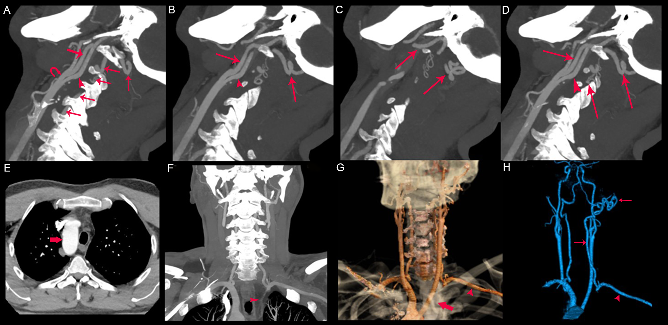
Figure 1. Long description
Panel A: A sagittal oblique maximum intensity projection (MIP) image showing the course of an anomalous left vertebral artery (arrows) arising from the left external carotid artery (curved arrow). The left internal carotid artery is indicated by arrowheads. Panel B: Another sagittal oblique MIP image showing the course of the anomalous left vertebral artery. Panel C: A sagittal oblique MIP image showing the course of the anomalous left vertebral artery. Panel D: A sagittal oblique MIP image showing the course of the anomalous left vertebral artery. Panel E: An axial CT angiographic image demonstrating a right-sided aortic arch (arrow). Panel F: A coronal MIP image showing the congenital absence of the left subclavian artery as a linear fibrous band (arrowhead). Panel G: A three-dimensional color-coded volume-rendered CT angiography showing the absence of the proximal segment of the left subclavian artery (arrow) and the continuity of the anomalous left vertebral artery with the left subclavian artery (arrowhead). Panel H: A color-coded three-dimensional vascular reconstruction illustrating the anomalous left vertebral artery arising from the left external carotid artery (arrows) and continuing as the left subclavian artery (arrowhead).
This unique vascular anatomy fully explains the retrograde vertebral flow observed on Doppler ultrasonography. Importantly, the reversed flow represented true steal physiology but resulted from a congenital developmental anomaly rather than the acquired proximal subclavian stenosis or occlusion that characterizes the classic subclavian steal syndrome.
Congenital anomalies of the vertebral and subclavian arteries with a right-sided arcus aorta are rare and are generally discovered incidentally. Reference Zhang, Cui, Ren, Chen and He1,Reference Nakata, Stahlfeld and Simone2 While the majority are clinically silent, uncommon developmental variants can cause hemodynamic alterations that mimic those of acquired vascular diseases. Reference Cai, Xie and Wang3,Reference Ballout, Schneider, Patel and Patsalides4 In particular, congenital anomalies involving collateral supply to the subclavian artery may generate retrograde vertebral flow identical to that seen in the classic subclavian steal syndrome, potentially leading to diagnostic confusion if evaluated by Doppler ultrasonography alone.
We describe an exceptionally rare congenital vascular configuration in which the left vertebral artery originated from the left external carotid artery and continued directly as the left subclavian artery, replacing the absent proximal subclavian segment. To the best of our knowledge, the combination of a right aortic arch, congenital absence of the proximal left subclavian artery and a left vertebral artery arising from the external carotid artery and continuing as the left subclavian artery has not been previously reported.
Although the Doppler findings were true for retrograde vertebral flow, the pathophysiology was entirely different from that of subclavian steal syndrome. Instead of being the result of an acquired subclavian occlusive disease, the retrograde flow was a result of a rare congenital collateral that perfused the left upper extremity. CT angiography was critical in defining the complex vascular anatomy, making the correct diagnosis and avoiding potential misdiagnosis as an acquired occlusion of the subclavian artery. Recognition of this unusual developmental variant is useful for preventing unnecessary vascular intervention and allowing for optimal clinical and surgical management.
Author contributions
MKD: Writing – original draft, Visualization, Validation, Supervision, Software, Resources, Methodology, Investigation, Formal analysis, Data curation, Conceptualization. AA, AA, TO: Writing – original draft, Visualization, Resources, Methodology, Investigation, Formal analysis, Data curation.
Funding statement
None.
Competing interests
None.
Ethical standard
Not required.
Declaration of patient consent
The authors certify that they have obtained all appropriate patient consent forms. In the form, the patient has given his consent for his images and other clinical information to be reported in the journal. The patient understands that his name and initials will not be published and due efforts will be made to conceal his identity, but anonymity cannot be guaranteed.

 Open access

