Introduction
Helminthiasis represents a major constraint to the development of livestock production as it is responsible for great economic losses, particularly in the regions where extensive grazing is practised (Waller, Reference Waller1997). Increased mortality, decreased meat production and reproductive fitness result from severe gastrointestinal nematode infection in ruminants (Hoste et al., Reference Hoste, Torres-Acosta, Paolini, Aguilar-Caballero, Etter, Lefrileux, Chartier and Broqua2005). In order to overcome the deleterious effects of helminths, conventional chemical anthelmintics are the first choice; however, the livestock production industry does not benefit through these control measures, due to higher cost and the non-availability of such compounds to small farmers. Moreover, these parasites have evolved resistance to various anthelmintics, such as benzimidazole, imidazothiazole and ivermectin, which has become a major problem worldwide (Singh et al., Reference Singh, Swarnkar and Khan2002; Waller, Reference Waller2003), making their management more difficult. The use of traditional botanicals has emerged as an environmentally safer alternative, proving successful against these parasites (Eguale et al., Reference Eguale, Tilahun, Debella, Feleke and Makonnen2007a; Kamaraj et al., Reference Kamaraj, Rahuman, Elango, Bagavan and Zahir2011; Kanojiya et al., Reference Kanojiya, Shankar, Sudan, Jaiswal and Parashar2015).
Haemonchus contortus, a predominant gastrointestinal nematode in the small ruminants of the tropical, sub-tropical and temperate countries, has been a targeted pathogen due to its rigorous damage to sheep and goats. Several studies have been conducted in various parts of the world, including India, depicting the anthelmintic activity of herbal extracts against H. contortus (Melo et al., Reference Melo, Reis, Bevilaqua, Vieira, Echevarria and Melo2003; Tandon et al., Reference Tandon, Lyndem, Kar, Pal, Das and Rao2004; Roy et al., Reference Roy, Lalchhandama and Dutta2008a, Reference Roy, Dasgupta and Tandonb; Buske et al., Reference Buske, Santurio, de Oliveira, Bianchini, da Silva and de la Rue2013). Evaluation of anthelmintic activity is based on the in vitro egg hatch assay, larval development inhibition (Hordegen et al., Reference Hordegen, Cabaret, Hertzberg, Langhans and Maurer2006; Costa et al., Reference Costa, Bevilaqua, Camurça-Vasconcelos, Maciel, Morais, Castor, Braga and Oliveira2008; Maphosa et al., Reference Maphosa, Masika, Bizimenyera and Eloff2010; Ademola & Eloff, Reference Ademola and Eloff2011; Gregory et al., Reference Gregory, Yoshihara, Ribeiro, Silva, Marques, Meira, Rossi, Sampaio, Louvandini and Hasegawa2015) and a few studies on adulticidal assays (Barrau et al., Reference Barrau, Fabre, Fouraste and Hoste2005; López-Aroche et al., Reference López-Aroche, Salinas-Sánchez, Mendoza de Gives, López-Arellano, Liébano-Hernández, Valladares-Cisneros, Arias-Ataide and Hernández-Velázquez2008; Akkari et al., Reference Akkari, Rtibi, B'chir, Rekik, Darghouth and Gharbi2014). In vitro studies are considered to be pilot screenings of herbal extracts, also assumed to be effective in in vivo trials.
In the past decade, silver nanoparticles (AgNPs) have emerged as one of the fastest growing materials, due to their unique physical, chemical and biological properties, small size and high specific surface area. Biological synthesis of nanoparticles has received attention due to the growing need to develop environmentally benign technologies in material synthesis. Several plant species have been utilized in this regard and only a few have been reported for their antiparasitic potential. Marimathu et al. (Reference Marimathu, Rahuman, Rajkumar, Santoshkumar, Kirthi, Jayaseelan, Bagavan, Zahir, Elango and Kamaraj2011) studied the effectiveness of AgNPs against larvae of Anopheles subpictus, Culex quinquefasciatus and Rhipicephalus microplus. Said et al. (Reference Said, Elsamed and Gohar2012) screened antigiardial activity of silver, chitosan and curcumin nanoparticles. Most recently, Kar et al. (Reference Kar, Murmu, Saha, Tandon and Acharya2014) studied the efficacy of gold nanoparticles fabricated from a phytopathogenic fungus Nigrospora oryzae against a model cestode Raillietina sp. However, no report exists to date on their anthelmintic properties against H. contortus.
The present study employs a green approach to synthesize AgNPs using Azadirachta indica (neem) leaf extract, which is well known for its medicinal properties in the Indian Ayurvedic system, and various parts of the neem tree have been recommended for use against gastrointestinal nematodes and related problems in many parts of the world (Biswas et al., Reference Biswas, Chattopadhyay, Banerjee and Bandyopadhyay2002; Subapriya & Nagini, Reference Subapriya and Nagini2005). The plant has been extensively reviewed and shown to possess antimalarial, antibacterial, antifungal, antitumour, antiviral and other biological activities (Tiwari et al., Reference Tiwari, Verma, Chakraborty, Dhama and Singh2014; More et al., Reference More, Raut and Taware2015); however, to the best of our knowledge, no reports are available on in vitro anthelmintic efficacy of green synthesized AgNPs using A. indica leaves against H. contortus. Therefore, the aim of the present study was to evaluate in vitro anthelmintic activity of biologically synthesized AgNPs against H. contortus.
Materials and methods
Synthesis of silver nanoparticles
Azadirachta indica leaves were collected from Dayalbagh, Agra (India) and a known amount (25 g) was weighed, washed with distilled water and finally chopped into small pieces in a 250-ml flask with 100 ml of Milli Q water. The aqueous extract was obtained by heating the suspension in a microwave, followed by filtration through Whatman no. 1 and 0.45 μm Millipore filters. This extract (25%) was interacted with freshly prepared 1.0 mm silver nitrate solution at a ratio of 9:1 and left overnight at room temperature (Shankar et al., Reference Shankar, Rai, Ahmad and Sastry2004). Synthesis of AgNPs was confirmed by periodic sampling of aliquots through ultraviolet–visible (UV-Vis) spectrophotometry, the broth containing AgNPs was centrifuged at 8000 rpm for about 15 min and redispersed in distilled water to remove any impurities, and the purified pellet so obtained was dried. Characterization of AgNPs was accomplished through transmission electron microscopy (TEM), scanning electron microscopy (SEM) and X-ray diffraction studies.
Egg hatch and worm in vitro assays
The egg hatch assay (EHA) was performed according to the guidelines of the World Association for the Advancement of Veterinary Parasitology (WAAVP) (Coles et al., Reference Coles, Bauer, Borgsteede, Geerts, Klei, Taylor and Waller1992). Adult female worms were collected from the abomasa of naturally infected goats being slaughtered at the slaughterhouse. The worms were placed in phosphate-buffered saline (PBS) and crushed to liberate eggs, which were washed repeatedly with distilled water. Aqueous A. indica leaf extract and biologically synthesized AgNPs were the test treatments, while albendazole was used as a positive control and untreated eggs in water served as the negative control. In the assay, approximately 100–200 eggs in 0.5 ml of water were placed in each test tube (5 ml). Serial concentrations of AgNPs (0.00001, 0.0001, 0.001, 0.01, 0.1 and 1 μg/ml) and aqueous A. indica leaf extract (10, 50, 100, 150, 200 and 250 μg/ml) were prepared in 0.5 ml of distilled water, which was made up to 1 ml of test volume together with water containing eggs. Albendazole, originally dissolved in dimethyl sulphoxide (DMSO), at concentrations of 0.03125, 0.0625, 0.125, 0.250 and 0.5 μg/ml, was used as the positive control. The test tubes were covered and left at room temperature for 48 h. To stop hatching after 48 h, a drop of Lugol's iodine solution was added in each test tube. Hatched larvae (dead and alive) and unhatched eggs were counted under a dissecting microscope at 40 × magnification and the experiment was run in triplicate for each concentration.
The in vitro effect of AgNPs and aqueous A. indica leaf extract on adult H. contortus was measured according to Hounzangbe-Adote et al. (Reference Hounzangbe-Adote, Paolini, Fouraste, Moutairou and Hoste2005). The adult worms were collected from the abomasa of freshly slaughtered goats. Immediately after slaughter, the abomasa were collected and transported to the laboratory in normal saline. The collected parasites were washed thoroughly and placed in PBS. Ten actively moving adult worms were placed in each Petri dish containing biologically synthesized AgNPs (1, 5, 10, 15, 20 and 25 μg/ml) and aqueous A. indica leaf extract (200, 400, 600, 800, 1000 and 1200 μg/ml). PBS alone served as the negative control group, whereas albendazole dissolved in DMSO and diluted in PBS at concentrations of 16, 32, 63, 125 and 250 μg/ml was used as the positive control. Each experiment was performed in triplicate. Inhibition of worm motility was the measure of the anthelmintic potential of test extracts. After 24 h, the test solution was washed away and the parasite was again suspended in PBS for about 30 min for possible recovery of parasite motility. The number of motile (alive) and immotile (dead) worms were counted under a dissecting microscope, and recorded for each concentration. Percent mortality (M) was calculated for each concentration using following formula:
 $$\eqalign{{\rm mortality}\,(\%) & = (\hbox{number of dead worms}/\hbox{total number} \cr & \qquad \hbox{of worms per test}) \times 100}$$
Data analysis
The egg hatch inhibition in the EHA and percent mortality in the adult worm motility inhibition test were analysed statistically and represented as mean ± SE. Probit analysis was conducted through the USEPA (2010) program version 1.5 software, based on Finney (Reference Finney1971), for determining the IC50 and IC90 inhibitory concentration for EHA, and LC50 and LC90 for in vitro anthelmintic activity of AgNPs against adult worms after correcting the mortality by Abbott's formula (Abbott, Reference Abbott1925).
Results
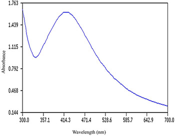
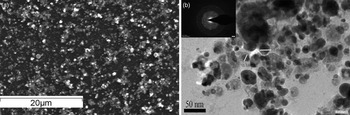
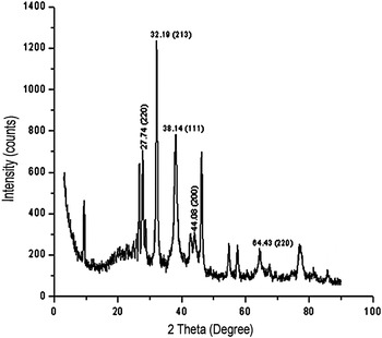
The synthesis of AgNPs using A. indica leaf extract was confirmed by UV-Vis spectral scanning at 300–700 nm. A change in colour of 1 mm silver nitrate solution from colourless to dark brown 24 h after the addition of A. indica leaf extract was the first indication of AgNPs synthesis. This bioreduction was completed at room temperature. UV-Vis spectral scanning demonstrated initiation of a peak at 336 nm with absorbance 0.957, and a sharp peak produced at 420 nm with absorbance 1.629 (fig. 1). The shape and size of the AgNPs were analysed after 24-h incubation through SEM and TEM imaging, where SEM depicted the surface topography of AgNPs (fig. 2a), whereas the spherical shape of synthesized AgNPs, measuring about 15–25 nm in size, has been revealed through TEM (fig. 2b, along with a selected area of a diffraction image of a randomly selected nanoparticle in the inset, indicating the presence of silver). This crystalline nature of synthesized AgNPs was further confirmed through XRD analysis, which is evident from distinct peaks at 38.14° and 44.08° corresponding to (111) and (200) planes of silver (International Center for Diffraction Data, ICDD no. 04-0783), although there were other, unassigned peaks which represented compounds formed on interaction with the glass slide (fig. 3).
UV-Vis spectrum of AgNPs synthesized using A. indica leaf extract.

(a) Scanning electron micrograph showing the morphological characteristics of synthesized AgNPs using A. indica leaf extract; and (b) transmission electron micrograph showing the shape and size of AgNPs, with an area of the diffraction image of randomly selected nanoparticles (inset).

X-ray diffraction patterns of biologically synthesized AgNPs using A. indica leaf extract, with characteristic crystalline silver peaks at 38.14° and 44.08°.

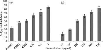
The mean egg hatch inhibition induced by green synthesized AgNPs after 48 h was calculated to be 85 ± 2.89% with the highest concentration of AgNPs (fig. 4). AgNPs were effective at much lower concentrations as compared to A. indica leaf extract. The highest concentration of albendazole (0.5 μg/ml) showed 100% egg hatch inhibition. Remarkable changes on egg hatching were induced by AgNPs, which is evident from shrinkage and disintegration of the embryo when compared to the control; however, comparatively less distortion was noticed in eggs following exposure to aqueous A. indica leaf extract.
Mean dose-dependent responses (±SE) of egg hatching of H. contortus exposed to (a) biologically synthesized AgNPs and (b) A. indica leaf extract.

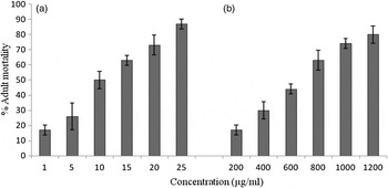
The mean percentage mortality induced by biologically synthesized AgNPs after 24 h showed that the highest concentration induced 87 ± 3.3% adulticidal activity (fig. 5). The mean percentage mortality induced by A. indica leaf extract after 24 h was recorded at fairly higher concentrations. Moreover, morphological alterations in H. contortus adult worms were also noted, including a remarkable coiling with AgNPs, which was more prominent in worms treated with A. indica leaf extract than in controls.
Mean dose-dependent responses (±SE) of adult mortality of H. contortus exposed to (a) biologically synthesized AgNPs and (b) A. indica leaf extract.

IC50 values were calculated as 0.001 μg/ml in the case of AgNPs treatment and 115.67 μg/ml with A. indica leaf extract. LC50 values were found to be 7.89 μg/ml in the case of AgNPs treatment and 588.54 μg/ml with A. indica leaf extract exposure (table 1).
Regression analysis of egg hatch inhibition and in vitro adult mortality activity of biosynthesized AgNPs and aqueous A. indica leaf extract against H. contortus; IC, inhibitory concentration; LC, lethal concentration; CL, 95% confidence limits.

Discussion
Haemonchus contortus is an important abomasal helminth of small ruminants, responsible for disease and major production losses worldwide. This blood-feeding parasite can cause severe anaemia and rapid death in livestock. The main objective of this work was to demonstrate ovicidal and adulticidal activity of biologically synthesized AgNPs using A. indica leaf extract. The mean percentage egg hatch inhibition induced by green synthesized AgNPs and A. indica leaf extract after 48 h was 85% and 82%, respectively, with the highest concentrations. The synthetic compound albendazole induced 84% mortality at a very low concentration, i.e. 0.5 μg/ml. The IC50 value for egg hatch assay was recorded as 0.001 μg/ml in the case of AgNPs treatment and 115.67 μg/ml in A. indica leaf extract exposure. It is evident from the study that AgNPs proved to be very effective in inhibiting egg hatching at very low concentrations and nano-sized AgNPs must have interfered with moulting and other physiological processes involved in the hatching of H. contortus eggs.
In the past decade various plants have been screened for their anthelmintic properties (Min et al., Reference Min, Pomroy, Hart and Sahlu2004; Githiori et al., Reference Githiori, Hoglund, Waller and Baker2004; Shaik et al., Reference Shaik, Terrill, Miller, Kouakou, Kannan, Kallu and Mosjidis2004; Costa et al., Reference Costa, Bevilaqua, Maciel, Camurca-Vasconcelos, Morais, Monteiro, Farias, da Silva and Souza2006; Lange et al., Reference Lange, Olcott, Miller, Mosjidis, Terrill, Burke and Kearney2006); however, in such studies, the inhibitory concentrations were reported to be quite high compared to the those in the present study. Later, Costa et al. (Reference Costa, Bevilaqua, Camurça-Vasconcelos, Maciel, Morais, Castor, Braga and Oliveira2008) assessed the ovicidal activity of ethyl acetate and ethanol extracts of A. indica on H. contortus eggs through EHA, and revealed that an ethanol extract was more effective, inhibiting 99.77% of egg hatching at 3.12 mg/ml. Adamu et al. (Reference Adamu, Naidoo and Eloff2013) evaluated the acetone extracts of 13 plants against H. contortus eggs and larval development. They observed that three plant species, i.e. Heteromorpha trifoliata, Maesa lanceolata and Leucosidea sericea, had EC50 values of 0.62 mg/ml, 0.72 mg/ml and 1.08 mg/ml, respectively, for the EHA, whereas H. trifoliata extract was the most effective, with an EC50 of 0.64 mg/ml, followed by L. sericea (1.27 mg/ml) in the larval developmental assay. Zhu et al. (Reference Zhu, Dai, Yang and Qiu2013) evaluated anthelmintic activity of various constituents of essential oils from Artemisia lancea against H. contortus larval development. Vargas-Magaña et al. (Reference Vargas-Magaña, Torres-Acosta, Aguilar-Caballero, Sandoval-Castro, Hoste and Chan-Pérez2014) studied the anthelmintic activity of acetone–water extracts of various foliage plants and their secondary compounds, and concluded that they blocked the eclosion of larvated eggs. Most recently, Kanojiya et al. (Reference Kanojiya, Shankar, Sudan, Jaiswal and Parashar2015) studied in vitro and in vivo efficacy of Eucalyptus globulus extract against this parasite and reported the methanolic extract to be more effective than the aqueous extract, as evident from an EHA. Hence, it is clear from the literature survey that findings of the present study showed the aqueous A. indica leaf extract to be more potent, and indirect heating through the microwave may account for this enhanced activity.
We have also observed anthelmintic properties of AgNPs and A. indica leaf extract against adult H. contortus and found that the highest concentration of AgNPs (25 ppm) induced 87% mortality in adults, whereas 80% mortality was caused by the highest concentration of A. indica leaf extract, i.e. 1200 μg/ml. LC50 values were recorded as 7.89 μg/ml in the case of AgNPs treatment and 588.54 μg/ml with A. indica leaf extract exposure. Suteky & Dwatmadji (Reference Suteky and Dwatmadji2011) have studied anthelmintic effects of Melastoma malabatricum on EHA, larval development and adult worms. Similarly, Marie-Magdeleine (Reference Marie-Magdeleine, Mahieu, D'Alexis, Philibert and Archimede2010) evaluated aqueous, methanol and dichloromethane extracts of leaf, fruit and root material of Tabernaemontana citrifolia on four developmental stages of the parasite, using EHA, a larval development assay, L3 migration inhibition assay (LMI) and adult worm motility assay. Most recently, Akkari et al. (Reference Akkari, Rtibi, B'chir, Rekik, Darghouth and Gharbi2014) evaluated the in vitro adulticidal effect of Artemisia campestris against H. contortus, and demonstrated that an ethanol extract was more effective over an aqueous extract, as evident from mortality data (91.3 and 3.2%, 8 h post-exposure with the ethanol and aqueous extract, respectively). In the present study, we also observed morphological alterations, which were characterized by coiling up of worms to avoid stress induced in the form of the test compounds used in the present study as potential anthelmintic agents.
The anthelmintic activity of diverse plants studied against H. contortus and other parasites is considered to be due to tannins or flavonoid molecules (Barrau et al., Reference Barrau, Fabre, Fouraste and Hoste2005; Hoste et al., Reference Hoste, Jackson, Athanasiadou, Thamsborg and Hoskin2006; Akkari et al., Reference Akkari, Dargouth and Ben Salem2008). The experimental plant in our study is regarded as the ‘wonder tree’ of India, and more than 300 compounds have been derived from various parts of this tree, which may account for its multi-faceted therapeutic effects (Tiwari et al., Reference Tiwari, Verma, Chakraborty, Dhama and Singh2014) and the effectiveness of the aqueous extract of A. indica leaves as an ovicidal and adulticidal agent in the current study.
AgNPs have been tested for the first time against H. contortus, and have shown excellent anthelmintic activity; however, their exact mode of action is not known. Past studies have shown that transcuticular diffusion is a common means of entry for non-electrolytic substances in nematodes. It has also been shown that this is the predominant route for the uptake of major broad-spectrum anthelmintics, as opposed to oral ingestion. Lipophilic anthelmintics, such as albendazole, are more able to cross the external surface of the helminths than hydrophilic compounds (Geary et al., Reference Geary, Sangster and Thompson1999; Eguale et al., Reference Eguale, Tilahun, Debella, Feleke and Makonnen2007b). However, in the present study a higher mortality rate of adult parasites following AgNPs treatment, as compared to treatment with the aqueous A. indica leaf extract, could be attributed to the extremely small size of the nanoparticles, i.e. 15–25 nm, which probably permitted transcuticular absorption of AgNPs by the parasite. This is the first report where the anthelmintic effects AgNPs have been evaluated against a gastrointestinal nematode, H. contortus, and further work is in progress on the possible cause of mortality effects of AgNPs on these parasites.
Acknowledgements
We are thankful to the Director, Dayalbagh Educational Institute, Dayalbagh, Agra for providing the necessary facilities to carry out this work. We further acknowledge the Advanced Instrumentation Research Facility (AIRF), JNU, New Delhi, for performing XRD; AIIMS, New Delhi, for TEM; and the National Institute of Oceanography, Goa, for SEM analysis.
Financial support
This research received no specific grant from any funding agency, commercial or non-profit sector.
Conflict of interest
None.