Introduction
Concurrent infections of helminths in vertebrate hosts are a common occurrence. The nature of the relationship between two or more parasite species that share a common environment has been extensively debated and studied in order to determine the extent of interactions that may be taking place (Cox, Reference Cox2001; Poulin, Reference Poulin2001; Janovy Reference Janovy2002). Such studies have shown synergistic, antagonistic or negligible levels of interactions (Christensen et al., Reference Christensen, Nansen, Fagbemi and Monrad1987; Cox, Reference Cox2001). Studies on interactions between helminths that occupy different sites within a host are more infrequent and are mainly restricted to mammalian hosts. In particular, concurrent infections of hookworm, intestinal or filarial nematodes with schistosomes that reside in the circulatory system suggest that both synergistic and antagonistic interactions can occur (Christensen et al., Reference Christensen, Nansen, Fagbemi and Monrad1987; Curry et al., Reference Curry, Else, Jones, Bancroft, Grencis and Dunne1995; Corrêa-Oliveira et al., Reference Corrêa-Oliveira, Golgher, Guilherme, Carvalho, Massara, Caldas, Colley and Gazzinelli2002). In fish, indirect interactions have been rarely studied in the field but available evidence would suggest that pairwise associations of larval helminths that share similar transmission routes can occur (Poulin & Valtonen, Reference Poulin and Valtonen2001), while the presence of specific species of larval cestodes in the body cavity or liver may influence the natural occurrence of intestinal helminths (Zhokhov & Pugacheva, Reference Zhokhov and Pugacheva2012; Izvekova & Tyutin, Reference Izvekova and Tyutin2014). For mammalian systems, such interactions could result from strong immunomodulatory mechanisms of one helminth species influencing the host's susceptibility to infection with others (Christensen et al., Reference Christensen, Nansen, Fagbemi and Monrad1987; Cox, Reference Cox2001).
However, other host-related factors may also be important when a helminth causes extensive pathology to a key organ, thereby inhibiting its normal physiological functions. One such organ is the liver, where glycogen synthesis and storage occur, thus providing a permanent or transient habitat for many parasite species (Cheng, Reference Cheng and Kennedy1976). In fish, due to deep interpenetration of the pancreas and liver these organs are considered collectively as the hepatopancreas, although more often referred to as just the ‘liver’. The fish liver therefore has a number of prominent functions, including digestion, immunity, detoxification, nitrogen catabolism, reproduction, blood turnover and storage (Bruslé & Anadon, Reference Bruslé, Anadon, Datta-Munshi and Dutta1996). Therefore any pathological changes in the liver have the potential to alter substantially the physiological environment of the intestine and its suitability as a habitat for parasites. In fish, both cestode and nematode infections of the liver can inhibit normal functioning (Margolis, Reference Margolis and Snieszko1970; Kuperman, Reference Kuperman1981), which may result in host mortalities under particularly high worm burdens.
Liver infections with plerocercoids of the cestode Triaenophorus nodulosus are particularly common in freshwater fish such as the European perch (Perca fluviatilis). Plerocercoids are especially long-lived and are capable of surviving in a fish for up to 3 years, without demonstrating any seasonal variation (Chubb, Reference Chubb1980). The burrowing activity of these parasites and subsequent lysis of liver cell membranes result in a range of pathological symptoms, including inflammation, atrophy, anaemia, leucocytosis, necrosis, structural disintegration, hyperanaemia and haemorrhage, ochre discolouration and dropsy (Kuperman, Reference Kuperman1981; Hoffman et al., Reference Hoffmann, Meder, Klein, Osterkorn and Negele1986; Brinker & Hamers, Reference Brinker and Hamers2007). Furthermore, a decrease in glycogen content and glucose levels in the liver, also associated with an increase in wet weight, influences carbohydrate metabolism (Izvekova, Reference Izvekova2001). Such extensive changes to the liver, in turn, can result in a substantial impact on the physiology of the intestine with, in particular, the activity of digestive enzymes being reduced (Izvekova & Izvekov, Reference Izvekova, Izvekov, Weiss and Faulkner2013). These enzymes have an important role in the establishment, development and site selection of many intestinal helminths (Lackie, Reference Lackie1975; Cheng, Reference Cheng and Kennedy1976; Macko, Reference Macko1980). Thus the intestinal distribution of the trematode Bunodera luciopercae occurring in T. nodulosus-infected perch from the Rybinsk reservoir in Russia was altered, with significantly more trematodes occurring in the posterior third of the intestinal tract compared with fish not infected with T. nodulosus (Izvekova & Tyutin, Reference Izvekova and Tyutin2014). These authors also suggested that, as other helminth species rarely occur in the intestine of perch in this habitat, changes in intestinal site selection by B. luciopercae may be related to the activity of enzymes and the influence that T. nodulosus in the liver has on their occurrence (Izvekova & Tyutin, Reference Izvekova and Tyutin2014).
This intriguing result, alongside the immunological studies undertaken on concurrent helminth infections in mammals, raises the question of whether the occurrence of all intestinal helminths in perch is influenced by the presence of T. nodulosus plerocercoids within the liver. The aim of the present study, using an existing large-scale dataset, is to determine the effects of T. nodulosus on the occurrence and site selection of four common intestinal helminths of perch.
Materials and methods
Collection and examination of fish
Perch were sampled from the Queen Mary reservoir in the lower Thames valley, England. This is a completely man-made circular embankment reservoir constructed to provide storage water for London, built up above the ground level of the surrounding land, with a surface area of 290 ha, depth 12 m and a volume of 30 × 106 m3. Further details are described by Morley & Lewis (Reference Morley and Lewis2010).
A total of 183 perch were trapped during the early summer using purpose-built cages, and transferred immediately to the laboratory before being examined within 24 h of capture. The total body length, weight, sex, liver and gonad weight were all recorded, and fish were dissected and examined for helminths, using standard parasitological techniques. Site selection for intestinal helminths was determined by measuring the entire length of the intestine, which was divided equally into upper, middle and lower sections, with parasites assigned to those sections during the post-mortem examination. The condition coefficient (K), hepatosomatic index (HSI) and gonadosomatic index (GSI) for each individual fish were calculated as follows
 $$\eqalign{& K = {\rm body}\,{\rm weight/body}\,{\rm length}^{\rm 3} \times 100 \cr & {\rm HSI} = {\rm liver}\,{\rm weight/body}\,{\rm weight} \times 100 \cr & {\rm GSI} = {\rm gonad}\,{\rm weight/body}\,{\rm weight} \times 100}$$
Data analysis
The prevalence, mean intensity and mean abundance of intestinal helminths between T. nodulosus-infected and non-infected perch were calculated according to Bush et al. (Reference Bush, Lafferty, Lotz and Shostak1997) and analysed using the Quantitative Parasitology 3.0 statistical package (Reiczigel & Rozsa, Reference Reiczigel and Rozsa2005). All other analyses were undertaken using SPSS version 21 statistical package (SPSS Inc., Chicago, Illinois, USA). One-way analysis of variance (ANOVA) was used to compare T. nodulosus infection levels between male and female fish, and also for comparison of K, HSI and GSI between T. nodulosus-infected and non-infected perch. Analysis of correlations between the intensities of T. nodulosus plerocercoid infections and the intensities of infections with each intestinal helminth species were undertaken using Pearson's correlation coefficient. For analysis of interactions between the general occurrence of intestinal helminths as well as their specific interactions along the length of the intestine, both within and between the two fish populations, data were normalized by transformation and zeros (uninfected fish) were removed from the dataset. The data were then analysed with linear regression for general occurrence, and multinomial logistic regression for intestinal site selection.
Results
The perch population in the Queen Mary reservoir was heavily infected with T. nodulosus plerocercoids, with a prevalence of 69.9% and mean intensity of 2.9 parasites in each liver, with the sex of the fish having no influence on infection levels (one-way ANOVA: P = 0.682, F = 0.169). Comparisons of the condition of T. nodulosus-infected and non-infected perch (table 1) showed that those harbouring this cestode were stunted, with a significantly lower length and weight (one-way ANOVA: P < 0.001, F = 32.654 and P < 0.001, F = 18.749, respectively). However, there was no significant difference in K, HSI and GSI values between the two perch populations (one-way ANOVA: P = 0.229, F = 1.455; P = 0.135, F = 2.253; P = 0.071, F = 3.292, respectively).
Condition of Trianophorus nodulosus-infected and non-infected perch, relative to body length and weight, condition coefficient (K), hepatosomatic (HSI) and gonadosomatic (GSI) indices.

The composition of the helminth fauna in the intestine of perch consisted of the acanthocephalan Acanthocephalus lucii, the nematode Camallanus lacustris, the cestode Proteocephalus percae and the trematode B. luciopercae. Analysis of the occurrence of these species between T. nodulosus-infected and non-infected fish (table 2) showed significantly lower prevalences for A. lucii (χ2 = 4.289, P = 0.038) and C. lacustris (χ2 = 5.728, P = 0.017) in perch harbouring plerocercoids in the liver, but no significant difference in the occurrence of the other two helminth species (P. percae: χ2 = 0.074, P = 0.786; B. luciopercae: χ2 = 0.162, P = 0.687). However, there were no significant differences in the mean intensities of the four intestinal helminth species compared between the T. nodulosus-infected and non-infected perch populations (A. lucii: P = 0.461, t = 0.765; C. lacustris: P = 0.227, t = 1.210; P. percae: P = 0.648, t = −0.468; B. luciopercae: P = 0.513, t = 0.699). On the other hand, there was a significantly lower mean abundance of C. lacustris in T. nodulosus-infected perch (P = 0.039, t = 2.253), but not of A. lucii (P = 0.204, t = 1.366), P. percae (P = 0.904, t = −0.131) and B. luciopercae (P = 0.442, t = 0.767). Within T. nodulosus-infected perch there were no significant correlations between the intensities of plerocercoids in the liver compared with intensities for the four intestinal helminth species (A. lucii: Pearson correlation = −0.027, P = 0.760; C. lacustris: Pearson correlation = 0.106, P = 0.232; P. percae: Pearson correlation = −0.094, P = 0.292; B. luciopercae: Pearson correlation = 0.110, P = 0.213).
Prevalence (%), mean intensity (± SD) and mean abundance (± SD) of infection of intestinal helminths in Triaenophorus nodulosus-infected and non-infected perch.

In samples of perch that were not infected with T. nodulosus, analysis of the occurrence of intestinal helminths demonstrated no significant correlation between most of the four species, with the exception of B. luciopercae which was positively correlated with the presence of P. percae (linear regression: P = 0.007, t = 2.794). There was also a similar lack of correlation within T. nodulosus-infected perch, except for the occurrence of B. luciopercae which, in these fish, was positively correlated with C. lacustris (linear regression: P < 0.001, t = 4.678).
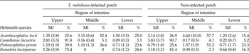
Analysis of site selection by the four helminth species along the intestine (table 3) showed no significant overall interactions within T. nodulosus-infected or non-infected perch (multinomial logistic regression: T. nodulosus-infected perch P = 0.965, χ2 = 0.271; non-infected perch P = 0.857, χ2 = 0.767). There were also no significant interactions between any individual species in either T. nodulosus-infected (multinomial logistic regression: P ≥ 0.644, χ2 ≤ 0.213) or non-infected perch (multinomial logistic regression: P ≥ 0.467, χ2 ≤ 0.530).
Site selection of intestinal helminths in Triaenophorus nodulosus-infected and non-infected perch to show mean intensity (MI ± SD) and percentage distribution (%) along the length of the intestine.

Discussion
Infections of perch with T. nodulosus plerocercoids are a common occurrence in the Queen Mary reservoir (Morley & Lewis, Reference Morley and Lewis2010); during the present sampling period prevalence levels of 69.9% and mean intensities of 2.9 were recorded. Infected fish were found to be stunted, a commonly reported observation, considered to be associated with the detrimental effect of T. nodulosus plerocercoids on fish growth (Kuperman, Reference Kuperman1981; Brinker & Hamers, Reference Brinker and Hamers2007). Any potential distorting effect of fish age on infection levels has been discounted as previous studies have found that young infected perch (< 700 days old) are generally larger than uninfected ones, with this trend being completely reversed as the infected fish age, resulting in as much as a 15.8% reduction in potential mass (Brinker & Hamers, Reference Brinker and Hamers2007). Nevertheless, the general condition of the fish in the present study remained unaffected, as no significant difference was observed in the K, HSI or GSI between infected and non-infected fish.
The four intestinal helminths recorded in this investigation are amongst the most common parasites of perch in Europe, typically reported from a range of still or flowing water habitats and generally unaffected by eutrophic or oligotrophic conditions (Andersen, Reference Andersen1978; Andrews, Reference Andrews1979; Lucky & Navratil, Reference Lucky and Navratil1984). The present study has shown that plerocercoids of T. nodulosus in the liver have the ability to influence the natural occurrence of these helminths in the intestine of perch. However, this effect appears to be selective, changing the prevalence, but not the intensity, of infections of the acanthocephalan A. lucii and the nematode C. lacustris, but with no effect on the cestode P. percae and the trematode B. luciopercae in the intestinal tract. It is unlikely that this effect is the result of an ecological factor, such as a change in selective feeding by T. nodulosus-infected fish. Although trophic specialization has been reported from many fish populations including perch (e.g. Svanbäck & Eklöv, Reference Svanbäck and Eklöv2002), in the present study this factor is not supported by the parasitological evidence. Within the Queen Mary reservoir, three of the four intestinal helminths recorded from perch, namely C. lacustris, P. percae and B. luciopercae, use planktonic crustaceans as intermediate hosts, with C. lacustris and P. percae sharing the same species of copepods (Moravec, Reference Moravec1994, Reference Moravec2004). Thus, if trophic specialization by T. nodulosus-infected perch was a significant factor, then the occurrence of these particular helminth species would be expected to change in a similar fashion, yet only C. lacustris was significantly affected. Furthermore, a change in feeding niche by T. nodulosus-infected fish should change the composition of the intestinal helminth community, favouring the introduction of generalist parasites associated with intermediate hosts that form part of any potentially changed diet. However, the intestinal community comprising the four helminth species remained unchanged between T. nodulosus-infected and non-infected perch, suggesting that the influence of this plerocercoid was due to a pathological or physiological effect rather than an ecological one.
Within mammals, concurrent laboratory infections of Schistosoma mansoni, in which eggs from female worms cause extensive granulomata in the liver and small intestine (Smyth, Reference Smyth1994), and intestinal nematodes and cestodes result in a reduction in the occurrence of these helminths in the intestine (Andreassen et al., Reference Andreassen, Odaibo and Christensen1990; Corrêa-Oliveira et al., Reference Corrêa-Oliveira, Golgher, Guilherme, Carvalho, Massara, Caldas, Colley and Gazzinelli2002). Intense intestinal inflammation induced by eggs of S. mansoni is likely to be responsible for this antagonistic effect (Andreassen et al., Reference Andreassen, Odaibo and Christensen1990). Similarly, extensive intestinal inflammation and hyperplasia occur in rats harbouring an encapsulated liver infection of the larval cestode Taenia taeniaeformis (Blaies & Williams, Reference Blaies and Williams1983; Lagapa et al., Reference Lagapa, Oku and Kamiya2008). These pathological effects, remote from the site of infection, are in this case considered to be the result of excretory/secretory (E/S) products being released by T. taeniaeformis. These cytotoxic larval products are likely to be absorbed into the body of the host and transported by the blood into the gastrointestinal mucosa. The degree of intestinal pathology induced is dependent on the number and size of developed larvae, with larger worms producing greater volumes of E/S products (Lapaga et al., Reference Lagapa, Oku and Kamiya2008).
Like T. taeniaeformis, T. nodulosus is also an encysted larval cestode occurring in the liver of its fish host. While extensive pathologies have been reported within the livers of fish harbouring this parasite (Kuperman, Reference Kuperman1981; Hoffman et al., Reference Hoffmann, Meder, Klein, Osterkorn and Negele1986; Brinker & Hamers, Reference Brinker and Hamers2007), no morphological effect on the intestine has been documented, although it is not known if these studies paid any particular attention to this organ. Only changes to the activity of intestinal enzymes in these infected fish have been reported so far (Izvekova & Izvekov, Reference Izvekova, Izvekov, Weiss and Faulkner2013). Therefore pathological changes to the intestine of perch as a result of T. nodulosus E/S products are likely and in vitro studies on other fish cestodes indicate that these products will provoke host cellular immune responses (Morley & Hoole, Reference Morley and Hoole1997; Scharsack et al., Reference Scharsack, Gossens, Franke and Kurtz2013). When combined with known T. nodulosus effects on the activity of host digestive enzymes (Izvekova & Izvekov, Reference Izvekova, Izvekov, Weiss and Faulkner2013), a profound alteration in the suitability of the fish intestine as an environment for the establishment and growth of other helminths is to be expected.
However, such changes appear to affect only some species of intestinal helminths, which may be associated not only with the different mechanisms of attachment to the intestinal mucosa, but also with the intensity of the infection. Usually trematodes and cestodes do not cause severe pathology to the intestine, generally due to their relatively superficial relationship with host tissues, while acanthocephalan and nematode parasites are regarded as much more pathogenic, capable of causing extensive damage to the alimentary canal (Dezfuli et al., Reference Dezfuli, Bosi, DePasquale, Manera and Giari2016). Thus, A. lucii is considered as a non-perforating species that occupies only a shallow attachment and regularly changes its point of contact, resulting in multiple localized lesions of the intestine. A necrotic belt of tissue encompasses the proboscis, with accompanying inflammation at the site of attachment (Taraschewski, Reference Taraschewski2000). Similarly, the camallanid nematode C. lacustris can penetrate deeply into the intestinal mucosa, not only sucking host blood and tissue fluids but also rasping the gut wall, resulting in localized inflammation and stimulating a cellular immune response (Moravec, Reference Moravec1994; Unger et al., Reference Unger, Brinker and Stich2008).
Therefore, both A. lucii and C. lacustris require a firm attachment to survive, and any decline in the structural integrity or increased inflammatory response of the intestinal wall may reduce their ability to remain and survive within the digestive tract, resulting in a lowered establishment success rate or increased frequency of spontaneous voiding. It would appear that the effect on these two species is not a gradually occurring one, as there is no significant change in the intensity of infections between T. nodulosus-infected and non-infected perch. Nevertheless, there was no correlation between the number of T. nodulosus present in each individual liver and the intensity of any intestinal species, suggesting that this aspect of the cestode's infection was not influential. Unfortunately, size and level of development of T. nodulosus was not recorded in this study and it therefore remains to be determined if, as found with T. taeniaeformis cysticerci in mammals (Lagapa et al., Reference Lagapa, Oku and Kamiya2008), the larval cyst size is an important factor in inducing intestinal pathology.
Interactions between helminths co-occurring in the intestine of perch have been demonstrated previously. Wootten (Reference Wootten1973) found a positive association between B. luciopercae and P. percae from a reservoir in south-east England, while a negative correlation between the presence of A. lucii and P. percae was reported by Anderson (Reference Andersen1978) from a small lake in Norway. Similarly, Lee (Reference Lee1981) found that the distribution, but not intensity, of A. lucii within the perch intestine was negatively affected by concurrent infections of P. percae or C. lacustris. In the present study there were positive correlations only between B. luciopercae and P. percae in non-infected perch and between B. luciopercae and C. lacustris in T. nodulosus-infected perch, but no changes were observed in intestinal site selection by any helminth species between the two fish populations. This is difficult to explain, as one would expect the correlations in non-infected perch to be carried over into those infected with T. nodulosus, especially as prevalence and intensity values of B. luciopercae and P. percae in non-infected perch were unaffected by the occurrence of T. nodulosus.
These results therefore suggest that little or no direct interactions between these helminth species in perch cause the positive correlation. When ingested by the target host larval helminths are typically ‘dormant’, requiring a number of optimal intestinal physiochemical conditions to be in place in order to be activated and initiate the process of establishment and development (Lackie, Reference Lackie1975; Macko, Reference Macko1980). In general, ingested parasite species respond to narrow or wide ranges of physiochemical parameters, including pH, bile and specific enzymes, and these in turn demonstrate natural variation between individual hosts. Other important activation factors include the morphological structure and microstructure of the intestinal tract, as well as mechanical pressure, intestinal microbial communities and the role of food movement (Macko, Reference Macko1980). Within T. nodulosus-infected perch, changing levels of intestinal enzymes (Izvekova & Izvekov, Reference Izvekova, Izvekov, Weiss and Faulkner2013), together with other altered physiochemical and morphological parameters, will change the intestinal environment and influence the conditions necessary to trigger activation of ingested stages of helminths. Such intestinal changes, as found with mammals (Lagapa et al., Reference Lagapa, Oku and Kamiya2008), are likely to vary with the intensity and level of development of T. nodulosus plerocercoids in the liver of perch, and thus the strength and range of physiochemical signals necessary to trigger parasite activation will narrow, depending on the sensitivity of the parasite species. Intraspecific variations in triggering activation could alter the nature of the relationship between helminth species, such as B. luciopercae, where correlations that were shown in non-infected perch had changed in T. nodulosus-infected fish. Helminths that respond to a broad range of intestinal conditions will, more than likely, achieve successful establishment in these altered environments. However, species requiring more specific or stronger triggering factors or intestinal conditions will probably fail to be activated, or will be voided soon after infection, even when ingested by a relevant target host.
The specific role that digestive enzymes play in structuring the establishment and distribution of helminths in the host intestine is difficult to determine and, in many infections, may be masked by more powerful immunological effects. No differences were apparent in the site selection of intestinal helminth species between T. nodulosus-infected and non-infected perch, unlike the observations of Izvekova & Tyutin (Reference Izvekova and Tyutin2014) who found an increased occurrence of B. luciopercae in the posterior sections of the intestinal tract of T. nodulosus-infected perch. These differences may be related to season, with Izvekova & Tyutin (Reference Izvekova and Tyutin2014) sampling in late summer (August–October) compared with early summer (June) in the present study. Alternatively, the effect reported by Izvekova & Tyutin (Reference Izvekova and Tyutin2014) may be an uncommon one, limited only to B. luciopercae and occurring in populations harbouring high prevalences and intensities, with few other species of intestinal helminths.
The present study has demonstrated that intestinal acanthocephalan and nematode infections may be detrimentally affected by concurrent infections with T. nodulosus plerocercoids. Although it has not been possible to designate the exact mechanisms involved, this seems likely to be associated, either individually or in combination, with the host's immune response, mechanical damage to the liver induced by T. nodulosus, and the release of E/S products by this parasite. These factors probably cause intestinal physiochemical and morphological changes that result in a less suitable environment for certain helminths. Indirect antagonistic and synergistic interactions between helminths in fish may be more common than previously reported, especially where highly pathogenic larval cestodes occur. Additional studies on these interactions would contribute further to our understanding of helminth population dynamics in fish.
Conflict of interest
None.