


Significant outcomes
-
Elevated HCC in patients with a current anxiety disorder and a within-range HCC in patients with recurrent anxiety disorders, at Baseline. These findings suggest potential desensitisation of HPA response to stress in patients who experience multiple episodes of anxiety disorders.
Limitations
-
The small number of participants represents a major limitation. Further research and resources are needed to examine HCC in a larger clinical sample.
Background
The hypothalamic–pituitary–adrenal (HPA) axis influences many body processes and haemostatic systems (including immune, metabolic, cardiovascular, reproductive and central nervous systems), and plays a major role in stress regulation and the sleep–wake cycle (Barrett et al., Reference Barrett and Ganong2016). When compared to the extensive literature on HPA axis dysfunction in depressive illness, evidence relating to HPA function in patients with anxiety disorders is limited. There is no unifying disturbance of the HPA axis function across anxiety disorders, and within each disorder, the findings of investigations using similar methodology have produced inconsistent findings (Elnazer et al., Reference Elnazer and Baldwin2014).
Assay of hair cortisol concentration (HCC) provides a measure of HPA function. Circulating cortisol is deposited from follicular capillaries, sweat glands and sebaceous glands into all layers of the growing hair shaft (Cook et al., Reference Cook and Spector1964; Russell et al., Reference Russell, Koren, Rieder and Van Uum2012; Kapoor et al., Reference Kapoor, Schultz-Darken and Ziegler2018), which lengthens by approximately 1 cm per month (with some ethnic variation), allowing determination of average HCC over a specific period (Schütz et al., Reference Schütz, Ahrens, Erdmann and Rochholz1993; Loussouarn et al., Reference Loussouarn2001). HCC ranges between 5 and 91 pg/mg (mean 18 pg/mg), and is not affected by hair colour or sex hormones (Raul et al., Reference Raul, Cirimele, Ludes and Kintz2004), hairdressing treatments or light exposure (Kristensen et al., Reference Kristensen, Larsen, Olsen, Fahrenkrug and Heitmann2017). HCC is strongly associated with salivary cortisol area under the curve (AUC) over 1 month (r = 0.61, p = 0.01) and maybe a more accurate reflection of overall cortisol output (Short et al., Reference Short, Stalder, Marceau, Entringer, Moog, Shirtcliff, Wadhwa and Buss2016).
HCC measurement facilitates the management of Cushing’s syndrome, tracking monthly cortisol levels and capturing periodic hypercortisolaemia (Manenschijn et al., Reference Manenschijn, Koper and van den Akker2012). Elevated HCC levels have been reported with ongoing stress (Herane Vives et al., Reference Herane Vives, De Angel, Papadopoulos, Strawbridge, Wise, Young, Arnone and Cleare2015), in athletes (Skoluda et al., Reference Skoluda, Dettenborn, Stalder and Kirschbaum2012), victims of natural disasters (Gao et al., Reference Gao, Zhong, Xie, Wang, Jin, Deng and Lu2014) and in hospitalised infants (Yamada et al., Reference Yamada, Stevens, de Silva, Gibbins, Beyene, Taddio and Koren2007). In children, HCC is associated with post-stress salivary cortisol AUC (Vanaelst et al., Reference Vanaelst, Huybrechts, Bammann, Michels, Vriendt, Vyncke and Henauw2012), AUC with respect to ground levels (AUCg) (Ouellette et al., Reference Ouellette, Russell, Kryski, Sheikh, Singh, Koren and Hayden2015) and AUC with respect to increase (AUCi) (Kao et al., Reference Kao, Doan, St. John, Meyer and Tarullo2018). HCC levels may be elevated in depressed patients (Dettenborn et al., Reference Dettenborn, Muhtz, Skoluda, Stalder, Steudte, Hinkelmann, Kirschbaum and Otte2012) and in patients with post-traumatic stress disorder (Steudte et al. Reference Steudte, Kolassa, Stalder, Pfeiffer, Kirschbaum and Elbert2011; Lou et al., Reference Luo, Hu, Liu, Ma, Guo, Qiu, Wang, Wang, Zhang, Zhang, Hannum, Zhang, Liu and Li2012). Findings from meta-analysis indicate a 22% higher median HCC in groups previously exposed to chronic stress, and 43% higher median HCC in groups experiencing ongoing stress, but no consistent correlations between HCC and self-reports of stress or depression (Stalder et al., Reference Stalder, Steudte-Schmiedgen, Alexander, Klucken, Vater, Wichmann and Miller2017).
Inflammation in the periphery is ‘communicated’ to the brain via humoral and neural pathways. Resident microglia are activated and release pro-inflammatory cytokines, which disrupt hippocampal neurogenesis. Peripheral cytokines activate the HPA axis, leading to increased levels of circulating glucocorticoids and suppression of neurogenesis, which may underlie some of the neurobehavioural changes associated with chronic inflammatory conditions (Chesnokova et al., Reference Chesnokova, Pechnick and Wawrowsky2016).
Some studies have found a positive effect for anti-inflammatory drugs on reducing depressive symptoms and levels of disrupted cytokines. A meta-analysis of clinical trials with non-steroidal anti-inflammatory drugs (NSAIDs) found beneficial effects in depressed patients (n = 1497) (Iyengar et al., Reference Iyengar, Gandhi, Aneja, Thorpe, Razzouk, Greenberg, Mosovich and Farkouh2013); and a randomised double-blind controlled trial (n = 40) of augmentation with the cyclo-oxygenase inhibitor-2 (COX-2) inhibitor celecoxib indicated it could enhance the response to antidepressants (Abbasi et al., Reference Abbasi, Hosseini, Modabbernia, Ashrafi and Akhondzadeh2012).
Less is known about circulating cytokines in anxiety disorders. Meta-analysis suggests differences in IL-10, TNF-α and IFN-α in patients with generalised anxiety disorder (GAD), when compared to controls (Costello et al., Reference Costello, Gould, Abrol and Howard2019). A case–control study of cytokine levels in patients with GAD found disturbances in the ratio of anti- and pro-inflammatory cytokines (with high ratios of TNF-α/IL-1, TNF-α/IL-4, INF-γ/IL-10 and INF-γ/IL-4) (Hou et al., Reference Hou, Garner, Holmes, Osmond, Teeling, Lau and Baldwin2017). As little is known about potential interactions between HPA markers and circulating cytokines in anxiety disorders, we examined cross-sectional and longitudinal relationships between HCC and circulating cytokines in patients with a range of anxiety disorders: at Baseline, after 6 weeks of antidepressant treatment (Week 6), and after a further 6 weeks of either augmentation with the COX-2 inhibitor celecoxib, or a further 6 weeks of ‘treatment as usual’ (Week 12).
Methods
Sample. Study participants were recruited from primary care (general practice) and secondary care mental health services. Potentially eligible patients were sent a participant information sheet, reply slip and stamped addressed envelope. When a potential participant contacted the research team, an appointment was arranged for screening with the Mini-International Neuropsychiatric Interview (MINI) (Sheehan et al., Reference Sheehan, Lecrubier, Sheehan, Amorim, Janavs, Weiller, Hergueta, Baker and Dunbar1998). Potential participants could ask questions about the study aims and procedures, and after providing consent underwent Baseline assessments. They were considered eligible for the study if aged between 18 and 70 years, with a primary diagnosis of an anxiety disorder or an anxiety-related disorder (post-traumatic disorder or obsessive-compulsive disorder), defined according to the Diagnostic and Statistical Manual of Mental Disorders, 5th Edition (DSM-5) criteria (American Psychiatric Association 2013), and competent to provide written consent. Patients were excluded from participation if they were outside the specified age range, had a primary diagnosis other than an anxiety disorder, if unable to provide written informed consent, had significant alcohol or substance use in the previous 3 months, had a physical illness that was unlikely to be stable over the course of the study or were pregnant or breastfeeding.
Participant questionnaire assessments. Patients were assessed using the Hospital Anxiety and Depression Scale (HADS) (Zigmond & Snaith Reference Zigmond and Snaith1983), Clinical Global Impression of Illness Severity (CGI-S) (Guy et al., Reference Guy1976), Warwick–Edinburgh Mental Well-Being Scale (WEMWEBS) (Tennant et al., Reference Tennant, Hiller, Fishwick, Platt, Joseph, Weich, Parkinson, Secker and Stewart-Brown2007), Arizona Sexual Experiences Scale (ASEX) (McGahuey et al., Reference McGahuey, Gelenberg and Cin2000) and Oxford Questionnaire of Emotional Side Effects of Antidepressants (OQuESA) (Price et al., Reference Price, Cole, Doll and Goodwin2012), at Baseline, Week 6 and Week 12. Treatment adherence was determined by patient report and tablet counts.
Hair sampling. Scalp hair samples (3 cm long) were collected from study participants at Baseline, Week 6 and Week 12. Hair strands were cut with fine scissors as close as possible to the scalp from a posterior vertex position in which the number of strands is differed in accordance with participant permission. Each hair strand was repeatedly washed using isopropanol, then dried for 6 h, and analyses were performed on non-pulverised hair.
Processing of hair samples. Each specimen was incubated for 18 h at room temperature in 1800 μl methanol. About 1600 μl of the resulting suspension was purged with nitrogen at 50 °C at a pressure of 0.1 bar for at least 40 min. Samples were spun in a centrifuge at 10,000 rpm for 2 min and 1 ml of the clear supernatant was transferred into a 2 ml tube. Methanol was evaporated at 65 °C under a constant stream of nitrogen until samples were completely dry (typically lasting around 20 min). Supernatants were re-suspended in 225 μl of distilled water and 50 μl were submitted to liquid chromatography coupled to tandem mass spectrometry (LC-MS/MS). The lower limits of quantification of this assay method were below 0.1 pg cortisol per mg hair. The median coefficient of variation of all replicates was 4.9% (interquartile range: 1.3–13.9%) (Gao et al., Reference Gao, Zhong, Xie, Wang, Jin, Deng and Lu2014).
Determination of HCC. Following milling of hair segments, two 50 mg aliquots of powdered hair from a single hair segment were processed in parallel. Twenty microlitres were removed from the vial and used for cortisol determination with a commercially available chemiluminescence immunoassay (CLIA, IBL-Hamburg, Germany). An unknown amount of antigen was present in the sample and a fixed amount of enzyme-labelled antigen that competes for the binding sites of the antibodies was coated onto the wells. After incubation, the wells were washed to stop the competition reaction. After the substrate reaction, the intensity of the developed colour is inversely proportional to the amount of the antigen in the sample. Results of samples were determined directly using the standard curve. The reference normal range for HCC using this method in healthy volunteers (reported by the same lab) is 1–30 pg/mg.
Blood sampling for pro- and anti-inflammatory cytokines. Venous blood samples (8–10 ml) were collected into a vacutainer containing no preservative at Baseline and Week 6, sampling being performed in accordance with National Health Service procedures, and observing sterile conditions with infection control by either a nurse trained in phlebotomy or a qualified medical practitioner. Blood samples were centrifuged at 1600× g (4000 rpm, setting 4) on a Capricorn 2000 bench centrifuge for 10 min. The plasma fraction was transferred using a Pasteur pipette to vial tubes, then placed in a Ziploc bag and airtight sealed container inside a lockable box for storage at −20oC prior to transfer to a − 80°C freezer for longer term storage.
Cytokine quantification. Luminex multi-analyte profiling (xMAP) technology employs proprietary bead sets distinguishable under flow cytometry. Each bead set is coated with a specific capture antibody, and fluorescence or streptavidin-labelled detection antibodies bind to the specific cytokine–capture antibody complex. Multiple cytokines in a fluid sample can be recognised and measured by the differences in both bead sets, with chromogenic or fluorogenic emissions detected using flow cytometric analysis: we used the Proinflammatory Panel 1 (human) kit, (V-PLEX, MESO SCALE DISCOVERY, Rockville, MD, USA) to measure IL-1β, IL-2, IL-4, IL-6, IL-8, IL-10, IL-12p70, IL-13 and TNF-α.
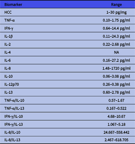
Data analysis. Some participants preferred not to provide blood samples at some test points, but were prepared to participate with other tests. Normal reference ranges determined by the lab where the study samples were analysed were used (Table 1). Data were analysed using IBM SPSS® Statistics 26.0.
Normal ranges and ratios

Ethical aspects and research governance. The project was approved by the Hampshire Research Ethics Committee (REC reference 16/SC/0038); UK Health Research Authority (HRA) (IRAS project ID 170365) and the Medicines and Healthcare Products Regulatory Agency (EudraCT number: 2016-000337-48), as part of a larger investigation of neuroinflammatory and endocrine factors.
Results
Study sample. A total of 170 referrals were received from primary or secondary health services: 35 individuals (23 women, 12 men; mean age 32.46 years [SD 12.87 years]) provided consent to participate in the study and underwent Baseline assessment. All participants had English as their first language: 29 were White British, 4 Asian British, 1 Black British and 1 self-identified as Other White Caucasian. Twenty participants (57%) were professionally active at the time of enrolment. Reasons for non-participation included: feeling too unwell to take part in the study (22%); concern about time commitments (15%); lack of financial compensation (14%); discomfort in talking about sexual matters (10%) and no reason (39%).
Treatment received. Two participants withdrew before the Week 6 review, and six participants before the Week 12 review. Among the 33 participants who underwent ‘treatment as usual’ between Baseline and Week 6, 19 received a selective serotonin reuptake inhibitor, 6 received a serotonin-norepinephrine reuptake Inhibitor, 3 received a noradrenergic and specific serotonergic antidepressant, 2 underwent cognitive behavioural therapy and a single participant received a beta blocker; 3 patients had not undergone any new treatment by the Week 6 review. At Week 12,18 participants had undergone celecoxib augmentation and 15 patients had undergone a further 6 weeks of continued antidepressant treatment.
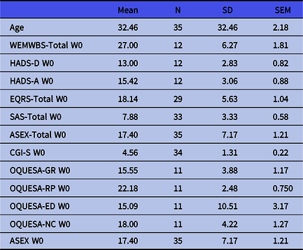
Clinical characteristics at Baseline. Interview with the MINI generated at least one DSM-5 diagnosis in each participant. Thirteen participants met diagnostic criteria for GAD, eight for panic disorder with agoraphobia, seven for social phobia, four for obsessive-compulsive disorder and three for panic disorder. Nineteen participants (54.3%) were within the range of healthy body mass index (18.5–24.9), but 13 were overweight and 3 were underweight. The mean scores (and standard error of the means) on rating scales at were as follows: WEMWBS 27 (1.81); HADS-A 15.42 (0.88); HADS-D 13 (0.82); CGI-1 4.56 (0.22); OQUESA-GR 15.5 (1.17); OQUESA-RP 22.18 (0.75); OQUESA-ED 15.09 (3.17); OQUESA-NC 18 (1.27) and ASEX 17.40 (1.21) (Table 2).
Rating scale and questionnaire scores at Baseline (Week 0)

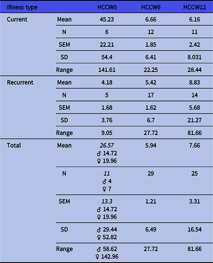
HCC at B aseline. Hair cortisol analyses were performed by chemiluminescence immunoassay on samples purified by LC-MS/MS from 11 participants: mean HCC was 26.66 pg/mg (median of 6.14 pg/mg). Mean HCC for male participants and female participants were 30.74 pg/mg [SEM 14.79] and 24.19 pg/mg [SEM 19.963], respectively. Mean HCC was higher than the normal range (1–30 pg/mg) in patients with panic disorder with agoraphobia (72.37 pg/mg, [SEM 71.48]), but mean HCC in participants with other diagnoses were unremarkable: GAD, 25.32 pg/mg[SEM 17.30]; social phobia 14.07 pg/mg [SEM 9.51]; panic disorder 1.24 pg/mg (single patient). Mean HCC was elevated in ‘current’ cases (45.23 [SEM 22.21]) but within the normal range in recurrent cases (4.18 pg/mg [SEM 1.68]) (Table 3).
HCC (pg/ml)

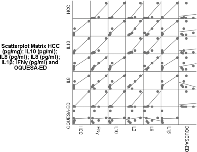
Cytokines at B aseline. Mean plasma cytokine concentrations were within normal ranges, other than a low mean concentration of IL-12p70 (0.11 pg/ml [SEM 0.04]) and high mean concentration of TNF-α (2.37 pg/ml [SEM 0.43]). Mean IL-1 β concentration was low in male participants (0.005 pg/ml [SEM 0.005]). HCC levels were significantly correlated with plasma concentrations of IFN-γ (p = 0.01), IL-10 (p = 0.00), IL-2 (p = 0.02) and IL-8 (p = 0.02) (Fig. 1). After adjusting for age, diagnosis and gender using logistic regression partial correlation, the correlation remained significant between HCC (pg/mg) and IL-10 mean concentration (pg/ml) (p = 0.00), and there was a significant correlation between mean HCC (pg/mg) and IL-1βa mean concentration (pg/ml) (p = 0.00). After adjustment for HADS-D score, there were no significant correlations between mean HCC and other parameters.
Correlations at Baseline (Week 0).

Effects of treatment on rating scale scores. Anxiety symptom severity (HADS-A) reduced from Baseline to Week 6 (mean 13.70 [SEM 0.73]), and quality of life (WEMWBS) improved from Baseline to Week 6 (mean 33.30 [SEM 1.75]). The severity of depressive symptoms (HADS-D) reduced from Baseline to Week 6 (mean 10.82 [SEM 0.68]). Mean OQUESA-ED declined from Baseline (20.25 [SEM 10.39]) to Week 6 (10.39 [SEM 0.947]), a non-significant change (p = 0.373); but there was no further change in Week 12 mean OQUESA-ED score (10.39 [SEM 1.067]). Mean ASEX increased from Baseline to Week 6 (20.18 [SEM 0.96]), a significant change (p = 0.01). Mean ASEX declined at Week 12 (17.26 [SEM 1.23]), a significant change (p = 0.013).
Celecoxib augmentation was associated with significant improvement in the quality of life (mean WEMWBS 7.50 increase [SEM 2.63, p = 0.01]), reductions in anxiety symptom severity (HADS-A 3.17 decrease [SEM 0.52, p = 0.00]) and depressive symptom severity (HADS-D 2.11 [SEM 0.80, p = 0.02]). A further 6 weeks of ‘treatment as usual’ was not associated with significant changes.
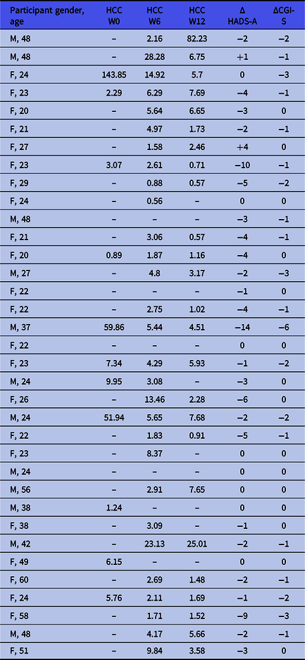
Changes in HCC. Chemiluminescence immunoassay of hair samples (n = 29) at Week 6 found a mean HCC of 5.94 pg/mg (SEM 1.21), within the normal range (1–30 pg/mg) and lower than the mean HCC at Baseline, although HCC values at Baseline and Week 6 were highly correlated (p = 0.00). Paired sample t-test for HCC concentration found a non-significant change from Baseline (Week 0) to Week 6 (26.51 pg/mg, p = 0.11). Celecoxib augmentation was associated with a non-significant reduction in HCC from Week 6 to Week 12 (−1.24 pg/mg, p = 0.80), whereas treatment as usual group was associated with a non-significant increase (1.72 pg/mg, p = 0.13). Post-treatment mean HCC (Week 6 and Week 12) were found to be within the normal range in both current and recurrent cases (Table 3).
We present HCC changes for individual participants at each test point (Table 4). The current/acute cases sub-group showed a reduction in mean HCC from Baseline to Week 12: by contrast, in the recurrent cases sub-group, mean HCC increased from Baseline to Week 12.
HCC for all participants at Baseline, Week 6 and Week 12

HCC pg/mg is approximated to two decimal points. M, male; F, female; –, missing value.
Changes in inflammatory markers. Analysis of cytokine levels (of IFN-γ, IL-1β, IL-10, IL-12p70, IL-13, IL-2, IL-4, IL-6, IL-8 and TNF-α) in participants who provided blood samples at Week 6 (n = 25) found a low mean concentration of IL-2 (0.22 pg/ml [median 0.17 pg/ml] and a high mean concentration of TNF-α (2.16 pg/ml [median 1.60 pg/ml]. Paired sample t-test for cytokine concentrations found that the changes from Baseline to Week 6 were not significant. Analysis of cytokine levels at Week 12 in the celecoxib augmentation group (n = 18), found a low mean concentration of IL-1β (0.04 pg/ml [SEM 0.024], low mean concentration of IL-12p70 (0.12 pg/ml [SEM 0.015], low mean concentration of IL-13 (0.11 pg/ml [SEM 0.05] and a high mean concentration of TNF-α (2.33 pg/ml [SEM 0.21]. Concentrations at Week 12 in the celecoxib augmentation group were correlated with Week 6 concentrations for IL-10 (p = 0.00); IL-13 (p = 0.00); IL-2 (p = 0.00); IL-6 (p = 0.00); IL-8 (p = 0.00) and TNF-α (p = 0.00). Paired sample t-test found a significant change from Week 6 to Week 12 in the mean concentration of IL-2 0.06 pg/ml (SEM 0.02, p = 0.00). Analysis of cytokine concentration in the ‘treatment as usual’ group at Week 12 (n = 4) found low mean concentrations of IL-1β (0.43 pg/ml [SEM 0.018]); IL-12p70 (0.102 pg/ml [SEM 0.04]); IL-13 (0. 27 pg/ml [SEM 0.11]) and IL-2 (0.21 pg/ml [SEM 0.03]). A paired sample t-test found no significant change from Week 6 to Week 12.
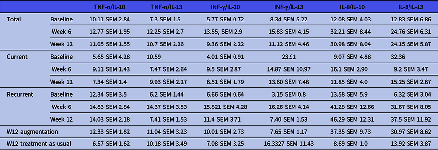
Pro- and anti-inflammatory cytokine ratio analysis (Table 5) found elevated mean TNF-α/IL-10, TNF-α/IL-13 and INF-γ/IL-13, at all testing points. The analysis also found elevated INF-γ/IL-10 at Week 6, low IL-8/IL-10 at Baseline and within range IL-8/IL-13 at all testing points. Current cases had low Baseline INF-γ/IL-10, within range INF-γ/IL-10 at Week 6 and low IL-8/IL-10 at Week 6 and Week 12. Recurrent cases had within range Baseline INF-γ/IL-10, elevated INF-γ/IL-10 at Week 12 and within range INF-γ/IL-13 at Baseline. Patients who underwent 6 weeks of augmentation by Week 12 had within range IL-8/IL-10. Patients who underwent 6 weeks of ‘treatment as usual’ by Week 12 had within range INF-γ/IL-10 and low IL-8/IL-10. Paired t-test analysis found no statistically significant change in cytokines ratios from Baseline to Week 6 and to Week 12, except in the current cases who did not opt for augmentation at Week 6 (and seemingly had a satisfactory response to treatment as usual) (n = 4). This sub-group had significant paired t-test change mean TNF-α /IL-10 from Week 6 to Week 12 (t 4.14, df 3, p = 0.026).
Cytokine ratios

Black, within reference range; red, higher than reference range; blue, below reference range.
Discussion
Recent evidence suggests that HCC is a reliable measure of cumulative cortisol concentration over a designated period (Short et al., Reference Short, Stalder, Marceau, Entringer, Moog, Shirtcliff, Wadhwa and Buss2016). We found some evidence of elevated HCC in patients with shorter duration (current) illness but not in longer duration (recurrent) illness. These findings might suggest a potential desensitisation of HPA response to stress in patients who suffer multiple episodes of anxiety disorders. We also found that changes in HCC were not correlated with changes in anxiety severity (HADS-A; CG-I) or mental well-being (WEMWBS).
The small number of participants represents a major limitation. Lack of financial compensation for the offered time was mentioned by many referred patients as a reason for declining to participate. Further research and resources are needed to examine HCC in a larger clinical sample.
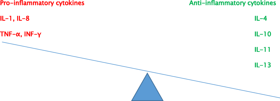
Pro-inflammatory and anti-inflammatory cytokines function in complementary and competing effects to produce a net-pattern response. The net-pattern response determines the onset, duration and severity of the inflammatory phase of healing. A net anti-inflammatory pattern is required for a less severe, shorter healing response (Fig. 2). We found elevated TNF-α/IL-10 and TNF-α/IL-13 ratios at Baseline, Week 6 and Week 12. We found elevated INF-γ/IL-10 at Week 6 (and elevated INF-γ/IL-13 at Week 12). Patients with current anxiety disorders had low Baseline INF-γ/IL-10 and low IL-8/IL-10 at Baseline, Week 6 and Week 12. Patients with recurrent anxiety disorders had elevated INF-γ/IL-10 at Week 6 and Week 12 and low IL-8/IL-10 at Baseline.
Cytokines response and inflammation.

Conclusion
This study provides some evidence for elevated HCC in patients with a current anxiety disorder and a within-range HCC in patients with recurrent anxiety disorders, at Baseline. Elevated HCC at Baseline normalised after 6 weeks of treatment. This continued to be the case after a further 6 weeks of treatment (Week 12). These findings may suggest a potential desensitisation of HPA response to stress in patients who experience multiple episodes of anxiety disorders. Patients with current anxiety disorders had an anti-inflammatory cytokine response pattern compared to patients with recurrent anxiety disorders.
Acknowledgements
The lead author (Dr Heham Elnazer) would like to acknowledge the advice and support of Professor Clemens Kirschbaum (Dresden, Germany).
Authors contributions
The authors meet the criteria of authorship described by The International Committee of Medical Journal Editors (ICMJE).
Financial support
This research received no specific grant from any funding agency, commercial or not-for-profit sectors.
Statement of interest
None.
Ethical standards
The authors assert that all procedures contributing to this work comply with the ethical standards of the relevant national and institutional committees on human experimentation and with the Helsinki Declaration of 1975, as revised in 2008.