Introduction
Undernutrition (UN) represents an important public health burden in underdeveloped countries. A considerable number of studies have been published, detailing the long-term consequences of inadequate maternal nutrition in adult offspring.Reference Brenseke, Prater, Bahamonde and Gutierrez1, Reference Gluckman, Hanson, Beedle and Spencer2 Individuals with a low birth weight are at a substantial risk of developing a range of cardiometabolic-related disorders later in life.Reference Hales and Barker3, Reference Barker, Osmond, Golding, Kuh and Wadsworth4 The concept of developmental programming proposes that these disorders arise due to fetal maladaptations arising from an altered maternal environment. Nutrient deprivation during gestation can result in permanent alterations to the structure and function of the organs and/or alterations to the metabolic and homeostatic systems, which in turn have an impact on subsequent health and eventually disease manifestation as adults.Reference Wadhwa, Buss, Entringer and Swanson5 Deficits in kidney nephron endowmentReference Hoppe, Evans and Moritz6–Reference Luyckx and Brenner8 and an altered renal renin–angiotensin system (RAS)Reference Langley-Evans, Sherman and Welham9–Reference Richter, Briffa, Moritz, Wlodek and Hryciw11 have been reported widely across a range of experimental models of developmental programming. However, little attention has been paid to renal inflammatory pathways as a potential mechanism underlying programmed disorders.
Inflammation has received increasing attention over recent years as is often found to accompany chronic diseases including diabetes, obesity and cancer. It is a tightly regulated process, with deviations from this process presenting a significant health risk with unresolved inflammation known to compromise tissue function.Reference Segovia, Vickers, Gray and Reynolds12 There are critical windows of innate immune vulnerability during prenatal and neonatal maturation of which developmental programming and the trajectory for childhood and adult inflammatory responses are largely established. Malnourishment during pregnancy using low protein exposure has previously been shown to alter renal inflammatory responses in offspring.Reference Stewart, Jung, Manning and Vehaskari13 Previous studies by our group have demonstrated the efficacy of pre-weaning growth hormone (GH) treatment on improving later life cardiometabolic outcomes in offspring following maternal UN.Reference Gray, Li, Reynolds and Vickers14–Reference Reynolds, Li, Gray and Vickers17 GH is known to be critical for normal growth, cardiac function and metabolic homeostasis as well as a key role in the regulation of normal kidney development and function.Reference Hammerman and Miller18 An adverse nutritional environment during fetal development has been linked with permanent alterations in the GH-Insulin-like growth factor axis.Reference Koch, Wilmoth and Wilson19–Reference Brennan, Olson and Symonds21 A number of clinical and experimental animal studies have shown GH administration increases renal mass, blood flow and glomerular filtration rate and decreases renal vascular resistance.Reference Hammerman and Miller22, Reference Kamenicky, Mazziotti, Lombes, Giustina and Chanson23 Perturbed GH concentrations have been linked with renal malfunction, hypertension and cardiovascular morbidity and mortality.Reference Hirschberg24–Reference Lombardi, Di Somma and Grasso26
Our group has previously demonstrated that pre-weaning GH treatment can ameliorate the programming effect of hypertension, vascular disorders as well as restoring the pro-inflammatory phenotype and insulin resistance in the adipose tissue in the UN offspring.Reference Gray, Li, Reynolds and Vickers14–Reference Reynolds, Li, Gray and Vickers17 This study aims to extend these previous observations, and we hypothesize that GH will normalize the renal pro-inflammatory phenotype, and other renal physiological and morphological changes, consequently conferring protection against adverse programming effects in terms of renal parameters in offspring.
Methods
All animal works were approved by the Animal Ethics Committee at the University of Auckland and use a cohort previously described by our group for which systolic blood pressure and body composition have been reported.Reference Gray, Li, Reynolds and Vickers14 Virgin Sprague-Dawley rats (n=30) were time mated at 110 days of age using a rat oestrous cycle monitor (EC-40; Fine Science Tools, Foster City, Canada). Upon confirmation of mating, animals were fed either a standard rodent control diet [CON; 2018 Teklad Global 18% Protein Rodent diet, 18.6% protein, 6.2% fat, 44.2% carbohydrate (by weight), energy density 3.1 kcal/g (24% kcals from protein, 18% kcals from fat, 58% kcals from carbohydrate)] or 50% of CON intake throughout pregnancy (UN) and were housed in a 12-h light/dark environment with a constant temperature of 25°C. Following birth, all dams were fed the standard chow ad libitum until the time of weaning (day 21). On postnatal day 2 (P2), litter size was randomly adjusted to 8 pups per litter (4 males and 4 females) to ensure adequate and standardized nutrition until weaning. From postnatal day 3 until weaning, CON and UN pups were treated with either saline (CON-S n=7 litters, UN-S n=7 litters) or GH (2.5 µg/g/day; rbGH Cyanamid, USA) by subcutaneous injection (CON-GH n=10 litters, UN-GH n=6 litters). After weaning, male offspring were fed a standard diet until the end of the study. Weight and food intake were measured every 3 days post-weaning. Female offspring were used in an independent study. Given that the mother is the biological replicate, sample size reported reflects one male analysed per litter per treatment group.
Blood and tissue collection
On day 150, rats were fasted overnight and killed by sodium pentobarbitone anaesthesia (60 mg/kg; IP) followed by decapitation. Plasma and tissue samples were collected and profiled as detailed below. Blood was collected into heparinized vacutainers and stored on ice until centrifugation and removal of plasma for analysis and stored at −20°C. Kidneys were weighed with one subsequently snap-frozen in liquid nitrogen and stored at −80°C and the other fixed in 10% neutral-buffered formalin until analysis.
Plasma analysis
Plasma from male offspring was analysed for interleukin (IL-1β), IL-6 and tumour necrosis factor (TNF)-α concentrations (Quantikine ELISA; R&D Systems, Minneapolis, USA) by commercial rat-specific enzyme-linked immunosorbent assays, according to manufacturer’s instructions. Plasma was also analysed for total protein (Colorimetric assay, Roche), urea (kinetic UV assay, Roche), uric acid (enzymatic colorimetric assay; Randox Laboratories Ltd, Ardmore, Crumlin, UK) and creatine phosphate using Hitachi 902 autoanalyser (Hitachi High Technologies Corporation, Tokyo, Japan).
Gene expression analysis
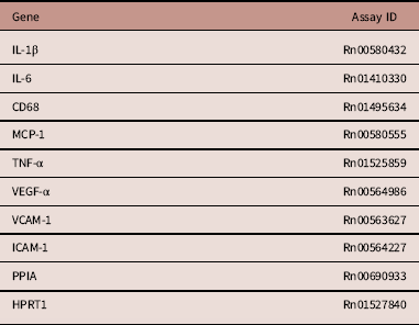
RNA was extracted from kidney tissue, using TRI-Reagent (100 mg tissue/ml) and stored at −80°C. Single-stranded complementary DNA (cDNA) was prepared using high-capacity cDNA archive kit (Applied Biosystems, Warrington, UK). Messenger RNA (mRNA) expression was quantified by real-time polymerase chain reaction on an ABI 7700 Sequence Detection System (Perkin-Elmer Applied Biosystems). Predesigned Taqman probes are outlined in Table 1. To control for between-sample variability, mRNA expression was normalized to the geometric mean of cyclophilin A and hypoxanthine phosphoribosyltransferase 1 for each sample by subtracting the Ct of controls from the Ct for the gene of interest, producing a ΔCt value. The ΔCt for each treatment sample was compared to the mean ΔCt for control samples using the relative quantification 2(−ΔΔCt) method to determine fold change.Reference Schmittgen and Livak27
Taqman predesigned probes (Applied Biosystems)

IL, interleukin; MCP-1, monocyte chemoattractant protein-1; TNF-α, tumour necrosis factor-α; VEGF; vascular endothelial growth factor; VCAM, vascular cell adhesion molecule; ICAM, intercellular adhesion molecular; PPIA, cyclophilin A; HPRT1, hypoxanthine phosphoribosyltransferase 1.
Nephron number and renal histological analysis
Kidneys fixed in 10% neutral-buffered formalin were paraffin embedded. Embedded kidney tissue blocks were cut into 5 µm thick sections and mounted on Superfrost® plus microscope slides. Sections were stained with Haematoxylin and Eosin (H&E) for light microscopic examination. Four microscopic fields of view of the renal cortex were taken per kidney at magnification 4×, using a Nikon ECLIPSE E800 Microscope camera (Shinjuku, Japan) using NIS-Elements F3.0 program (Nikon). The images were then processed using ImageJ software to count all glomeruli present in the four fields of view and a mean glomerular number was calculated for each group.
Periodic Acid-Schiff (PAS) and Masson’s trichrome (MTS) staining was performed as appropriate using standard histological protocols and mounted onto Superfrost® plus microscope slides with coverslips. Sections from each treatment group were first examined to identify pathophysiological features such as monocyte infiltration, glomerular interstitial nephritis, glomerulosclerosis or basement membrane or mesangial cell thickening.Reference Huang, Haylor and Hau28
Statistical analysis
All statistical analysis was performed using SigmaPlot for Windows version 12.0 (Systat Software Inc., San Jose, CA, USA). Data were analysed by two-way factorial ANOVA, with maternal diet and GH treatment as factors. Holm-Sidak post-hoc tests were performed to detect any differences between groups. Differences were considered significant at P<0.05. All data are presented as means±s.e.m., unless otherwise indicated. Where data failed normality testing, log transformation was performed.
Results
Previous work from our group has demonstrated that UN-S offspring are significantly heavier and have greater fat mass and blood pressure in adulthood compared to all other groups.Reference Gray, Li, Reynolds and Vickers14–Reference Reynolds, Li, Gray and Vickers16 Systolic blood pressure was significantly increased (P<0.001) in UN-S offspring compared to all other groups (CON-S 120.9±2.2 mmHg, CON-GH 115.8±2.7, UN-S 145.7±2.9, UN-GH 127±2.0), with a maternal diet×GH treatment interaction (P<0.005) highlighting the reduction in systolic blood pressure in the UN-GH group as compared to the UN-S group.
Effect of maternal UN and GH treatment on body weights and absolute and relative kidney weights
Absolute body weights were significantly increased in UN-S animals compared to the CON-S group and were normalized in the UN-GH group compared to controls (Table 2). There were no effects of maternal diet or pre-weaning GH treatment on adult absolute kidney weights (Table 2). However, kidney weight/body weight ratio, expressed as a percentage, was significantly reduced in UN-S offspring and normalized to that of controls in the UN-GH group (Table 2).
Effect of maternal UN and pre-weaning GH treatment on body weights and absolute/relative kidney weights at postnatal day 150

GH, growth hormone; NS, not significant; UN, undernutrition.
Body weights during adulthood (P150) and absolute adult kidney weights and kidney weight/body weight expressed as a percentage. Data were analysed by two-way ANOVA with maternal diet (MD) and growth hormone (GH) treatment as factors. Holm-Sidak post-hoc tests were performed to detect any differences between groups, +indicates significantly different from CON-S; ^ indicates significantly different from UN-S (P<0.05); NS denotes non-significance. Data presented as mean±S.E.M., n=minimum 6 litters represented per group.
Effect of maternal UN and GH on plasma parameters
Total protein concentration was increased in offspring from CON-GH, UN-S and UN-GH groups compared to CON-S (Table 3). There was a small but significant overall effect of GH treatment on total protein concentrations with a significant maternal and GH interaction, whereby the GH-mediated increase in total protein was higher in CON groups than for the UN groups. Plasma urea concentrations were significantly higher in the UN-S offspring compared to CON-S offspring. Although pre-weaning GH treatment had no effect on plasma urea in the CON group, plasma urea concentrations were normalized in the UN-GH group as reflected in a significant maternal diet and GH interaction (Table 3). Maternal UN and pre-weaning GH treatment had no effect on plasma uric acid concentrations. Creatine phosphate concentrations were significantly increased in UN-S offspring compared to CON-S offspring (Table 3). Pre-weaning GH treatment reduced creatine phosphate concentrations in the UN-GH group compared to UN-S but was not changed with treatment in the CON groups as reflected in a maternal diet×GH treatment interaction (Table 3).
Effect of maternal UN and pre-weaning GH treatment on plasma concentrations of total protein, urea, uric acid and creatine phosphate at postnatal day 150

Effects of maternal UN and pre-weaning GH treatment on plasma parameters: total protein (A), urea (B), uric acid (C), and creatine phosphate (D).
GH, growth hormone; NS, not significant; UN, undernutrition.
Data presented as mean±s.e.m. n=minimum 6 litters per group. Data were analysed by two-way ANOVA with maternal diet and GH treatment as factors. Holm-Sidak post-hoc tests were performed to detect any differences between groups; +indicating significantly different from CON-S (P<0.05); ^ indicating significantly different from UN-S (P<0.05).
Effect of maternal UN and pre-weaning GH on renal macrophage markers
Renal gene expression of CD68 was significantly reduced in UN-GH compared to UN-S rats but was not different between CON-S and CON-GH groups. This was reflected in a maternal diet×GH treatment interaction (Fig. 1a). Renal expression of monocyte chemoattractant protein-1 (MCP-1) was significantly elevated in the UN-S group as compared with CON-S. There was no effect of GH treatment in the CON groups, though MCP-1 expression was normalized in the UN-GH group as compared to UN-S and reflected in a maternal nutrition and GH treatment interaction (Fig. 1b).
Effects of maternal undernutrition (UN) and pre-weaning growth hormone (GH) treatment on CD68 (a) and MCP-1 gene expression (b) as determined by real-time polymerase chain reaction (RT-PCR). Data are presented as means±s.e.m. (n=6–10 litters represented per group).+indicates significantly different from CON-S (P<0.05); ^ indicates significantly different from UN-S (P<0.05).

Effect of maternal UN and pre-weaning GH treatment on inflammatory markers
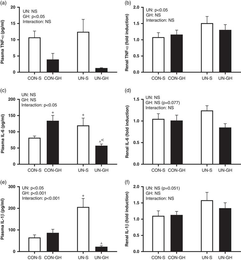
There was no effect of maternal UN on plasma TNF-α concentrations, but there was a significant overall effect of GH treatment on reducing plasma TNF-α concentrations in both UN and CON groups (Fig. 2a). Renal TNF-α gene expression was significantly higher in the UN-S group when compared to the CON-S group (Fig. 2b), but there was no effect of GH treatment. Plasma IL-6 concentrations were significantly increased in the plasma of the CON-GH and UN-S offspring compared to CON-S and UN-GH offspring (Fig. 2c). This was reflected in a significant maternal nutrition and GH interaction with plasma IL-6 concentrations increased with GH treatment in CON animals but decreased following GH in UN animals. Renal IL-6 gene expression was not different across any of the groups (Fig. 2d). Plasma IL-1β was significantly higher in the UN-S group compared to CON-S and was normalized to that of controls in the UN-GH group (Fig. 2e). There was no effect of maternal diet or GH treatment on IL-1 β gene expression in the kidneys (Fig. 2f). IL-10 and IL-4 expression were not detectable in the kidney.
Effects of maternal undernutrition (UN) and pre-weaning growth hormone (GH) treatment on plasma and renal inflammatory cytokines; TNF-α (a,b); IL-6 (c,d); IL-1β (e,f). Data presented as mean±s.e.m. (n=6–10 litters represented per group). ^indicates significantly different from UN-S (P<0.05).+indicates significantly different from CON-S (P<0.05). < indicates significantly different from CON-GH (P<0.05).

Effect of maternal UN and pre-weaning GH treatment on angiogenic and adhesion markers
There was no significant difference in vascular endothelial growth factor (VEGF)-a expression between any of the groups (Fig. 3a). Intercellular adhesion molecular (ICAM)-1 was significantly upregulated in the UN-S compared to the CON-S offspring groups and was normalized following GH treatment in the UN-GH group as shown in a maternal diet×GH treatment interaction (Fig. 3b). Vascular cell adhesion molecule (VCAM)-1 was upregulated in the UN-S group when compared with CON-S and normalized in the UN-GH group as reflected in a maternal diet and GH treatment interaction (Fig. 3c).
Effects of maternal undernutrition (UN) and pre-weaning growth hormone (GH) treatment on vascular endothelial growth factor (VEGF)-a (a), vascular cell adhesion molecule (VCAM)-1 (b) and intercellular adhesion molecular (ICAM)-1 (c). Data are presented as means±s.e.m. (n=6–10 litters represented per group). +indicates significantly different from CON-S (P<0.05); ^ indicates significantly different from UN-S (P<0.05).

Effect of maternal UN and pre-weaning GH treatment on relative glomerular number and renal histology
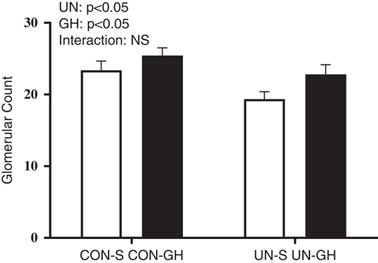
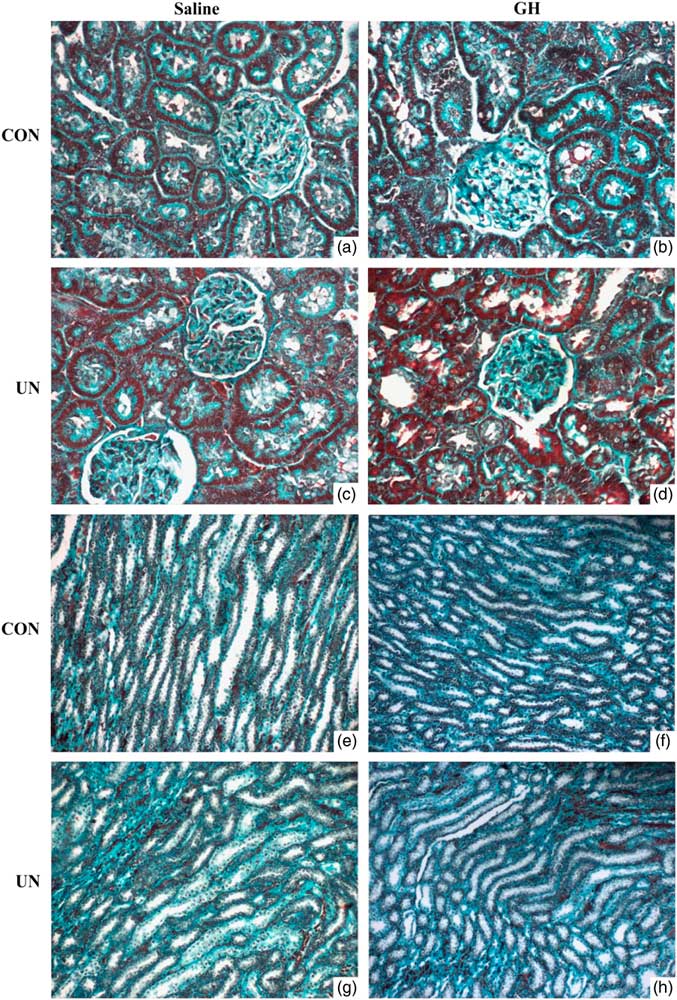
There was a significant effect of maternal diet and pre-weaning GH treatment on glomerular number. Glomerular number was decreased in UN groups compared to CON and was increased with GH treatment in both CON and UN groups (Fig. 4). Histological examination of glomeruli in all treatment groups indicated no obvious morphological or structural abnormality according to representative kidney histologic sections stained with H&E, PAS and MTS (Fig. 5). H&E staining of CON-S glomerulus at 40× magnification shows normal glomeruli with intact Bowman’s capsule and mesangium and cellularity (Fig. 5a). No prominent glomerular lymphocyte infiltration or any evidence of glomerulosclerosis was observed in maternal UN and/or pre-weaning GH treated offspring (Fig. 5b–5d). Glomerular basement membrane is of normal thickness in all groups following PAS staining (Fig. 5e–5h). MTS staining showed increased presence of collagen fibres in the UN-GH group, and this can be indicative of increased glomerular fibrosis (Fig. 6a–6d) although there was no evidence of tubulointerstitial fibrosis (Fig. 6e–6h).
Glomerular number. Data were analysed by two-way ANOVA with maternal diet and growth hormone (GH) treatment as factors. P<0.05 for effect of maternal diet and GH treatment. Holm-Sidak post-hoc tests were performed to detect any differences between groups. Data are presented as means±s.e.m., n=6–10 litters represented per group.

Representative glomerular histologic sections of CON-S (a,e), CON-GH (b,f), UN-S (c,g) and UN-GH (d,h) offspring [Haematoxylin and Eosin (H&E) stain (a–d), Periodic Acid-Schiff [PAS] stain (e–h), magnification 40×].

Representative glomerular (a–d) and tubular (e–h) histologic sections of CON-S (a,e), CON-GH (b,f), UN-S (c,g) and UN-GH (d,h) offspring [Masson’s trichrome (MTS) stain, magnification 40×].

Discussion
The present study examined potential renal pathogenic mechanisms in the developmental programming of hypertension following maternal UN. Our previous work in this experimental paradigm has shown that male offspring born to UN mothers have reduced birth weight followed by catch-up growthReference Li, Reynolds, Gray and Vickers29 and, as adults, in addition to hypertension are characterized by significant increases in body weight and fat mass as well as impaired insulin sensitivity.Reference Reynolds, Li, Gray and Vickers16
Inflammation in the kidneys plays an important role in the aetiology and pathogenesis of prenatally programmed hypertension.Reference Stewart, Jung, Manning and Vehaskari13, Reference Landgraf, Martinez and Rastelli30, Reference Khraibi31 Spontaneous hypertensive rats have identifiably altered immune function which is associated with high blood pressure with immune intervention conferring restorative effects on immune function but also normalizing increases in blood pressure.Reference Khraibi31 While there was an increase in systemic IL-6 and IL-1β concentrations in the UN-S group compared to CON-S, the inflammatory profile of the kidney did not follow the same trend, with significance reached only with TNF-α expression and a trend towards an overall increase in UN animals in renal IL-1β. TNF-α plays an important role in mediating the pro-inflammatory response in the kidney and plays an integral role in the pathogenesis of renal disease and its production is positively correlated with disease severityReference Donate-Correa, Martin-Nunez, Muros-de-Fuentes, Mora-Fernandez and Navarro-Gonzalez32 including that related to diabetic nephropathy.Reference Donate-Correa, Martin-Nunez, Muros-de-Fuentes, Mora-Fernandez and Navarro-Gonzalez32, Reference Ho, Wong and Lam33 Furthermore, TNF-α has the ability to modulate the expression of other inflammatory mediators, and it has been reported that TNF-α could upregulate the expression of the chemokine MCP-1 and the expression of cell surface adhesion molecules ICAM and VCAM-1 in human renal cells and endothelial cells.Reference Ho, Wong and Lam33, Reference Frank and Lisanti34 Infiltrating leukocytes can also produce pro-inflammatory cytokines including TNF-α which further induce the expression of chemokines such as MCP-1 in resident tissue cells forming a positive feedback loop.Reference Rudemiller and Crowley35 The current study provides some evidence of this with increased MCP-1 and adhesion molecule expression in the UN-S group which is associated with an increase in plasma TNF-α concentrations as well as renal mRNA expression. MCP-1 is associated with hypertensive conditions, and inhibiting its action reduces macrophage infiltration, normalizes blood pressure, prevented chronic renal damage and vascular inflammation in the hypertensive rat.Reference Kashyap, Warner and Hartono36–Reference Usui, Egashira and Tomita38
In the present study, ICAM-1 and VCAM-1 were significantly upregulated in the kidneys of UN-S. Cellular adhesion molecules along with MCP-1 are induced by shear stress and angiotensin II.Reference Miravete, Dissard and Klein39, Reference Hilgers, Hartner and Porst40 It is probable that altered glomerular hemodynamics in hypertensive UN-S offspring may also contribute to the induction of MCP-1 and adhesion molecules. This is in concordance with other reports that demonstrate that pro-inflammatory gene expression is significantly upregulated in both kidneys of deoxycorticosterone acetate-salt and aldosterone/salt hypertensive rats and also rat remnant kidney.Reference Taal, Zandi-Nejad and Weening41, Reference Blasi, Rocha and Rudolph42
Use of immunosuppressive drugs following exposure to a low-protein diet in utero prevented immune cell infiltration and the development of programmed hypertension in the offspring.Reference Stewart, Jung, Manning and Vehaskari13 However, in the current study, the expression of CD68, a marker of macrophage infiltration, was not significantly different despite increased MCP-1 and adhesion molecule expression. Furthermore, as we did not observe any prominent tubulointerstitial abnormality in the UN-S offspring as shown by our histological findings, nor did we see any evidence of prominent macrophage infiltration, this is consistent with the notion that macrophage infiltration is causative of intrarenal inflammatory damage. This finding also indirectly suggests that macrophage infiltration is crucial in the development of renal injury. These observations may arise due to a number of factors. First, slow rolling along the walls of the vessel maybe a prerequisite for subsequent firm adhesion. Increased rolling velocity and shear stress as with hypertension in the UN offspring may serve as one possible explanation in which inefficient conversion from rolling to adhesion led to decreased leukocyte infiltration. Thus, although ICAM-1 and VCAM-1 are critical for leukocyte adhesion and transendothelial migration, haemodynamic changes may have hindered subsequent adhesion and migration through the vascular wall.Reference Perry and Granger43 In agreement with this hypothesis, it was shown in another study that UN offspring had a lower numbers of rolling leukocytes.Reference Landgraf, Martinez and Rastelli30 Upregulation of ICAM-1 and VCAM-1 may serve as a compensatory mechanism to increase the adhesiveness of endothelial cells, thus to increase the likelihood of leukocyte recruitment into the tissue when exposed to chronic high blood pressure.Reference Barreiro, Martin, Gonzalez-Amaro and Sanchez-Madrid44 Furthermore, we should recognize that there is significant chemotactic redundancy among chemokines during disease and there are highly divergent and context-specific effects of these chemokines on blood pressure and tissue injury. We should bear in mind that inflammation is not a localized response but a systemic change, rather than considering inflammation based on an organ level, we should also consider the probability that these changes may be brought about by sustained inflammatory state of the endothelium following hypertension and altered immune function. Despite having an insignificant CD68 result, we cannot rule out the possibility of a subtle yet progressive change occurring inside the kidney as a consequence of inflammation and hypertension.Reference Ojeda, Grigore and Alexander45 There is modest renal hypotrophy as indicated by the kidney weight to body weight ratio in the UN-S offspring; and these changes, corrected with GH treatment, may have become more pronounced over a longer time period as the animals in the current study were still relatively young. This speculation is supported by elevated plasma urea and creatine concentrations, which are indicators of impaired renal function. The altered glomerular number in this study also likely contributed to impaired renal function as reported by others.Reference Luyckx and Brenner8, Reference Langley-Evans, Welham and Jackson46
There is little data on the effect of early GH treatment on reversing the adverse programming effect in the kidney following maternal UN. It is well established that maternal UN can reduce glomeruli number in the ratReference Chen and Chou47; and in the present study, we also observed a reduced number of glomeruli in UN offspring. Unlike the human, whereby nephrogenesis is complete before birth, 80% of nephrons in the rat kidney form in the first 7 to 10 days after birth.Reference Schreuder, Nyengaard, Remmers, van Wijk and Delemarre-van de Waal48 Thus, given the potential for GH to impact on renal development, we examined the efficacy of neonatal GH treatment and did observe a small but significant increase in glomeruli number, concomitant with normalization of renal mass, that paralleled the observed beneficial changes in systolic blood pressure. GH treatment also normalized plasma inflammatory markers including IL-6, TNF-α and IL-1β and also significantly reduced the levels of CD68, MCP-1, ICAM-1 and VCAM-1 in the kidney, suggesting that GH treatment may exert its effect by way of the inflammatory pathway. Serri et al. showed that adults with GH deficiency have elevated plasma concentrations of TNF-α and IL-6 and increased adhesiveness of their blood monocytes to endothelial cells in vitro.Reference Serri, St-Jacques, Sartippour and Renier49 As was previously demonstrated by our group, the increases in circulating pro-inflammatory cytokines was attributable not only to leukocytes but also to the adipose tissue.Reference Reynolds, Li, Gray and Vickers16 The significant downregulation of VCAM-1 and ICAM-1 with GH treatment may be related to GH’s ability to decrease plasma TNF-α concentrations. However, the linkage between GH activity and mediation of inflammatory processes remains poorly defined, with data to date suggesting a complex relationship with GH able to exert both pro- and anti-inflammatory effects, while inflammation itself can influence GH release and function.Reference Andreassen, Frystyk, Faber and Kristensen50–Reference Chen, Sood, Krishnamurthy, Rotwein and Rabkin52
Increased blood pressure in children born small for gestational age (SGA) is associated with changes in cardiac morphology that are altered following GH treatment.Reference Aurensanz Clemente, Ayerza Casas, Samper Villagrasa, Ruiz Frontera and Bueno Lozano53 Work by Sas et al. indicated that GH treatment to children born SGA had at least up to 6 years positive instead of negative effects on body composition, blood pressure and lipid metabolism.Reference Sas, Mulder and Hokken-Koelega54 However, some studies have reported side effects associated with GH use such as increased incidence of fluid retention, renal dysfunction, impaired glucose intolerance and intracranial hypertension,Reference Pawlikowska-Haddal55 but these may relate in part to dose, length of treatment and timing of treatment as many such studies are undertaken in adults.Reference Stochholm and Johannsson56, Reference Carroll and Van den Berghe57 In the present study, we noted histological evidence of glomerular fibrosis in the UN-GH group although this was not mirrored in any of the outcome measures made. It has previously been reported that GH treatment can exacerbate glomerulosclerosis and fibrosis in male rats in the setting of diabetic renal injury,Reference Whitney, Bilkan, Sandberg, Myers and Mulroney58 and GH has been shown to worsen the progression of kidney damage in models of marked renal disease. However, these are severe disease models including streptozotocin-induced diabetes and nephrectomyReference Flyvbjerg and Bennett59, Reference Flyvbjerg60 or animals model of GH gene disruptionReference Bellush, Doublier and Holland61 and outside of histological evidence for fibrosis in the present study, most of the observed effects of GH treatment on renal inflammatory markers were beneficial.
In conclusion, this study has provided evidence that renal immune mechanisms are associated with prenatally programmed hypertension and cardiovascular disease in the UN offspring. Pro-inflammatory markers and adhesion molecules were significantly upregulated in the kidneys in the UN offspring. The present study also highlighted adverse renal consequences related to maternal UN in offspring as illustrated by significant changes in plasma urea, creatine and total protein profiles. Furthermore, this study has provided further support to the protective effects of pre-weaning GH treatment in ameliorating some of the adverse cardiovascular disturbances observed in adult UN offspring including hypertension. These data strongly suggest that anti-inflammatory properties of GH may have contributed to the beneficial effects observed in UN offspring following early life GH treatment. The long-term benefits of GH treatment demonstrated by the current study might be due to its administration early in development in which critical windows of developmental plasticity exist, with GH intervention during this window normalizing the adverse effects of maternal UN on glomeruli structure and number and immune system regulation. Further, the current study was undertaken in male offspring, and further work is required in females to examine the potential for sex-specific effects of early GH treatment on renal function as reported by others.Reference Whitney, Bilkan, Sandberg, Myers and Mulroney58 A better understanding of maternal nutrition and the in utero nutritional environment on renal development and later progression to hypertension would potentially enable us to develop therapeutic strategies aimed at reducing the burden of later cardiovascular disease.
Acknowledgements
The authors acknowledge the technical support from Angelica Bernal and Rachna Patel (Liggins Institute) and the VJU, University of Auckland. Xiaoyuan Zhang, Clint Gray, Minglan Li and Clare Reynolds were supported by Gravida: National Centre for Growth and Development.
Disclosure statement
The authors have nothing to disclose.