Introduction
The concept of narcissism originates in psychodynamic theory and is also central in clinical psychiatry. Clinically, the concept of narcissism was developed by Kohut (Reference Kohut1971) and Kernberg (Reference Kernberg1975), and has been the basis for the incorporation of the narcissistic personality disorder into the DSM-III (APA, 1980) (see discussion for more details about the concept of narcissism). Narcissism is also regarded as a trait of the healthy personality (Emmons, Reference Emmons1984) and a prominent behavior pattern in western cultures (Lasch, Reference Lasch1979). Today, narcissism is widely seen as a continuum from healthy self-esteem to severe narcissistic pathology (Watson et al. Reference Watson, Sawrie, Greene and Arredondo2002; Ritter & Lammers, Reference Ritter and Lammers2007).
Patients with narcissistic personality disorder have been reported to show abnormalities not only in their self but also in empathy in terms of reduced affective resonance with other persons (Dimaggio et al. Reference Dimaggio, Nicolò, Popolo, Semerari and Carcione2006). Although, conceptually and clinically, the relationship between self and non-self (i.e. the other) in narcissism has been widely discussed, the psychological and neuronal manifestations of empathy and its relationship to the narcissistic self remain unclear. Empathy describes the sharing and understanding of the emotional and cognitive states of others; refined neuroimaging techniques have enabled the identification of specific brain regions involved in empathy (Decety & Lamm, Reference Decety and Lamm2006; Singer & Lamm, Reference Singer and Lamm2009). Core regions identified in the neural network underlying empathy are the anterior insula, the left inferior frontal cortex (including the mirror neurons), and other regions such as the premotor cortex and the dorsolateral prefrontal cortex (DLPFC; Decety & Lamm, Reference Decety and Lamm2006; Singer & Lamm, Reference Singer and Lamm2009). Although the involvement of these regions in empathy has been demonstrated in several studies, the relationship between this empathy network and narcissism remains unclear. Among these regions implicated in empathy, the anterior insula is most notable because it has been shown to be involved not only in empathy with others but also in focusing on the own self (Enzi et al. Reference Enzi, de Greck, Prosch, Tempelmann and Northoff2009; Modinos et al. Reference Modinos, Ormel and Aleman2009). Because subjects with high narcissism show a high self-focus, we can preliminarily hypothesize altered activity in the anterior insula in these subjects when compared to those with low narcissism.
The aim of this exploratory study was to investigate the psychological, behavioral and neuronal manifestations of empathy in a non-clinical sample with high and low narcissism respectively. Self-ratings of narcissism, empathy and clinical symptoms (e.g. somatization, obsessive–compulsive, interpersonal sensitivity, depression, anxiety, anger–hostility, phobic anxiety, paranoid ideation and psychoticism) were applied. These were complemented by a functional magnetic resonance imaging (fMRI) study of empathic perception of emotional faces that have been shown to induce an internal affective representation of the observed face in the observer (Preston & de Waal, Reference Preston and de Waal2002; Singer & Lamm, Reference Singer and Lamm2009). Based on theoretical conceptualizations and clinical observations, it was hypothesized that non-clinical subjects with high narcissism differ in psychological, behavioral and neuronal measures of empathy when compared to those with low narcissism.
Method
Subjects
Thirty-four subjects (21 females, 13 males, age 40.09±8.71 years, four left-handed) were investigated. Each of them received €30 compensation for voluntary participation in the study, other than the amount they obtained in the reward trials. None of the subjects reported a history of head injury or trauma, central nervous system disease, developmental disorder, medical disorder, substance abuse or psychiatric hospitalization. None were receiving psychotropic medication. All subjects gave written informed consent and the study was approved by the local review board.
Psychological measures
We included a scale on narcissism and also, with regard to our emotional focus, scales for empathy and emotions. In addition, we included a measure for depression, the Beck Depression Inventory (BDI), so as to exclude depression as a possible confounding variable, and also accounted for general non-specific clinical symptoms with the Symptom Checklist-90 – Revised (SCL-90-R). The German versions of the following questionnaires were used: (a) the Narcissism Inventory (NI; Denecke & Hilgenstock, Reference Denecke and Hilgenstock1989), a 163-item questionnaire that covers four dimensions of narcissism: (1) the threatened self, (2) the ‘classical’ narcissistic self, (3) the idealist self, and (4) the hypochondriac self; (b) the 20-item Toronto Alexithymia Scale (TAS-20; Bach et al. Reference Bach, Bach, de Zwaan, Serim and Bohmer1996); (c) the Interpersonal Reactivity Index (Paulus, Reference Paulus2009) to assess empathy on the four subscales fantasy, empathic concern, perspective-taking, and personal distress; (d) the SCL-90-R (Franke, Reference Franke1995), and (e) the BDI (Hautzinger et al. Reference Hautzinger, Bailer, Worall and Keller1994).
Experimental task
We presented emotional pictures of faces and instructed the subjects to empathize with the respective person. After the presentation of the pictures, the subjects were instructed to give an online judgment of their degree of empathy on a visual analogue scale presented on the screen. Following a recent hypothesis (Preston & de Waal, Reference Preston and de Waal2002), the mere perception of an emotional face should be sufficient for inducing an empathic response. Hence, we presented no faces (i.e. smoothed faces as the non-empathic control condition; see Supplementary method online for details), although this required additional investigations to control for various confounding factors (discussed later in detail). Every empathy trial began with the 5 s display of an emotional face or the presentation of a control stimulus. Subjects were instructed to empathize with the emotional face presented. Immediately after the presentation of the emotional face, a visual analogue scale was presented and subjects were asked to subjectively judge and evaluate their extent of affective sharing. In consideration of excluding the motor confound associated with the judgment and evaluation phase, the empathy task was preceded by a short finger-tapping task, which identifies motor regions for further exclusive masking analysis. Moreover, to exclude possible confounding variables, such as face and emotion perception, general evaluation and reward (Singer et al. Reference Singer, Seymour, O'Doherty, Stephan, Dolan and Frith2006), in our imaging results of empathy, we conducted additional and complementary studies focusing on these factors (see online Supplement). This allowed us to control for statistical independence, a recently highlighted methodological issue of imaging studies (Kriegeskorte et al. Reference Kriegeskorte, Simmons, Bellgowan and Baker2009). The main fMRI experiment consisted of six blocks, each 630 s in duration. Blocks 2, 4 and 6 were empathy blocks, and blocks 1, 3 and 5 were reward blocks where we used a reward paradigm, a slightly modified version of the Monetary Incentive Delay Task (Knutson et al. Reference Knutson, Adams, Fong and Hommer2001a, Reference Knutson, Fong, Adams, Varner and Hommerb).
Before entering the scanner, each subject read detailed information of the paradigm and completed a few trials to familiarize themselves with the task. While lying in the scanner, the stimuli were displayed using the ‘Presentation’ software package (Neurobehavioral Systems, USA) and were projected onto a matt screen using an LCD projector, visible through a mirror mounted on the headcoil (see online Supplement).
fMRI data acquisition and analysis
The fMRI data were collected in a 1.5-T MR scanner (General Electric Sigma Horizon, USA) using the standard circular polarized headcoil. Using a midsagittal scout image, a stack of 23 slices was aligned parallel to the bicomissural plane. During each functional run, 320 whole-brain volumes were acquired [gradient echo planar imaging (EPI), repetition time (TR)=2 s, echo time (TE)=35 ms, flip angle=80°, field of view=200×200 mm2, slice thickness=5 mm, interslice gap=1 mm, spatial resolution=3.125×3.125×5 mm3].
Image processing and statistical analyses were carried out according to the general linear model approach using the SPM2 software package running on MATLAB 6.5.1 (The Mathworks Inc., USA). The first five volumes were discarded due to saturation effects. All functional images were slice-time corrected with reference to the first slice acquired, corrected for motion artifacts by realignment to the first volume, and spatially normalized to a standard T1-weighted SPM template. The images were resampled to 2×2×2 mm3 and smoothed with an isotropic 6-mm full-width half-maximum Gaussian kernel. The time-series fMRI data were filtered using a high-pass filter with a cut-off at 128 s.
The first-level analysis modeled data of each subject using an event-related design by convolving the blood oxygen level-dependent (BOLD) signal with a canonical SPM hemodynamic response function. The movement parameters (translation and rotation) resulting from realignment were included as regressors to account for any residual effects of head motion. Regionally specific condition effects were tested by performing linear contrasts for each subject between conditions of interest.
To identify the empathy network as such we performed whole-group analysis with all 34 subjects. We thus compared the contrast ‘empathy>non-empathy’ and elucidated signal changes for the whole group. The contrast images resulting from the first-level analysis were submitted to a second-level random-effects analysis by applying a one-sample t test to the images acquired for all subjects. False discovery rate (FDR) correction was applied to control for multiple comparison using a threshold of FDR-corrected p<0.01, cluster size >50 voxels. The anatomical localization of significant activations was assessed with reference to the standard stereotactic atlas by superimposition of the SPM maps on a standard brain template [Montreal Neurological Institute (MNI)] provided by SPM2. To exclude possible movement confound associated with the judgment phase, activation in the whole-group analysis was then exclusively masked with the motor region found in the finger-tapping contrast. Furthermore, the activation found in this group analysis of empathy contrast was compared with the results of a meta-analysis of previous fMRI studies on empathy (see online Supplement).
The whole-group analysis was followed by group comparison between high and low narcissism groups using a two-sample t test. To investigate the neural difference between high and low narcissism groups in their empathic perception, the contrast images of the two groups for the first-level analysis ‘empathy>non-empathy’ were submitted to a second-level group comparison by applying an independent-sample t test. The threshold level was first set to 0.001 uncorrected, and then followed by a small volume correction with an independent region of interest (ROI) in the right anterior insula defined by the meta-analysis results of previous fMRI studies on empathy (see online Supplement). To further reveal the hemodynamic process during empathy, which differs in high and low narcissism groups, the results of this group comparison were selected as ROIs, in which the original BOLD signal changes were extracted and analyzed with the Marseille Region of Interest Toolbox software package (MarsBaR 0.42, http://marsbar.sourceforge.net/) (see online Supplement).
These analyses were supplemented by several analyses to account for possible confounding functions. To independently validate activations of the whole-group analysis for empathy, our results were compared with previous findings of empathy that resulted from a recent meta-analysis (see online Supplement). The possible impact of mere emotion processing was accounted for by comparing an emotional face with a neutral face in the signal changes of ROIs resulting from the group comparison (see online Supplement). Possible differences between high and low narcissism in face processing were tested by comparing signal changes extracted from the bilateral fusiform face area, which were elucidated in the whole-group analysis (see online Supplement). Unspecific task-related effects were controlled for by an additional empathy measure in fMRI in a different group of healthy subjects. There we compared the judgment of empathy with the judgment of skin color, which allowed us to control for unspecific task-related effects of judgment, that is general evaluation. Percentages of signal changes were extracted from this control experiment in ROIs defined by the results of our group comparison, and were compared between empathy and skin-color judgments. It was hypothesized that, if these activations are due to a general evaluation process, they would not differ between the judgment of empathy and the judgment of skin color; however, if they are indeed related to empathy, differences in ‘empathy versus skin color’ would be expected (see online Supplement and the results section for further details and reasoning). Finally, because previous data (Singer et al. Reference Singer, Seymour, O'Doherty, Kaube, Dolan and Frith2004, Reference Singer, Seymour, O'Doherty, Stephan, Dolan and Frith2006) showed a strong overlap of empathy with reward, we also excluded possible reward effects. We therefore tested for the difference between ‘anticipation of reward >anticipation of no outcome’ between high and low narcissism in the regional coordinates resulting from the high versus low narcissistic group comparison (see online Supplement).
Results
Psychological and behavioral data
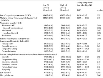
Eleven subjects scored beyond the 66th percentile of the NI (Denecke & Hilgenstock, Reference Denecke and Hilgenstock1989) and were regarded as highly narcissistic (six females, five males, age 40.09±10.34 years), the medium group consisted of 11 subjects (eight females, three males, age 40.18±10.08 years), and the low narcissism group had scores below the 33rd percentile (seven females, four males, age 40.18±6.42 years) (two subjects were excluded from the group comparison because of missing data). The high and low narcissism subgroups differed significantly in their narcissism total score (Table 1). No significant differences between the groups occurred with regard to age [T(20)=−0.025, p=0.98], gender (χ2=0.188, p=0.67) and general intelligence (Table 1). High narcissism subjects showed significantly higher alexithymia scores than low narcissism subjects (Table 1). No significant differences occurred in the ability to detect emotions in faces or spoken sentences (the Florida Affect Battery) and empathy (the Interpersonal Reactivity Index). This was confirmed by the lack of group differences in the post-scanning results of empathy and other dimensions (Table 1). High narcissism subjects yielded significantly higher psychopathology including depression (Table 1).
Comparison between high and low NI groups in psychological and behavioral data

LPS-3, Achievement Measure System (Leistungsprüfsystem; Horn, Reference Horn1983); MWT-B, the Multiple Choice Vocabulary Intelligence Test–B (Mehrfachwahl-Wortschatz-Intelligenztest–B; Lehrl et al. Reference Lehrl, Triebig and Fischer1995); SCL-90-R, Symptom Checklist-90 – Revised; GSI, Global Severity Index; BDI, Beck Depression Inventory; s.d., Standard deviation; df, degrees of freedom.
Neuronal data
We first performed a one-sample t test across the whole sample (n=34) to detect regions activated in the contrast ‘empathy>non-empathy’. This analysis yielded several regions typically associated with empathy (Supplementary Table 1) that were not confounded by movement associated with the judgment phase (Supplementary Fig. 1 a), and that were further confirmed by a meta-analysis on previous fMRI studies of empathy (Supplementary Fig. 1 b).
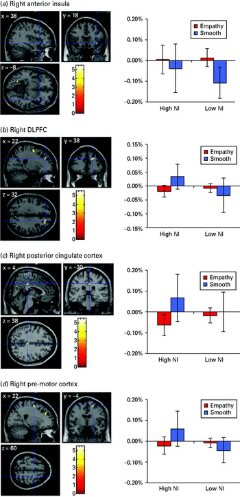
Signal changes in those regions that differ significantly between high and low narcissistic subjects in empathy versus non-empathy: (a) right anterior insula; (b) right dorsolateral prefrontal cortex; (c) right posterior cingulate cortex; (d) right premotor cortex. The results are based on a two-sample t test with p<0.001 uncorrected, k>10; and a small volume correction for the right anterior insula with a 10-mm-radius sphere that showed false discovery rate-corrected p value=0.017 and familywise error-corrected p value=0.033. Note that high Narcissism Inventory (NI) subjects differ from low NI subjects, especially in the non-empathy condition, showing less task-induced deactivation or even activation.

Subjects with high and low narcissism were then compared with each other. Using an SPM-based two-sample t test on the contrast ‘empathy>non-empathy’, regional signal changes differed significantly between the groups in the right anterior insula, right DLPFC, posterior cingulate cortex, and right lateral premotor cortex (Fig. 1 and Table 2). Signal change analysis for these regions in both conditions revealed that high narcissism subjects showed either decreased deactivation, as in the right anterior insula, or increased activation, as in the right DLPFC, the posterior cingulate cortex and the premotor cortex, during the control (that is, the non-empathic task; Fig. 1).
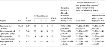
Brain activation contrast ‘empathize with emotional faces >view control faces’ for ‘low NI >high NI’

BA, Brodmann area; MNI, Montreal Neurological Institute.
Two-sample t test: p<0.001 uncorrected; k>10.
a Small volume correction with region of interest (ROI) in right anterior insula [centered at (38, 22, −1), volume=216 voxels] defined independently from meta-analysis results of previous functional magnetic resonance imaging (fMRI) studies on empathy (see online Supplement), which showed false discovery rate (FDR)-corrected p value=0.036 and familywise error (FWE)-corrected p value=0.013.
*** p<0.001, ** p<0.01.
Methodologically, it is important to note that our fMRI paradigm and our experimental settings were developed for measuring ‘affect sharing’, a distinct component of the multi-level concept of ‘empathy’ (Singer & Lamm, Reference Singer and Lamm2009). More specifically, we relied here on empathic face perception, which, according to a recent model of empathy (Preston & de Waal, Reference Preston and de Waal2002), should be sufficient for inducing an internal affective representation in the observer, which forms the basis of empathic response (Hooker et al. Reference Hooker, Verosky, Germine, Knight and D'Esposito2008). Although we aimed to control for possible confounding factors such as emotion processing, unspecific task-related effects, reward effects and face processing, we are aware of the experimental and conceptual difficulties in this field and refer the reader to a review highlighting the above-mentioned concept of empathy and its differentiation from the closely related concept of mentalizing (Singer, Reference Singer2006).
To rule out the impact of possible confounding factors, such as general face perception (as distinguished from face recognition), general evaluation and reward in these regions, we conducted a series of consecutive analyses (Table 2). First, to exclude differences in face processing, we compared the two groups with regard to neural activity in the fusiform face area with the coordinates for this region taken from the whole-group analysis. This revealed no significant difference between high and low narcissism groups in the activation of the left fusiform face area (−18, −92, −22) [F(1, 20)=1.093, p>0.3] and the right fusiform face area (46, −70, −16) [F(1, 20)=0.481, p>0.4].
To account for the confounding effect of unspecific and general emotion processing (as distinct from affective sharing and thus empathy), the signal changes from four ROIs resulting from ‘high narcissism versus low narcissism’ were entered as functional localizers for the analysis of the same data with regard to empathy for emotional and neutral faces (in half of the subjects who were also subjected to neutral stimuli, i.e. neutral faces). BOLD signal changes in the right anterior insula and the right posterior cingulate cortex showed no significant difference between emotional and neutral empathy conditions. However, signal changes in both regions, that is the right anterior insula and right posterior cingulate cortex (unlike in the right DLPFC and premotor cortex), during emotional and neutral empathy differed from the non-empathic condition, that is the smooth faces (Supplementary Fig. 2 a). This means that the factor emotion did not play a significant role in the observed activity changes in the right anterior insula and the right posterior cingulate cortex during empathy (because of the lack of difference between emotional and neutral faces). To further explore whether any difference between emotional and neutral empathy correlated with narcissism, the contrast ‘emotion >neutral’ was correlated with NI score in a second-level regression analysis, with these subjects who had been subjected to neutral stimuli (see online Supplement). Activations in the right ventromedial prefrontal cortex [r VMPFC, Brodmann area (BA) 10 (18, 60, −4)], the pre-supplementary motor area [pre-SMA, BA 8 (8, 14, 58)] and the left lateral prefrontal cortex [l LPFC, BA 10 (−32, 52, 4)] were found to correlate negatively with NI score (Supplementary Fig. 2 b), suggesting a difference in general emotion processing associated with narcissism. However, the right anterior insula and the right posterior cingulate cortex were not found to be involved in this comparison between emotional and neutral faces.
To exclude unspecific task-related effects of general evaluation (Singer, Reference Singer2006; Kriegeskorte et al. Reference Kriegeskorte, Simmons, Bellgowan and Baker2009) in the empathy task, we applied the coordinates differing between the high and low narcissism groups to a data set, where we directly compared empathy perception/judgment with perception/judgment of skin color (see online Supplement). The comparison of empathy versus skin color judgment yielded a significant difference for the coordinates from the right anterior insula whereas the other regions from the high versus low narcissism comparison did not show any significant difference (Table 2). This supports the assumption that the difference in the right anterior insula between high and low narcissism is due to empathy rather than to some non-specific task-related effect of a general evaluation function.
We also tested for possible rewarding effects of empathy. Singer et al. (Reference Singer, Seymour, O'Doherty, Kaube, Dolan and Frith2004, Reference Singer, Seymour, O'Doherty, Stephan, Dolan and Frith2006) showed a strong neural and psychological overlap between reward and empathy. It is not clear whether the differences between high and low narcissistic groups regarding their empathic response are due to empathy or are related to the rewarding effects of empathic stimuli (i.e. that emotional faces have a significant meaning to the perceiver). Thus we included a reward task to control for possible effects of reward related to the empathic stimuli. Using a reward task (Knutson et al. Reference Knutson, Adams, Fong and Hommer2001a, Reference Knutson, Fong, Adams, Varner and Hommerb) enabled us to test whether the neuronal activity differences between the two groups during empathy might be due to reward effects in the same regions. Using the coordinates from the group difference during empathy to the data acquired during reward did not show any significant interaction between the two groups with regard to the anticipation of reward (compared to the anticipation of no outcome) (Table 2).
Finally, to further confirm the association between narcissism and the empathy-related activity in the right anterior insula, we examined whether the medium NI group showed intermediate findings in the right anterior insula (see online Supplement). The percentage of signal changes of all three groups of subjects, extracted from the ROI of the right anterior insula defined independently by the meta-analysis results of previous fMRI studies on empathy, were found to correlate negatively with the NI scores [r(30)=−0.370, p<0.05] while controlling for the impact of age and intelligence [partial correlation r(30)=−0.374, p<0.05] (Supplementary Fig. 3).
Taken together, our neuroimaging results demonstrate neuronal differences between high and low narcissism subjects, most notably in the right anterior insula, that cannot be traced back to confounding functions such as emotion processing, face perception, general evaluation and reward.
Discussion
We have investigated the relationship between emotions, empathy and narcissism in an exploratory study on a non-clinical sample that combined psychological, behavioral and neuronal measures. Psychologically, highly narcissistic subjects yielded higher scores of alexithymia, general psychopathology and depression. By contrast, no significant psychological differences were observed with regard to empathy and post-hoc ratings for emotional reaction to the stimuli. Neuronally, highly narcissistic subjects showed differences predominantly in the right anterior insula that, as they were carefully controlled for, could not be traced back to other possible confounding functions. Taken together, these results indicate psychological and neuronal group differences in highly narcissistic subjects when compared to low narcissistic subjects. This may contribute to a better understand of the physiological basis of narcissism in general.
On a continuum from non-clinical narcissism to narcissistic personality disorder (Watson et al. Reference Watson, Sawrie, Greene and Arredondo2002), our study sample is comparable to that of a control sample of healthy volunteers described by Denecke & Hilgenstock (Reference Denecke and Hilgenstock1989). The mean narcissism total score in our sample was 22.5, compared to 23.0 in the above-mentioned group. Our high narcissism group with a mean score of 26.2 is located in the middle between the controls and a group of patients with narcissistic personality disorder (mean=29.2) (Denecke & Hilgenstock, Reference Denecke and Hilgenstock1989). Daig et al. (Reference Daig, Burkert, Fischer, Kienast, Klapp and Fliege2010) reported a mean score of 27.8 for a group of psychosomatic out-patients. Thus, our low narcissism group can be regarded as narcissistic below average, whereas the high narcissism group is above average but still below clinical severity.
Our high narcissism subjects exhibited more alexithymia, which parallels the clinically often described emotional abnormalities in narcissistic subjects (Kohut, Reference Kohut1971; Kernberg, Reference Kernberg1975; Dimaggio et al. Reference Dimaggio, Nicolò, Popolo, Semerari and Carcione2006). The co-occurrence and interdependency of narcissistic traits and alexithymia in eating-disordered patients have been reported by Lawson et al. (Reference Lawson, Waller, Sines and Meyer2008). The correlation of narcissism and lack of empathy is even more established, as documented in clinical–conceptual accounts (Kohut, Reference Kohut1971; Kernberg, Reference Kernberg1975), experimental investigation (Watson et al. Reference Watson, Grisham, Trotter and Biderman1984) and the DSM-IV (APA, 1994) diagnostic criteria for narcissistic personality disorder.
Our main neural finding concerns the observation of neuronal differences in the right anterior insula in particular and also in other regions such as the DLPFC, the posterior cingulate cortex, and the lateral premotor cortex. The right anterior insula has indeed been typically associated with empathy (Singer et al. Reference Singer, Seymour, O'Doherty, Kaube, Dolan and Frith2004, Reference Singer, Seymour, O'Doherty, Stephan, Dolan and Frith2006; Singer & Lamm, Reference Singer and Lamm2009; Lamm & Singer, Reference Lamm and Singer2010). The fact that high narcissistic subjects show differences in this region when compared to low narcissistic subjects further supports the assumption of changes in empathy in narcissism. However, the exact mechanisms associated with insula activity in empathy remain unclear. One prominent theory assumes simulation of the other's affective state to be crucial in empathy (Singer & Lamm, Reference Singer and Lamm2009; Lamm & Singer, Reference Lamm and Singer2010); but whether the differential insula activity in our groups may be related to different degrees of simulation of the other's affective state remains unclear.
Unlike in the other regions (i.e. the DLPFC, lateral premotor cortex and posterior cingulate cortex), neuronal activity changes in the right anterior insula in high narcissism subjects could not be traced back to related functions such as general evaluation, face perception or reward. This indicates that the right anterior insula must play a specific role in the constitution of empathy in people with narcissistic personality traits, which is in line with the characterization of the psychological function of the right anterior insula by Craig (Reference Craig2009). Based on its connectivity pattern, Craig assumes that the right anterior insula, as distinguished from the left one, is involved in representing the self, more specifically the bodily self or the ‘material me’ as he calls it. Because our high narcissism subjects show increased preoccupation with the self, as indicated by the narcissism scores, their altered activity in the insula may thus result from increased focus on the own self as, for instance, during mind-wandering in the non-empathic control condition. This is compatible with recent imaging studies showing recruitment of the right anterior insula during tasks focusing on the self (Enzi et al. Reference Enzi, de Greck, Prosch, Tempelmann and Northoff2009; Modinos et al. Reference Modinos, Ormel and Aleman2009).
We should also consider the fact that the main signal changes in the insula and the other regions stem from differences, for instance lower deactivation, in the non-empathic control condition rather than the empathy condition itself. However, the origin of the difference in the non-empathic control condition remains unclear. Because of the absence of an identifiable external stimulus in the empathic control condition, it is possible that activity differences in this condition might be related to differences in internal activity rather than externally stimulus-induced activity. This interpretation should, however, be considered with caution because our design did not include a true resting-state period. Moreover, the psychological correlates of the resting state in general and in narcissism remain unclear. Daydreaming (Singer, Reference Singer1966) and mind-wandering (Smallwood & Schooler, Reference Smallwood and Schooler2006) may be central; how these are involved in narcissism is a subject for future investigation. To lend further support to our speculative hypothesis, future investigation of the neuronal relationship between empathy, mind-wandering and self-related fantasies in narcissism is necessary.
Questions may also be raised concerning how our psychological and neuronal findings relate to the concept of narcissism as presupposed in psychoanalytic theory and clinical psychiatry. The concept of narcissism went through transference from pure intra-psychic nature of the self (and the ego as originally postulated by Freud, Reference Freud1905, Reference Freud and Freud1910) into a more relational or intersubjective ‘self-objects’ framework (as reviewed in Kohut, Reference Kohut1971, Reference Kohut1977, Reference Kohut1984; Gehrie, Reference Gehrie2009; Hartmann, Reference Hartmann2009) that includes the other (non-self) as a constitutive element (Kohut, Reference Kohut1984; Fosshage, Reference Fosshage2009), which, on the one hand, reflects its character as a continuum from ‘mature narcissism’ to ‘archaic or immature narcissism’, such as the narcissistic personality disorder in DSM-IV (APA, 1994; see also Russ et al. Reference Russ, Shedler, Bradley and Westen2008), and, on the other hand, is in accordance with recent accounts that link narcissism to emotions and empathy (Hartmann, Reference Hartmann2009). Both our psychological and neural findings lend further support to such a relational view of narcissism, which may consequently be closely related to changes in intersubjective relationships to other persons. The apparent difficulty in accessing the own emotions, as suggested by our finding of increased alexithymia, may lead to problems in simulating the other's affective states as our neural findings may be interpreted. The lack of accessing the other's emotions may in turn induce a shift from inter- to intra-subjective relationships, where the subject aim to relate to themselves through their narcissistic fantasies. This, however, is somewhat speculative at this point and requires further more detailed empirical investigation.
Finally, our findings also lend support to the concept of narcissism as a continuum between healthy and pathological forms. Narcissism is regarded as a trait of the healthy personality (Emmons, Reference Emmons1984) and a prominent behavior pattern in western cultures (Lasch, Reference Lasch1979). Our findings indicate that, even within a healthy sample, different degrees of narcissism as a personality trait seem to go along with neural and psychological differences. What may be of interest in the future is to investigate pathological forms of narcissism. Based on our findings reported here, we may expect even higher degrees of alexithymia and lower deactivation in, for instance, the right anterior insula during empathy. This remains to be investigated in the future.
Note
Supplementary material accompanies this paper on the Journal's website (http://journals.cambridge.org/psm).
Acknowledgments
We thank the staff from the Department of Neurology at the University of Magdeburg for their generous support. This study was supported financially by grants from the Hope of Depression Research Foundation (HDRF), the German Research Foundation (SFB-779, A6), the CIHR and the EJLB-Michael Smith Foundation to G.N.
Declaration of Interest
None.