INTRODUCTION
Cognitive and neuroimaging studies have demonstrated an association between the frontal lobes and cognitive set-shifting (e.g., McDonald et al., 2005; Moll et al., 2002; Stuss et al., 2001). One of the most commonly used measures of set-shifting is the traditional Trail Making Test (TMT), which has become a standard neuropsychological assessment instrument (Army Individual Test Battery, 1944; Brown & Partington, 1942; Partington & Leiter, 1949). The TMT has been shown to be sensitive to the presence of brain injury in general, and frontal lobe injury in particular (Demakis, 2004; Johnstone et al., 1995; Lange et al., 2005; Stuss et al., 2001).
Some researchers have suggested isolating the “executive” component of Part B of the traditional TMT from its perceptual and motor demands by using either a difference score, which subtracts Part A from Part B, or by using a ratio score of Part B over Part A (Arbuthnott & Frank, 2000; Lamberty et al., 1994). However, others have found that these methods did not add to the diagnostic utility of the test (Lange et al., 2005; Martin et al., 2003). One possible reason is that Part A and Part B of the test have somewhat different perceptual and motor demands, such as different numbers of stimuli and different trail lengths (Arnett & Labovitz, 1995; Gaudino et al., 1995) and that other cognitive skills may be tapped by Part B but not Part A (e.g., letter sequencing). Another possible reason is that brain injury may not lead to a true set-shifting deficit above and beyond a more basic sequencing deficit. The question arises then, whether frontal lobe patients' difficulty on the TMT is because of a set-shifting deficit per se or to a more basic visuo-motor sequencing deficit. For example, Martin et al. (2003) did not find disproportionate impairment in set-switching beyond a sequencing impairment on the TMT, in a sample of patients with traumatic brain injuries (TBI).
This study explored whether frontal lobe damage leads to simply a sequencing deficit or a higher-level switching deficit. This was investigated with a new version of the TMT. The Trail Making Test of the Delis-Kaplan Executive Function System (D-KEFS; Delis et al., 2001) was created in part to isolate set-shifting from other component skills such as letter sequencing and visual scanning. The test accomplishes this by including four baseline conditions (Visual Scanning, Number Sequencing, Letter Sequencing, and Motor Speed) and by placing equal numbers of stimuli in the three sequencing conditions. The D-KEFS Trail Making Test has been shown to detect executive functioning impairment in children with fetal alcohol syndrome (Mattson et al., 1999) and in adolescents and adults with autistic and Asperger's disorder (Kleinhans et al., 2005). Patients with frontal lobe epilepsy were disproportionately impaired compared to patients with temporal lobe epilepsy on the Number-Letter Switching condition (McDonald et al., 2005). A patient with focal ventromedial prefrontal cortex damage displayed subtle deficits on the Number-Letter Switching condition, which became apparent only when analyzing errors (Cato et al., 2004). To our knowledge, Cato et al.'s case study has been the only report of D-KEFS Trail Making Test performance in any patient with a focal frontal lobe lesion. Further studies are needed to examine the effects of focal frontal lobe lesions on the D-KEFS Trail Making Test.
This study compared performance on the D-KEFS Trail Making Test by patients with focal lateral prefrontal cortex lesions to matched control participants. We predicted that lateral prefrontal cortex patients would be disproportionately impaired on the set-shifting condition of the D-KEFS Trail Making Test relative to controls.
METHOD
Participants
The current study included 12 patients (8 men and 4 women) with focal lesions in the lateral prefrontal cortex (LPC) and 11 age- and education-matched control participants. Patients were included based on the following criteria: a single lesion, no prior neurologic or psychiatric history, and no history of substance abuse. Nine of the patients were European American, and the other three were African American, Philippine American, and mixed Native and Hispanic American. All participants were native English speakers. Patients were tested at least one year post-lesion. Lesion etiologies included embolic and hemorrhagic stroke (n = 8), and surgery for meningioma, cyst, aneurysm, and arterio-venous malformation (n = 4; see Table 1).
Demographic and descriptive variables of sample

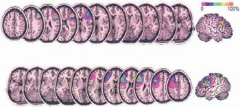
CT and/or MRI scans were obtained on patients at least five weeks post-onset and generally within a few months of testing. Our patients' health status is followed either by VA medical records or by patient and caregiver reports. If a subsequent neurologic event occurs during the testing period, the patient is re-scanned, and patients with secondary events are excluded from study. Patients' lesions were reconstructed by a board-certified neurologist onto an 11-slice axial template based on the atlas of DeArmond et al. (1976). These templates were then digitized, using in-house lesion reconstruction software (Frey et al., 1987). This software also allowed for an estimation of patients' lesion volume, which ranged from 12.9 to 200.4 cubic centimeters (see Table 1). Figure 1 shows lesion reconstructions for 11 of the 12 patients. (It was not possible to reconstruct one patient's lesion because of technical problems, but lesion site was verified on MRI.) As can be seen, patients' lesions were relatively focal and were confined to either the right or left LPC. Patients with lesions that extended significantly into other portions of frontal cortex (e.g., orbital regions) and/or involved other lobes were excluded from the study.

Lesion reconstructions of patients with right hemisphere (top) and left hemisphere (bottom) lateral prefrontal cortex lesions. The color bar shows the degree of lesion overlap, with the greatest overlap (100% of lesions) shown in red. The far right figure shows the lesions projected on to the lateral surface of each hemisphere.
Eleven age- and education-matched controls (7 men and 4 women) were recruited from the same community. The controls were neurologically-normal and had no history of neurologic, psychiatric, or substance abuse issues. Ten of the control participants were European American, and one was Hispanic American. Patients and controls did not differ with respect to age, t (21) = 0.74, p = .47, or education, t (21) = .61, p = .55. Patients and controls were all right-handed. However, due to right-sided weakness, two patients (1 and 4) used their non-dominant hand to perform the tasks. Analyses were completed with and without these two participants, and results were equivalent with one exception: after removing the two participants, groups were no longer significantly different on the Motor Speed completion time (described below).
All participants read and signed consent forms prior to participation. The study was approved by the VA Institutional Review Board, and was conducted in compliance with the Helsinki declaration.
Materials and Procedures
The D-KEFS Trail Making Test involves a series of 5 conditions: visual scanning, number sequencing, letter sequencing, number-letter switching, and motor speed. In all five conditions, the stimuli are spread over an 11 × 17-inch area, which provides longer trails and more interference stimuli than the traditional TMT (Delis et al., 2001). In the Visual Scanning condition, examinees cross out all the 3s that appear on the response sheet. In the Number Sequencing condition, examinees draw a line connecting the numbers 1–16 in order; distractor letters appear on the same page. The Letter Sequencing condition requires examinees to connect the letters A through P, with distractor numbers present on the page. In the Number-Letter Switching condition, examinees switch back and forth between connecting numbers and letters (i.e., 1, A, 2, B, etc., to 16, P). Last, a Motor Speed condition is administered in which examinees trace over a dotted line connecting circles on the page as quickly as possible, in order to gauge their motor drawing speed. Each condition is preceded by a short practice trial. In all conditions, examinees are told to work as quickly and as accurately as possible. In all but the visual scanning condition, the examiner corrects mistakes by placing an “X” over a wrong connection, and examinees are asked to continue from the last correct connection. The stopwatch remains running during such corrections.
RESULTS
Completion Time
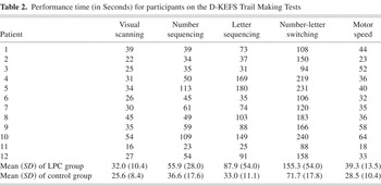
Raw scores for all LPC patients and the mean LPC patient and control scores are presented in Table 2. Data from each subtest of the D-KEFS Trail Making Test were entered into a series of five univariate analyses of variance (ANOVAs) to compare performances by patients with LPC lesions and controls. Performance on the Visual Scanning subtest was equivalent between groups, F(1,21) = 2.58, p = .12, η2 = .11. The difference between groups on the Number Sequencing subtest approached significance, F(1,21) = 3.83, p = .06, η2 = .15. Patients with LPC lesions took significantly longer to complete the Letter Sequencing subtest, F(1,21) = 10.93, p < .01, η2 = .34. There was a large difference between the groups on the Number-Letter Switching condition, with the LPC patients taking longer to complete it, F(1,21) = 23.86, p < .001, η2 = .53. Lastly, a smaller difference was found between groups on the Motor Speed subtest, F(1,21) = 4.50, p < .05, η2 = .18. To summarize, the largest significant differences between patients and controls were found on the Number-Letter Switching condition, followed by the Letter Sequencing condition, followed by the Motor Speed condition.
Performance time (in Seconds) for participants on the D-KEFS Trail Making Tests

To determine whether the set-shifting performance in LPC patients on the D-KEFS Trail Making Test was significantly different over and above the other observed differences, we also conducted a one-way analysis of covariance (ANCOVA) between LPC patients and controls with Number-Letter Switching as the dependent variable, and the other four conditions as covariates. The difference between LPC patients and controls on Number-Letter Switching was still significant, F(1,16) = 11.02, p < .01, η2 = .39, suggesting that the Number-Letter Switching condition posed a disproportionate challenge to LPC patients that did not simply stem from deficits on letter sequencing or reduced motor speed, for example.
Errors
Patients with LPC lesions committed significantly more errors (both in set-switching and sequencing) on the Number-Letter Switching condition, t (21) = 2.06, p = .05. Nine of the 12 patients with LPC lesions committed one or more errors, compared with 4 of the 11 control participants. No significant differences were found in the number of errors committed on the Visual Scanning and Letter Sequencing subtests. Neither group made any errors in the Number Sequencing and Motor Speed subtests.
DISCUSSION
The current study investigated performance on the D-KEFS Trail Making Test in patients with focal lesions in the lateral prefrontal cortex (LPC) and in age- and education-matched control participants. This test allows for the isolation of set-shifting deficits by controlling for variables such as visual scanning, number sequencing, letter sequencing, and motor speed. We found that LPC patients were disproportionately slower compared to control participants on the Number-Letter Switching condition of the D-KEFS Trail Making Test, even when performances on the baseline conditions were controlled. In addition, patients with LPC lesions also made more errors than controls on the Number-Letter Switching condition.
These results suggest that damage to the LPC may lead to deficits in cognitive set-switching, above and beyond a deficit in sequencing. Our findings expand on the work by Stuss et al. (2001) who found that performance on Part B of the traditional TMT was significantly worse in patients with dorsolateral frontal lobe lesions, compared to patients with lesions in other locations and control patients. We found similar results with the new D-KEFS Trail Making Test. In addition, we found that patients with frontal lesions were significantly slower on the Number-Letter Switching condition even after controlling for performance on Number Sequencing, Letter Sequencing, Visual Scanning, and Motor Speed subtests. This suggests that damage to the LPC may lead to impairment in cognitive set-switching, above and beyond a basic deficit in number or letter sequencing. Whereas Martin et al. (2003) did not find disproportionate impairment in set-switching beyond a sequencing impairment on the traditional TMT in a sample of TBI patients, our results suggest that LPC damage does indeed lead to a set-switching deficit. This deficit may be elicited particularly clearly on the D-KEFS Trail Making Test.
The current results are consistent with those found by McDonald et al. (2005), who compared patients with frontal-lobe epilepsy to patients with temporal-lobe epilepsy and controls on the D-KEFS Trail Making Test. Likewise, Cato et al. (2004) found that a patient with ventromedial prefrontal cortex damage showed deficits on the set-switching conditions of the D-KEFS Trail Making and Color-Word Interference Tests. Cato et al.'s findings along with the present study suggest that the medial and lateral prefrontal cortex may be involved with set-shifting, but more research is needed before this conclusion can be drawn. Our results are also consistent with earlier studies using Part B of the traditional TMT in patients with dorsolateral prefrontal cortex damage (Stuss et al., 2001), and in normal participants in a fMRI study (Moll et al., 2002). The LPC may thus be highly involved when engaging in tasks that require a large degree of cognitive flexibility.
Taken together, these findings suggest that the D-KEFS Trail Making Test may tap into frontal-lobe functioning by affording a method for parsing higher-level set-shifting from more fundamental component skills. That is, deficits in frontal-lobe functioning may become most apparent on the Number-Letter Switching condition, when increasingly higher-level cognitive processing and flexibility are required. These results offer some support for the construct validity of the D-KEFS Trail Making Test, particularly its ability to assess set-shifting over and above component skills and to be sensitive to focal frontal-lobe lesions. Whereas past studies with the traditional TMT have not found evidence of added diagnostic utility in parsing out Part A from Part B (Lange et al., 2005; Martin et al., 2003), the present results suggest that focal frontal patients do show evidence of disproportionate deficits in set-shifting relative to the four baseline abilities on the D-KEFS Trail Making Test. In addition, the LPC patients also performed significantly more slowly than controls on the Letter Sequencing condition, which suggests that letter sequencing per se may be a more effortful task than number sequencing for patients with frontal-lobe lesions.
Current findings are suggestive of further research in at least three directions. First, future research should explore the specificity of the observed deficits with respect to the LPC. Larger patient samples are needed to assess how patients with lesions restricted to subregions within the frontal lobes as well as in nonfrontal areas perform on these measures. For example, studies could explore whether the lateral or medial prefrontal cortex is more heavily involved in cognitive set-shifting. Also, it is possible that our results are due merely to an increase in task complexity when moving from scanning, to number or letter sequencing, to simultaneous number and letter sequencing. Further experimental manipulations are needed to tease out this issue. In addition, it is important to explore whether these findings generalize to non-language, or non-visual measures of set-shifting.
ACKNOWLEDGMENTS
Disclosures: Dr. Dean Delis, the fourth author of this manuscript, has a financial interest in the measure that is the focus of this study. He is the lead author of the Delis-Kaplan Executive Function System (D-KEFS), published by The Psychological Corporation. In addition, the participants received compensation for their time from The Psychological Corporation. At the time the data were collected, Dr. Baldo was supported by NIH Grant #NS17778 and a National Research Service Award.