INTRODUCTION
Schistosomiasis has been eradicated from a number of areas of the world through valiant integrated control measures based on snail control, drug treatment and environmental modification. In many other endemic areas morbidity has been dramatically reduced in recent times by treatment with praziquantel, the only drug now in widespread use (Fenwick and Webster, Reference Fenwick and Webster2006). In such areas the snail host populations remain unaffected, transmission persists and reinfection is common. Relaxation of vigilance in ‘eradicated’ areas or of control efforts in endemic areas or development of resistance to praziquantel would rapidly lead to resurgence of the disease. Despite the potential of regular chemotherapy to control morbidity, the desirability of a vaccine to supplement and sustain chemotherapy-based control efforts remains as high as in 1970–1990 (Taylor, Reference Taylor1994) when schistosome vaccine research was reasonably well funded. A particular focus of attention during this time was on vaccination with radiation-attenuated larval infections which were demonstrated to be highly effective in many experimental hosts (Taylor, Reference Taylor1994) and this encouraged belief in the feasibility of developing a defined schistosome vaccine for humans. Further studies of irradiated vaccines yielded considerable understanding of the schistosome life-cycle stages which are the optimal inducers and targets of such immunity but limited progress was made into defining and validating the protective antigens involved. Other, parallel approaches to vaccine development yielded several recombinant–derived schistosome proteins which showed promise in their labs of origin but less so when tested independently (Bergquist and Colley, Reference Bergquist and Colley1998). With this, the impetus for vaccine development waned, particularly since there was a general perception in the public health community at this time that the schistosomiasis problem could be ‘solved’ globally using population-scale praziquantel treatment.
However, there is now a growing appreciation of the limitations of what chemotherapy campaigns alone can achieve, and recent application of proteomic and genomic techniques has facilitated the identification and characterization of membrane and secreted antigens, likely candidates for the protection induced by irradiated vaccines. So it is timely to review some of the lessons learned from the irradiated vaccine work in the hope of encouraging the new generation of those who share the belief that schistosome vaccines are both feasible and needed (Bergquist et al. Reference Bergquist, Leonardo and Mitchell2005; Bethony et al. Reference Bethony, Diemert, Oliveira and Loukas2008). More exhaustive reviews of irradiated vaccines (Coulson, Reference Coulson1997; Dean, Reference Dean1983) and of approaches to schistosome vaccine development (e.g. Hewitson et al. Reference Hewitson, Hamblin and Mountford2005; Wilson and Coulson, Reference Wilson and Coulson2006; McManus and Loukas, Reference McManus and Loukas2008) are available.
IRRADIATED VACCINES – ESTABLISHMENT OF EFFECTIVE PROTOCOLS
Use of radiation-attenuated helminth infections to induce protection was first tested in the 1950s and, notably, led to development of the commercial live vaccine Dictol® against Dictyocaulus viviparus, a nematode of cattle (Jarrett et al. Reference Jarrett, Jennings, McIntyre, Mulligan and Urquhart1958). The vaccine comprised oral infection with larvae irradiated with 40 krad (kilorads) such that they did not reach the adult pathogenic stage but survived long enough to stimulate cells in the mesenteric lymph nodes. Prompted by this success irradiated vaccines against other helminths were soon being tested e.g. Miller (Reference Miller1978). In the case of schistosomes, doses of 3–4 krad of X- or γ-radiation were shown to prevent developing schistosome worms surviving to the adult pathogenic egg-laying stage while doses of 1–3 krad resulted in persistence of a small proportion of stunted and generally sterile adults (Smithers, Reference Smithers1962). There followed various protection studies against S. mansoni and S. japonicum in mice and repeatedly exposed rhesus monkeys using cercariae exposed to relatively low levels of radiation (Hsu et al. Reference Hsu, Hsu and Osborne1962; Radke and Sadun, Reference Radke and Sadun1963; Sadun et al. Reference Sadun, Bruce and Macomber1964; Smithers, Reference Smithers1962; Villella et al. Reference Villella, Gomberg and Gould1961) and most studies reported higher levels of partial protection to challenge with very low doses of radiation which allowed a small proportion of stunted worms to survive. These studies were carried out at a time when the prevailing notion, based on studies in rhesus monkeys harbouring unattenuated S. mansoni infections (Smithers and Terry, Reference Smithers and Terry1965, Reference Smithers and Terry1967), was that the adult worm is the prime stimulus to protection and that the schistosomula stages contribute little to the development of resistance. Thus, with respect to these early irradiated infections Smithers (Reference Smithers, Cohen and Sadun1976) suggested that ‘… the immunizing effect is predominantly due to the few stunted worms which survive the lethal effects of radiation’. However, Hsu et al. (Reference Hsu, Hsu and Osborn1969), wishing to avoid inflammatory foci in the lung and liver due to death of immunizing larvae in these organs, tested highly irradiated (24 or 48 krad) cercariae of S. mansoni and S. japonicum which would arrest in the lung/skin. Both radiation doses resulted in high levels of protection in rhesus monkeys (>80% fewer worms than controls) especially after repeated (>3) infections.
In the early 1970s studies were initiated using the schistosome species of domestic animals, S. mattheei and S. bovis, which meant that vaccination efficacy could be assessed in natural hosts i.e. sheep and cattle. Initial studies in sheep using 1 or 2 exposures to S. mattheei larvae irradiated at 3 or 6 krad were ineffective but high levels of partial protection >70% could be induced following 4 vaccinations and comparable protection could be induced with percutaneously applied cercariae and intramuscularly injected in vitro-transformed schistosomula (Taylor et al. Reference Taylor, James, Nelson, Bickle, Dunne and Webbe1976b). Similar protection could be induced against S. bovis in cattle with a single vaccination (Bushara et al. Reference Bushara, Hussein, Saad, Taylor, Dargie, Marshall and Nelson1978). These studies culminated in a field trial in cattle of an S. bovis vaccine comprising a single intramuscular injection of schistosomula irradiated at 3 krad (Majid et al. Reference Majid, Bushara, Saad, Hussein, Taylor, Dargie, Marshall and Nelson1980). High partial protection to natural exposure was demonstrated and was followed by similar successful field testing of an irradiated, cryopreserved, schistosomula vaccine for S. japonicum in cattle and buffaloes, key hosts of this zoonotic schistosome species (Hsu et al. Reference Hsu, Xu, He, Shi, Shen, Hsu, Osborne and Clarke1984).
In the case of the human schistosome species, S. mansoni, a similar approach was applied using baboons, regarded as a suitable model for human schistosomiais since these primates, unlike rhesus monkeys, are natural hosts of both S. mansoni and S. haematobium. However, repeated exposure to larvae given low doses of radiation did not induce significant protection (Taylor et al. Reference Taylor, James, Nelson, Bickle, Andrews, Dobinson and Webbe1976a). This failure prompted studies in mice and rats aimed at defining the key parameters for optimal vaccination protocols. It was soon established that for resistance induced by a single exposure of mice the dose of radiation had a significant effect. Both low doses of radiation, allowing parasite survival through to stunted adult worms, and very high doses, arresting the larvae in the skin, were poorly effective whereas significantly higher protection could be induced with optimal doses (56 krad (Minard et al. Reference Minard, Dean, Jacobson, Vannier and Murrell1978a) or 20 krad (Bickle et al. Reference Bickle, Taylor, Doenhoff and Nelson1979c)). In both cases the effects of these optimal doses of radiation on the larvae were similar, resulting in a slight delay in the skin but eventual migration to and death in the lungs (Minard et al. Reference Minard, Dean, Vannier and Murrell1978b; Bickle et al. Reference Bickle, Dobinson and James1979b; Mastin et al. Reference Mastin, Bickle and Wilson1983; Mangold and Dean, Reference Mangold and Dean1984). The reasons for the numerical difference in these optimal doses is not clear but since at this early time most subsequent studies in the USA, although not all (Reynolds and Harn, Reference Reynolds and Harn1992), employed doses around 50 krad and in the UK, 20 krad. In rats, 20 krad also arrested S. mansoni larvae in the lungs and similarly proved to be an optimal dose (Ford et al. Reference Ford, Bickle and Taylor1984a).
The superior protective efficacy of highly irradiated cercariae in mice led to renewed testing in baboons and, similarly, somewhat greater protection could be induced with optimally-irradiated infections. Thus, studies using S. haematobium (Sturrock et al. Reference Sturrock, James, James and Webbe1980; Webbe et al. Reference Webbe, Sturrock, James and James1982; Harrison et al. Reference Harrison, Bickle, Kiare, James, Andrews, Sturrock, Taylor and Webbe1990) demonstrated high levels of protection (~90%) with multiple (2–3) exposures to 20 krad cercariae whilst lower protection was obtained with 3 krad or 60 krad and significantly less with a single vaccination (~30%) (Harrison et al. Reference Harrison, Bickle, Kiare, James, Andrews, Sturrock, Taylor and Webbe1990). Repeated exposure to highly irradiated cercariae was also protective against S. mansoni (Stek et al. Reference Stek, Minard, Dean and Hall1981b) and could be boosted by repeated exposures (Kariuki et al. Reference Kariuki, Farah, Yole, Mwenda, Van Dam, Deelder, Wilson and Coulson2004). Significant partial protection (~45%) was also demonstrated in chimpanzees, our closest primate relative (Eberl et al. Reference Eberl, Langermans, Frost, Vervenne, Van Dam, Deelder, Thomas, Coulson and Wilson2001).
So, high levels of protection have been demonstrated in a wide variety of hosts against various schistosome species including the three major human species. Choice of optimal radiation dose and use of multiple vaccinations are more crucial for some species than others, notably primates.
INDUCERS AND TARGETS OF IMMUNITY
Nearly all of our further understanding of the mechanisms of immunity induced by irradiated infections has come from studies of S. mansoni in mice, and to some extent from rats. The fact that optimal doses of radiation result in migration to and death in the lungs focused attention on the immunogenicity of the lung-stage schistosomula (LS) and led to experiments showing that irradiated LS introduced into the lungs via intravenous injection (Dean et al. Reference Dean, Cioli and Bukowski1981; Ford et al. Reference Ford, Bickle and Taylor1984a), or into the skin (Coulson and Mountford, Reference Coulson and Mountford1989) could confer immunity. Emphasis on the larval stages was further supported by the relative failure of irradiated 3- or 4-week-old worms (Dean et al. Reference Dean, Cioli and Bukowski1981) or unirradiated adult worm (single sex) infections to induce protection in mice (Bickle et al. Reference Bickle, Bain, McGregor and Doenhoff1979a).
The LS was also shown, in the majority of studies, to be the principal target of immunity in challenged animals. By recovery of parasites following mincing and incubation of tissue (Minard et al. Reference Minard, Dean, Vannier and Murrell1978b; Stek et al. Reference Stek, Dean and Clark1981a) by quantitative histology (Mastin et al. Reference Mastin, Bickle and Wilson1983; Von Lichtenberg et al. Reference Von Lichtenberg, Correa-Oliveira and Sher1985) and most accurately by autoradiographic tracking in compressed organs (Dean et al. Reference Dean, Mangold, Georgi and Jacobson1984; Wilson et al. Reference Wilson, Coulson and Dixon1986; Dean and Mangold, Reference Dean and Mangold1992) it was shown that migration of percutaneously applied challenge larvae to the lungs is delayed in vaccinated compared with naïve mice but otherwise comparable. Subsequently, a greater proportion of schistosomula are retained in the lungs of vaccinated mice and these gradually disappear between 2–5+ weeks post-infection (Wilson et al. Reference Wilson, Coulson and Dixon1986; Dean and Mangold, Reference Dean and Mangold1992). LS introduced by i.v. injection into the lungs of vaccinated rats (Ford et al. Reference Ford, Bickle, Taylor and Andrews1984b; McLaren et al. Reference McLaren, Pearce and Smithers1985) and mice (Mangold et al. Reference Mangold, Dean, Coulson and Wilson1986) were also highly susceptible to attrition although, in some experiments in mice, somewhat higher levels of resistance followed percutaneous challenge (Smythies et al. Reference Smythies, Betts, Coulson, Dowling and Wilson1996; Coulson and Wilson, Reference Coulson and Wilson1997) which the authors suggested indicates that the skin stage may help to initiate a recall response prior to lung attrition.
Histological studies of the response elicited by percutaneously applied challenge infections in vaccinated mice showed pronounced inflammatory foci around schistosomula in the skin and subsequently in the lungs (Von Lichtenberg et al. Reference Von Lichtenberg, Correa-Oliveira and Sher1985), for example, by day 8 post-challenge there were >10-fold more lung foci in vaccinated compared with control mice. The cellular infiltration, comprising predominantly mononuclear cells, starts as soon as the larvae reach the lungs and peaks around 8 days after infection (Smythies et al. Reference Smythies, Betts, Coulson, Dowling and Wilson1996). Cells accumulate within the blood vessel and between the vascular endothelium and the alveolar epithelium resulting, it is suggested, in the intravascular migration of the larvae being impeded. Certainly, with time, an increasing proportion of larvae occur within alveoli where they induce even more pronounced foci (Crabtree and Wilson, Reference Crabtree and Wilson1986). Although Von Lichtenberg et al. (Reference Von Lichtenberg, Correa-Oliveira and Sher1985) described damaged LS in vaccinated mice, Crabtree and Wilson (Reference Crabtree and Wilson1986) found no evidence of parasite damage and this led to the notion that LS may not be killed within the lung but rather diverted from their intravascular migration into the alveoli and subsequently lost via the trachea and eventually the GI tract. This was supported by later studies showing that challenge LS recovered from lungs of vaccinated mice over the extended period of attrition showed normal viability when injected into naïve mice (Coulson and Wilson, Reference Coulson and Wilson1988). Furthermore, radio-isotope labelled, challenge organisms in compressed lungs always appeared as discrete autoradiographic foci of equal intensity, unlike foci caused when radio-isotope labelled, heat-killed LS were injected into the lungs which became smaller and fainter with time (Dean and Mangold, Reference Dean and Mangold1992). The lack of evidence of in vivo death of LS is consistent with the demonstration that they are also insusceptible to immune damage in vitro by either antibody- or cell-mediated mechanisms (Moser et al. Reference Moser, Wassom and Sher1980; Sher et al. Reference Sher, James, Simpson, Lazdins and Meltzer1982b; Bickle and Ford, Reference Bickle and Ford1982; Ahmed et al. Reference Ahmed, Oswald, Caspar, Hieny, Keefer, Sher and James1997).
MECHANISMS OF IMMUNITY
Initial investigations into the mechanisms of vaccine-induced resistance in rodents implicated the immune response since B cell-depleted mice and athymic mice and rats failed to develop resistance (Sher et al. Reference Sher, Hieny, James and Asofsky1982a; Ford et al. Reference Ford, Bickle and Taylor1987a). Both antibody and CD4+ T-cell-mediated, IFN-γ-dependent effector mechanisms have been demonstrated, depending on the host species and the number of vaccinations.
In mice, larvae from a percutaneuosly applied, optimally irradiated infection undergo a protracted migration in the skin (Mangold and Dean, Reference Mangold and Dean1984), specifically in the skin draining lymph nodes (SLN), and release greater amounts of parasite antigen than unirradiated larvae (Mountford et al. Reference Mountford, Coulson and Wilson1988). This results in marked increases in cell number (Constant et al. Reference Constant, Mountford and Wilson1990) and prolonged in vitro antigen-specific CD4 +ve cell proliferation which is followed some time later by similar events in the lungs and draining mediastinal lymph nodes as larvae migrate there (James et al. Reference James, Labine and Sher1981; Lewis and Wilson, Reference Lewis and Wilson1982; Pemberton et al. Reference Pemberton, Smythies, Mountford and Wilson1991). Initial percutaneous exposure to either normal or irradiated cercariae induces both Th1 (IFN-γ and IL-2) and Th2 (IL-5 and IL-4) cytokine responses in the SLN (Caulada-Benedetti et al. Reference Caulada-Benedetti, Al-Zamel, Sher and James1991) but these are more protracted with the irradiated infection and Th1 cytokines responses tend to increase preferentially at later time points (Pemberton et al. Reference Pemberton, Smythies, Mountford and Wilson1991). Th1-polarized cytokine production is more prominent in the spleen (Caulada-Benedetti et al. Reference Caulada-Benedetti, Al-Zamel, Sher and James1991; Pearce et al. Reference Pearce, Caspar, Grzych, Lewis and Sher1991; Pemberton et al. Reference Pemberton, Smythies, Mountford and Wilson1991) and the lungs (Smythies et al. Reference Smythies, Pemberton, Coulson, Mountford and Wilson1992b). Based on an extensive series of studies (reviewed by Coulson, Reference Coulson1997) Wilson and colleagues determined that the cells primed in the SLN enter the systemic circulation and are recruited as effector/memory cells to the lung airways and draining mediastinal lymph nodes, this recruitment coinciding with cell accumulation around irradiated larvae reaching the lungs (Mastin et al. Reference Mastin, Bickle and Wilson1985). This cell recruitment to the lung is dependent on the vaccinating larvae reaching the lungs and is essential for optimal immunity since naïve mice parabiosed to vaccinated partners show transfer of antigen-specific cells to the spleen but not to the lung, and manifest lower resistance (mean 35% lower) than their vaccinated partners (Coulson and Wilson, Reference Coulson and Wilson1997). In contrast to the latter, the parabiosed mice are also unable to resist an intravenous challenge with LS which is consistent with the concept that optimally irradiated larvae result in ‘arming’ of the lung with cells able to respond rapidly to migrating LS (Wilson and Coulson, Reference Wilson and Coulson1989). These experiments and others (Mountford et al. Reference Mountford, Coulson, Pemberton, Smythies and Wilson1992) highlight the fact that the high levels of immunity which can be induced in mice by a single exposure to attenuated larvae crucially depend on a proportion of the larvae surviving to reach and die in the lungs.
Cell-mediated immunity (CMI)
A role for Th1-mediated immunity in mice exposed once to irradiated cercariae was first indicated by the failure to induce resistance in CMI-deficient P strain mice (James and Sher, Reference James and Sher1983) and later confirmed by the observation that significantly lower resistance occurred if CD4+ cells were depleted at the time of challenge (Kelly and Colley, Reference Kelly and Colley1988; Vignali et al. Reference Vignali, Crocker, Bickle, Cobbold, Waldmann and Taylor1989a). After challenge of vaccinated mice, bronchoalveolar lavage (BAL) cells increase markedly in the lung and IFN-γ production from CD4+ cells dominates, peaking at day 14 as foci develop around challenge parasites (Smythies et al. Reference Smythies, Pemberton, Coulson, Mountford and Wilson1992b). Marked increases in IFN-γ, IL-12 p40, TNF-α, and iNOS mRNA expression occur (Wynn et al. Reference Wynn, Oswald, Eltoum, Caspar, Lowenstein, Lewis, James and Sher1994, Reference Wynn, Jankovic, Hieny, Cheever and Sher1995) and high levels of iNOS mRNA occur specifically in the parasite-associated inflammatory foci (Wynn et al. Reference Wynn, Oswald, Eltoum, Caspar, Lowenstein, Lewis, James and Sher1994). BAL cells also show highly elevated nitric oxide (NO) production to a challenge of LS administered i.v. (Coulson et al. Reference Coulson, Smythies, Betts, Mabbott, Sternberg, Wei, Liew and Wilson1998). However, experiments designed to determine the contribution of IFN-γ and NO to protection in singly vaccinated mice have reached somewhat varied conclusions. Depletion of IFN-γ during challenge with neutralizing monoclonal antibody has been reported to dramatically (Smythies et al. Reference Smythies, Coulson and Wilson1992a) or modestly (Wynn et al. Reference Wynn, Oswald, Eltoum, Caspar, Lowenstein, Lewis, James and Sher1994; Jankovic et al. Reference Jankovic, Wynn, Kullberg, Hieny, Caspar, James, Cheever and Sher1999) reduce protection. Similarly IFN-γ Receptor KO mice show marked reduction (~55%) in protection (Wilson et al. Reference Wilson, Coulson, Betts, Dowling and Smythies1996) whereas IFN-γ KO mice show a modest reduction (25%) (Jankovic et al. Reference Jankovic, Wynn, Kullberg, Hieny, Caspar, James, Cheever and Sher1999). Use of iNOS KO mice or inhibitors of NO production have led to the conclusion that NO plays a modest role (reducing protection by ~30%) (James et al. Reference James, Cheever, Caspar and Wynn1998; Wynn et al. Reference Wynn, Oswald, Eltoum, Caspar, Lowenstein, Lewis, James and Sher1994) or no significant role (Coulson et al. Reference Coulson, Smythies, Betts, Mabbott, Sternberg, Wei, Liew and Wilson1998) in protection. Overall, these studies support a significant if not dominant role for CMI in singly vaccinated mice but Hewitson et al. (Reference Hewitson, Hamblin and Mountford2007) have recently questioned this since mice lacking CD154, which are deficient in both Th1 and IgG responses, fail to develop protection when the Th1 component is reconstituted by administration of IL-12.
Antibody-mediated immunity
Antibody contributes to immunity following single vaccination in mice since protection is significantly (~30–50%) lower in B-cell-deficient (μMT) compared with WT mice (Anderson et al. Reference Anderson, Coulson, Ljubojevic, Mountford and Wilson1999; Jankovic et al. Reference Jankovic, Wynn, Kullberg, Hieny, Caspar, James, Cheever and Sher1999) and transfer of serum from singly vaccinated WT donors restores μMT immunity to that in WT mice (Jankovic et al. Reference Jankovic, Wynn, Kullberg, Hieny, Caspar, James, Cheever and Sher1999). Following repeated exposure of mice CMI becomes less important and antibody more so. CD4+ cell depletion has less effect on protection after double vaccination (Vignali et al. Reference Vignali, Crocker, Bickle, Cobbold, Waldmann and Taylor1989a) and, following multiple (5×) vaccination, depletion has no effect which, together with much enhanced antibody levels, indicates that any CMI requirement is fully replaced by antibody-mediated protection (Kelly and Colley, Reference Kelly and Colley1988). This is consistent with the demonstration by Caulada-Benedetti et al. (Reference Caulada-Benedetti, Al-Zamel, Sher and James1991) that following repeated (3×) exposure Th2 cytokines are upregulated at the expense of Th1 cytokines. Repeat vaccination also leads to somewhat enhanced protection (Minard et al. Reference Minard, Dean, Jacobson, Vannier and Murrell1978a; Richter et al. Reference Richter, Incani and Harn1993a; Anderson et al. Reference Anderson, Coulson, Ljubojevic, Mountford and Wilson1999; Jankovic et al. Reference Jankovic, Wynn, Kullberg, Hieny, Caspar, James, Cheever and Sher1999) and to boosting of antibody titres (Delgado and McLaren, Reference Delgado and McLaren1990; Caulada-Benedetti et al. Reference Caulada-Benedetti, Al-Zamel, Sher and James1991; Anderson et al. Reference Anderson, Coulson, Ljubojevic, Mountford and Wilson1999; Jankovic et al. Reference Jankovic, Wynn, Kullberg, Hieny, Caspar, James, Cheever and Sher1999). This boosting of the antibody response explains the increased protection since both occur in IFN-γ KO but not in B-cell-deficient mice (Anderson et al. Reference Anderson, Coulson, Ljubojevic, Mountford and Wilson1999; Jankovic et al. Reference Jankovic, Wynn, Kullberg, Hieny, Caspar, James, Cheever and Sher1999).
The ability of attenuated larval infections to induce protective antibody was demonstrated directly by passive transfer of protection with serum from rabbits vaccinated with 20 krad-irradiated cercariae (Ford et al. Reference Ford, Bickle and Taylor1984b; Bickle et al. Reference Bickle, Andrews, Doenhoff, Ford and Taylor1985). Homologous passive transfer of protection using sera from singly or multiply vaccinated rats indicated that antibody is the principal mediator of protection in rats (Ford et al. Reference Ford, Bickle, Taylor and Andrews1984b, Reference Ford, Dissous, Pierce, Taylor, Bickle and Capron1987b). Such serum transfer is equally or more effective at day 5, when the larvae are in the lungs, than at the time of challenge, but less effective when the larvae reach the liver (Ford et al. Reference Ford, Bickle, Taylor and Andrews1984b; Bickle et al. Reference Bickle, Andrews, Doenhoff, Ford and Taylor1985; Mangold and Dean, Reference Mangold and Dean1992). This demonstrates that irradiated vaccine-induced antibody is effective against LS which were first shown to be a target of immunity in passive transfer experiments using serum from rats infected with unattenuated larvae (Mangold and Knopf, Reference Mangold and Knopf1981). Using a different strain of rat McLaren and Smithers (Reference McLaren and Smithers1985) found that passive transfer of vaccinated rat serum was effective at day 5 post-challenge but not at day 0 and was also effective against a challenge of LS delivered into the lung via tail vein injection if given on the same day but not 4+ days later. This demonstration of the unique susceptibility of the lung stage is consistent with the observed lung-stage attrition of the challenge infection in vaccinated rats (Ford et al. Reference Ford, Bickle, Taylor and Andrews1984b). The protection was shown to be mediated by IgG2a (Ford et al. Reference Ford, Dissous, Pierce, Taylor, Bickle and Capron1987b), the major isotyope in rats (Bazin et al. Reference Bazin, Beckers and Querinjean1974) and by IgG in rabbit serum (Mangold and Dean, Reference Mangold and Dean1992).
Homologous passive transfer in mice was first shown by Mangold and Dean (Reference Mangold and Dean1986) (50–85% transfer of the resistance of the donors). Successful transfer depended on the donor mice receiving repeat (2 or more) vaccinations and, for optimal protection, for the serum to be given at the lung stage (6–10 days post-challenge) rather than at the time of challenge. Similar results were obtained by Richter et al. (Reference Richter, Incani and Harn1993a) and for S. japonicum by Moloney and Webbe (Reference Moloney and Webbe1990). However, McLaren and Smithers (Reference McLaren and Smithers1988) who, in contrast to other workers (Mastin et al. Reference Mastin, Bickle and Wilson1983; Von Lichtenberg et al. Reference Von Lichtenberg, Correa-Oliveira and Sher1985; Dean et al. Reference Dean, Mangold, Georgi and Jacobson1984; Wilson et al. Reference Wilson, Coulson and Dixon1986; Dean and Mangold, Reference Dean and Mangold1992) had described challenge parasite attrition in the skin of vaccinated mice (Kamiya et al. Reference Kamiya, Smithers and McLaren1987) reported markedly higher protection when serum was transferred at the skin rather than at the lung stage. Various studies have shown that IgG1, IgG2a and IgG2b are boosted following multiple vaccination of mice although IgG1 is the most prominent isotype (Delgado and McLaren, Reference Delgado and McLaren1990; Caulada-Benedetti et al. Reference Caulada-Benedetti, Al-Zamel, Sher and James1991; Wynn et al. Reference Wynn, Reynolds, James, Cheever, Caspar, Hieny, Jankovic, Strand and Sher1996). By passive transfer, IgG (Mangold and Dean, Reference Mangold and Dean1986) and specifically IgG1 (Delgado and McLaren, Reference Delgado and McLaren1990) have been identified as protective isotypes but the non-IgG serum fraction has also been implicated (Jwo and LoVerde, Reference Jwo and LoVerde1989).
In primates, repeated exposures are needed to induce modest to high levels of protection and result in boosting of antigen-specific IgG, levels of which correlate with protection (Harrison et al. Reference Harrison, Bickle, Kiare, James, Andrews, Sturrock, Taylor and Webbe1990); Soisson et al. Reference Soisson, Reid, Farah, Nyindo and Strand1993; Yole et al. Reference Yole, Reid and Wilson1996). This indicates that antibody plays a major role in irradiated vaccine immunity in primates.
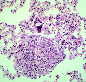
Regarding the mechanism of antibody-mediated attrition, mice vaccinated with repeat (2 or 3) exposures and in which antibody is believed to play a key role, show essentially similar mononuclear cell-rich foci around challenge LS in the lung (Vignali et al. Reference Vignali, Bickle and Taylor1988a; Kassim et al. Reference Kassim, Dean, Mangold and Von Lichtenberg1992) as have been described for singly vaccinated mice (Crabtree and Wilson, Reference Crabtree and Wilson1986). Again the larvae appear undamaged and a proportion of them occur within alveoli, in which case they induce marked inflammatory responses (Kassim et al. Reference Kassim, Dean, Mangold and Von Lichtenberg1992). McLaren and Smithers (Reference McLaren and Smithers1988) reported that transfer of immune serum at the skin stage elicited inflammatory reactions of mononuclear cells and eosinophils around schistosomula in the skin which, nevertheless, appeared morphologically similar to LS and so even in this model of apparent skin phase attrition, lung-form larvae appear to be the target. In rats, where there is consensus that challenge attrition occurs in the lungs of vaccinated animals (Ford et al. Reference Ford, Bickle, Taylor and Andrews1984b; McLaren et al. Reference McLaren, Pearce and Smithers1985) passive transfer of immune serum to naïve rats 5 days post-infection, when the challenge parasites are in the lungs, elicits an enhanced and accelerated inflammatory mononuclear cellular infiltration around the larvae compared with normal serum (Vignali et al. Reference Vignali, Klaus, Bickle and Taylor1989b) as shown in Fig. 1. Because in mice there is no evidence of parasite damage or of intimate cellular attachment, cellular accumulation interfering with parasite migration (Crabtree and Wilson, Reference Crabtree and Wilson1986) is a plausible cause of parasite loss. How IgG is able to mediate such inflammatory foci is unknown. Protection occurs in FcγR KO mice (Jankovic et al. Reference Jankovic, Wynn, Kullberg, Hieny, Caspar, James, Cheever and Sher1999) and so Fc-receptor-mediated activation of cells for ADCC in not required. In rats, complement depletion during passive transfer at the lung stage significantly reduces protection (Vignali et al. Reference Vignali, Bickle, Taylor, Tennent and Pepys1988b) and so generation of the chemoattractants C3a and C5a by antibody/antigen fixation of complement may play a role in focus formation, at least in the rat. Other possible roles of antibody in interfering with parasite migration/viability by binding to surface receptors or signalling molecules remain to be assessed.
Antibody-mediated mononuclear cell infiltration around a lung-stage schistosomulum induced by homologous passive transfer of vaccinated rat serum.

THE ANTIGENS INVOLVED
Although not all studies agree, the weight of evidence from mice and rats is that the lung schistosomula alone can induce high levels of protection and that they are the only necessary targets eliciting cell- or antibody-mediated reactions during lung transit. It follows that the protective antigens are those responsible for the induction of lung foci such as shown in Fig. 1. Antigens responsible for T cell-mediated focus formation would be soluble or secreted antigens available to antigen presenting cells. Antibody could bind to such secreted antigens but could also bind to exposed epitopes of integral membrane antigens. Although the LS shows minimal surface antigenicity as judged by immunofluorescence (Pearce et al. Reference Pearce, Basch and Sher1986) sera from vaccinated animals can mediate transient cell adherence (Bickle and Ford, Reference Bickle and Ford1982; Lawson et al. Reference Lawson, Bickle and Taylor1993) indicating that antibody does have access to exposed larval antigens.
It follows that investigation of the vaccine potential of the secreted and membrane antigens of LS is a rational approach and soluble extract of in vitro-derived LS is indeed able to induce significant protection in mice (Mountford et al. Reference Mountford, Anderson and Wilson1996). To characterize the LS antigens Harrop et al. (Reference Harrop, Coulson and Wilson1999) used antisera against products released by such larvae in culture to screen Western blots of cercarial, LS and adult antigens. No LS-specific antigens were detected and when the sera were used to screen cDNA libraries most clones corresponded to previously described immunodominant antigens whose representation in the LS culture supernatant may have been due to release from damaged larvae. Cercariae, in vitro-derived schistosomula and adult worms are much more ready sources of antigen for study than in vivo-derived LS, but whether these stages share the protective LS antigens involved in irradiated vaccine immunity or whether these are transiently expressed during larval development in vivo is uncertain. However, strong protection (~90%) can be induced in mice with repeated (5×) exposure to very highly irradiated (e.g. 100 krad) cercariae which die in the skin (Hsu et al. Reference Hsu, Hsu and Burmeister1981) and comparable levels of protection to those demonstrated with LS (Mountford et al. Reference Mountford, Anderson and Wilson1996) can be induced with cercarial, in vitro-derived schistosomula or adult antigens given with BCG intradermally (James et al. Reference James, Pearce and Sher1985). So the early larval and adult stages clearly manifest antigens which can induce protection. Recently, newer technologies for characterizing membrane and secreted antigens have been applied to analysis of schistosome adult worm proteins. Rigorous proteomic analysis has led to identification and characterization of a large number of adult tegumental (Braschi et al. Reference Braschi, Borges and Wilson2006) and surface-associated (Braschi and Wilson, Reference Braschi and Wilson2006) proteins. Cloning of genes with signal sequences has allowed identification of adult proteins targeted for secretion or surface expression (Smyth et al. Reference Smyth, McManus, Smout, Laha, Zhang and Loukas2003). Application of such methods has recently led to identification of tegumental proteins able to induce protection in mice e.g. the tetraspanins, TSP-1 and TSP-2 (Tran et al. Reference Tran, Pearson, Bethony, Smyth, Jones, Duke, Don, McManus, Correa-Oliveira and Loukas2006) and Sm29 (Cardoso et al. Reference Cardoso, Macedo, Gava, Kitten, Mati, De Melo, Caliari, Almeida, Venancio, Verjovski-Almeida and Oliveira2008). Their involvement in irradiated vaccine immunity and exposure on the surface of living worms remains to be established although Sm29 has been shown to be expressed by the LS tegument (Cardoso et al. Reference Cardoso, Macedo, Gava, Kitten, Mati, De Melo, Caliari, Almeida, Venancio, Verjovski-Almeida and Oliveira2008).
Various approaches have been applied to identify antigens specifically recognized by vaccinated animals e.g. characterizing larval surface antigens recognized by vaccine serum (Simpson et al. Reference Simpson, James and Sher1983); generating monoclonal antibodies against larval surface antigens using vaccinated donors (Bickle et al. Reference Bickle, Andrews and Taylor1986; Dalton et al. Reference Dalton, Tom and Strand1987); testing known recombinant-derived antigens for reactivity with antibody or cells from vaccinated mice (Richter and Harn, Reference Richter and Harn1993; Richter et al. Reference Richter, Reynolds and Harn1993b); identification of recombinant-derived antigens uniquely or preferentially recognized by irradiated vaccine serum (compared with infection serum) (Soisson et al. Reference Soisson, Masterson, Tom, McNally, Lowell and Strand1992; Francis and Bickle, Reference Francis and Bickle1992). Some of these antigens were included in the independent vaccine testing (Bergquist and Colley, Reference Bergquist and Colley1998) but others remain to be characterized and tested. Amongst these are larval surface antigens uniquely recognized by vaccine serum but not by infection serum, e.g. 15 kDa (Simpson et al. Reference Simpson, James and Sher1983) and 16 kDa (Bickle et al. Reference Bickle, Andrews and Taylor1986) antigens and which appear to have transient expression in the larval stages (Bickle and Oldridge, Reference Bickle and Oldridge1999). Definition of such larval surface antigens will likely need application of those techniques so successfully applied to the adult tegument. It should be noted that it is not yet clear whether any of the above antigens or the many other schistosome antigens identified by other means, play a significant role in irradiated vaccine-induced immunity and specifically in mediating lung focus formation.
PROSPECTS FOR A DEFINED VACCINE
The evidence from the irradiated vaccine models suggests that provided the immunogenic moieties can be produced, protective immune responses may not be particularly challenging to induce. Thus, although WT mice, which make a balanced Th1/Th2 response, develop optimal immunity, high partial protection occurs in cytokine KO mice whether Th1- or Th2-polarized responses are induced (Hoffmann et al. Reference Hoffmann, James, Cheever and Wynn1999).
To simulate the antibody-mediated protection induced by irradiated vaccines IgG antibodies will be needed rather than IgE antibodies which are implicated in naturally-acquired resistance in humans (Hagan et al. Reference Hagan, Ndhlovu and Dunne1998). Longevity of sufficiently high antibody titre will be a requirement. Based on irradiated vaccine studies in various host species, immunizing regimens which are believed to induce antibody-mediated immunity have been shown to lead to long-lived protection (Dean Reference Dean1983; Hsu et al. Reference Hsu, Hsu and Burmeister1981; Bickle et al. Reference Bickle, Taylor, James, Nelson, Hussein, Andrews, Dobinson and Marshall1979d; Ford et al. Reference Ford, Bickle and Taylor1984a) although some decline in protection with time has been shown in baboons (Harrison et al. Reference Harrison, Bickle, Kiare, James, Andrews, Sturrock, Taylor and Webbe1990).
A vaccine aimed at reproducing the CMI mechanisms which help mediate protection in singly vaccinated mice faces a number of challenges. It would need to induce the lung sensitization required for optimal immunity in this model and this is likely to require targeting vaccine delivery to the lung as proposed, for example, for TB vaccines (Kallenius et al. Reference Kallenius, Pawlowski, Brandtzaeg and Svenson2007). It would need to induce Th1 responses but avoid over-induction of regulatory cytokines e.g. IL-10, since this can prevent manifestation of resistance (Oswald et al. Reference Oswald, Caspar, Wynn, Scharton-Kersten, Williams, Hieny, Sher and James1998). Challenge exposure downregulates the Th1 response in mice (Pemberton and Wilson, Reference Pemberton and Wilson1995) and in the likely event of a vaccine inducing only partial protection the ensuing egg production is likely to profoundly alter the response from a Th-1 to a Th2-dominated response, as occurs when schistosome eggs are injected into vaccinated mice (Pearce et al. Reference Pearce, Caspar, Grzych, Lewis and Sher1991). Furthermore, in endemic areas, vaccinees are likely to have an underlying Th2 biased response due to other worm infections or to previously treated schistosome infections that could interfere with the generation of Th1 responses. Regarding the influence of current/previous infection on vaccine efficacy it is encouraging that studies in baboons have shown that multiple irradiated vaccine regimens, likely to induce antibody-mediated protection, are equally effective in naïve and in infected or infected and previously treated animals (Kariuki et al. Reference Kariuki, Van Dam, Deelder, Farah, Yole, Wilson and Coulson2006).
Given the availability of a new crop of promising protective antigens and the enthusiastic application of new technologies for indentifying membrane and secreted antigens a schistosome vaccine modelled on the irradiated vaccine seems increasingly technically feasible. However, as Loukas et al. (Reference Loukas, Tran and Pearson2007) urged, development of this promise into a human vaccine will require the wheel to turn and significant investment to return to schistosome vaccine development.
I am very grateful to Martin Taylor and Helena Helmby for critical review of this manuscript.