Introduction
Nasal polyps originate from the mucosa of the nasal cavity or paranasal sinuses. Most originate from the lateral nasal wall. Approximately three-quarters of all polyps are related to the sinus outlets. The middle meatal mucosa, uncinate process, infundibulum, superior meatal mucosa and sphenoethmoidal recess are the most common sites of origin. Polyps originating from the mucosa of the nasal septum are rare.Reference Ozgirgin, Kutluay, Akkuzu and Gungen1–Reference Stammberger4
The aetiology of polyp formation is multifactorial. The most common aetiological factors reported for polyp formation include allergy, infection, immunological factors, metabolic disease and autonomic dysfunction.Reference Drake-Lee, Lowe, Swanston and Grace5 The clinical appearance of polyps varies; they may be glassy, oedematous, cystic, coarse or fibrotic. The most common clinical presentations of nasal polyps are nasal obstruction, sinusitis and anosmia.Reference Ozgirgin, Kutluay, Akkuzu and Gungen1, Reference Stammberger4, Reference Drake-Lee, Lowe, Swanston and Grace5 Here, we present a 77-year-old man with bilateral nasal septal polyps that caused nasal obstruction.
Case report
A 77-year-old man presented to the otolaryngology department of Baskent University with a nasal obstruction of two years' duration. The condition had become progressively worse over time. There was no history of allergy or recurrent rhinosinusitis.
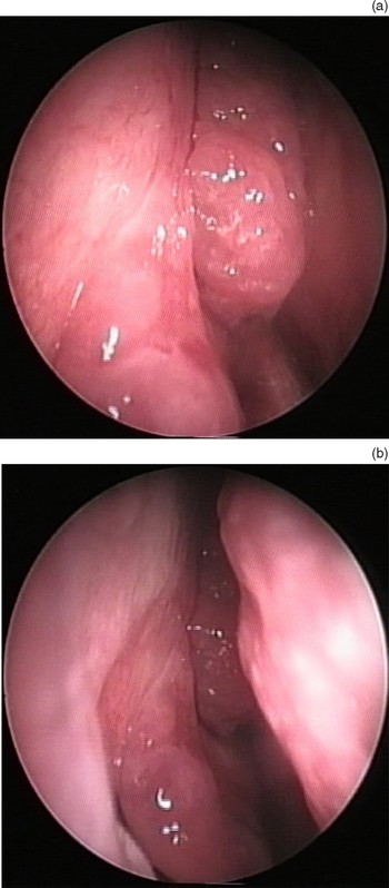
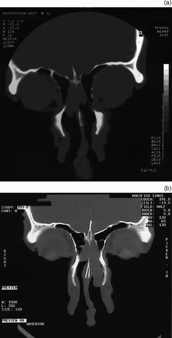
Endoscopic nasal examination revealed bilateral polyps originating from the nasal septum. A polyp was seen to protrude into the nasal passage, bilaterally, causing the nasal obstruction (Figure 1). Computed tomography scanning demonstrated bilateral nasal polyps originating from the nasal septum and the cribriform plate. The paranasal sinuses were clear (Figure 2).
Endoscopic appearance of a polyp inside the right nasal passage. (a) Close up view of the nasal polyp in the right nasal cavity. (b) Polyp in the right nasal cavity between nasal septum and middle turbinate.

Two different coronal computed tomography sections showing polyps originating from the nasal septum.

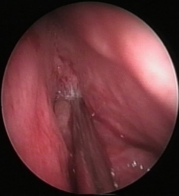
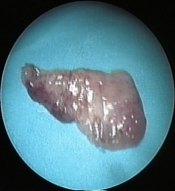
Endoscopic surgery was performed with the patient under local anaesthesia. The lesions was excised (Figures 3 and 4). Histopathological examination of the lesions demonstrated chronic inflammation.
Intra-operative, endoscopic appearance of the left nasal polyp, showing the origin of the polyp.

The polyp excised from left nasal passage.

At six-month follow up, there was no evidence of recurrence.
Discussion
Nasal polyps are inflammatory growths of the nasal or paranasal mucosa. The condition is poorly understood, but most theories of aetiology involve allergy and inflammation. The frequency of nasal polyps in the general population remains unclear. Most small polyps do not become large enough to produce symptoms. Therefore, symptomatic polyps may represent only a small proportion of all nasal polyps.Reference Ozgirgin, Kutluay, Akkuzu and Gungen1, Reference Larsen and Tos2, Reference Drake-Lee, Lowe, Swanston and Grace5, Reference Tos, Larsen, Kennedy, Bolger and Zinreich6
Nasal polyp development starts with submucosal oedema. Over time, and with the effect of gravity, the oedematous site enlarges and protrudes from its base, eventually forming a polyp.Reference Ogawa7, Reference Yanagisawa and Klenoff8
Histopathologically, nasal polyps are composed of fibrous tissue, vessels, inflammatory cells and glands. The surface of the polyp is covered most commonly with pseudostratified columnar epithelium. However, transitional and squamous epithelium may also be present on the polyp surface. The size and number of mucous glands varies. Various inflammatory cells have been described in polyps, but the predominant cell type is eosinophils.Reference Ozgirgin, Kutluay, Akkuzu and Gungen1, Reference Tos, Larsen, Kennedy, Bolger and Zinreich6, Reference Ogawa7
Most polyps originate from the lateral walls of the nasal passage. Larsen and Tos noted from autopsy studies that the most common site of origin was the middle meatus; the superior meatus was the second most common site of origin.Reference Larsen and Tos2, Reference Larsen and Tos3 The maxillary sinus and sphenoid sinus are seldom involved.Reference Stammberger4 Conducive microenvironmental conditions may be caused by air turbulence; because the lateral walls of the nasal passages are exposed to more air turbulence than are other structures in the nose, irritants can cause inflammation more easily.Reference Ozgirgin, Kutluay, Akkuzu and Gungen1 Also, the anatomical properties of the lateral nasal wall predispose it to polyp formation; it is thinner, less vascular and more loosely adherent to the underlying structures.Reference Bernstein, Gorfien and Noble9
Because of these anatomical and microenvironmental properties, nasal polyp formation in the medial nasal wall is rare. In a series of 200 patients, Stammberger reported only three patients with medially based polyps that originated from the posterior nasal septum.Reference Stammberger4 Yanagisawa and Klenoff reported only three cases of septal polyps, but only one of these polyps was isolated.Reference Yanagisawa and Klenoff8 Ozgirgin and colleagues reported one case of an isolated septal polyp that protruded to the choana.Reference Ozgirgin, Kutluay, Akkuzu and Gungen1
Nasal polyps present with multiple symptoms, being associated with rhinorrhoea, postnasal drip, anosmia, hyposmia, nasal obstruction and facial pain.Reference Drake-Lee, Lowe, Swanston and Grace5 Isolated septal polyps are rare but can cause such symptoms as nasal obstruction and olfactory disorders. The best treatment for symptomatic septal polyps is endoscopic excision, as performed in our patient.