

Introduction
One of the outstanding physical characteristics of Thoroughbreds is their high capacity for maximal exercise lasting for short periods of a few minutes. It has been reported that Thoroughbreds show high plasma and muscle lactate concentrations after strenuous exerciseReference Snow, Harris and Gash1, Reference Weber, Parkhouse, Dobson, Harman, Snow and Hochachka2. Lactate is not merely a waste product of glycolysis and glycogenolysis, but it is also a substrate for oxidation, and oxidation is the major pathway of lactate metabolism during exerciseReference Brooks3. Lactate is produced in fast fibres, and it can be utilized in slow fibres and heart muscles. In this predominant pathway of lactate metabolism, known as the cell to cell lactate shuttle, lactate is transported through the plasma membrane via monocarboxylate transporters (MCTs)Reference Juel and Halestrap4, Reference Bonen5. Fourteen different proteins have been identified in the MCT familyReference Bonen, Heynen and Hatta6. MCT1 and MCT4 are two of the major MCTs present in the skeletal muscles. The protein expression of the first isoform MCT1 is highly correlated with the oxidative fibre composition of the muscles and other indices of oxidative metabolismReference Juel and Halestrap4, Reference Bonen5. MCT4, which is abundant in glycolytic fibres, is related to the release of lactateReference Wilson, Jackson, Heddle, Price, Pilegaard and Juel7, Reference Manning Fox, Meredith and Halestrap8. Collectively, these suggest that MCT1 and MCT4 are primarily responsible for the extrusion of lactate out of the muscles and for the uptake of lactate into the muscles. Studies in humans and rodents have shown that the effects of exercise or muscle stimulation on lactate transport capacity are associated with changes in MCT1 and MCT4 in musclesReference Bonen, Heynen and Hatta6, Reference Yoshida, Hatta, Kato, Enoki, Kato and Bonen9. High-intensity training has been reported to increase muscular expression of both MCT1 and MCT4Reference Bonen5, Reference Pilegaard, Domino, Noland, Juel, Hellsten and Halestrap10. However, there are only a few studies investigating the expression of MCTs in horses. Koho et al. Reference Koho, Vaihkonen and Poso11 reported that MCT1 and MCT2, another isoform of MCTs, but not MCT4, were present in the red blood cells of Standardbred horses. They also reported the presence of MCT1 and MCT4 in the muscles of Standardbred horsesReference Koho, Hyyppa and Poso12. However, there are very limited data regarding the training response of MCTs and the relationship between MCTs and exercise performance in horses. Therefore, the purpose of this study was to investigate training-induced changes in lactate metabolism and protein expression of MCT1 and MCT4 in Thoroughbred horses. Since high intensity of training is known, through studies in humans and rodents, to be necessary to increase MCT1 and MCT4 in muscles we employed high-intensity-type trainingReference Pilegaard, Domino, Noland, Juel, Hellsten and Halestrap10, Reference Baker, McCullagh and Bonen13. The gluteus medius muscle is one of the main muscles producing thrust for running in horses, and is often used experimentally in muscle biopsy sampling. Muscle biopsy sampling from the gluteus medius muscle was done at rest before and after the training.
Materials and methods
Animals
Five Thoroughbred horses (three males and two geldings; 3–10 years old; 478 ± 13 kg) were used in this study. They were all pedigreed and raised for investigation following the standard procedures of the Equine Research Institute of the Japan Racing Association. Ethical approval for the experiment was obtained from the Animal Welfare and Ethics Committee of the Japan Racing Association.
Training and incremental all-out test protocol
All training and tests were performed on a treadmill with a 10% incline (Mustang 2200, Kagra AG, Fahrwangen, Switzerland). After a warming-up exercise, horses ran at 100% maximal oxygen uptake (VO2max) for 1 min (3 days per week, 3 weeks) or 2 min (2 days per week, 4 weeks) for a total of 7 weeks.
Thoroughbreds were subjected to an incremental all-out test before and after the 7 weeks of training. In the test, after 2 min of walking (1.8 m s− 1) and 2 min of trotting (3.6 m s− 1), the speed was increased to 6 m s− 1 for a minute, then to 8 m s− 1 for a minute, and thereafter the speed was increased by 1 m s− 1 every minute until exhaustion. Exhaustion was defined as the point at which Thoroughbreds could not keep pace with the treadmill belt.
Measurement of VO2max and duration in the incremental all-out test
The measurement of VO2 was performed using an open flow system as described previouslyReference Yamano, Eto, Hiraga and Miyata14. O2 and CO2 concentrations (Vice Medical, Chiba, Japan) and temperature and relative humidity (Takara Instruments, Tokyo, Japan) were measured continuously (sampling rate 40 times s− 1). The average VO2 and VCO2 over the last 15 s of each running speed were determined as the VO2 and VCO2 for that speed. VO2max was determined at a levelling-off point by regression line analysis.
The duration of the incremental all-out test was measured as total seconds from the start of running at 10 m s− 1 to the end of all-out running, as the first 6 min of treadmill running before the speed of 10 m s− 1 were considered to be a warming-up period. For example, a horse that was exhausted after running for 30 s at 11 m s− 1 would be said to have a duration of 90 s, as the horse ran 60 s at 10 m s− 1 plus 30 s at 11 m s− 1.
Biochemical analysis
Blood samples for lactate measurements were collected from the jugular vein at rest before the incremental all-out test, at the end of the 6 and 9 m s− 1 of running, and immediately after exhaustion using an indwelling catheter as we described previouslyReference Mukai, Ohmura, Hiraga, Eto, Takahashi and Asai15. Blood lactate concentrations were measured with an automated blood lactate analyzer (1500 SPORT, Yellow Springs Instruments, Yellow Springs, OH, USA) as reported previouslyReference Eto, Yamano, Mukai, Sugiura, Nasu and Tokuriki16.
Muscle samples were obtained from the same portion of the gluteus medius muscle and at the same depth (5 cm from the skin surface) from all the horses as described previouslyReference Yamano, Eto, Hiraga and Miyata14. The sampling was carried out at rest the day before the incremental all-out test. Protein content of MCT1 and MCT4 in the gluteus medius muscle was detected using Western blotting. Antibodies for MCT1 and MCT4 were raised in rabbits against the oligopeptide corresponding to each C-terminus region of human MCT1 (CQKDTEGGPKEEESPV) and MCT4 (CEPEKNGEVVHTPETSV) (Operon Biotechnologies, Huntsville, AL, USA)Reference Wilson, Jackson, Heddle, Price, Pilegaard and Juel7, Reference Fishbein, Merezhinskaya and Foellmer17. Since several amino acids differ in the human and equine MCT1 sequencesReference Wilson, Jackson, Heddle, Price, Pilegaard and Juel7, Reference Reeben, Koho, Raekallio, Hyyppa and Poso18, the specificity was verified with peptide blocking. The procedures for the detection of MCT1 and MCT4 for whole muscle samples have been described in detail by our laboratory previouslyReference Yoshida, Holloway, Ljubicic, Hatta, Spriet and Hood19, Reference Enoki, Yoshida, Hatta and Bonen20. Briefly, middle gluteus muscles (15–20 mg) were homogenized in buffer A (210 mM sucrose, 2 mM ethylene glycol-bis-(β-aminoethyl ether)-N, N, N′, N′-tetraacetic acid, 5 mM EDTA and dimethylsulphoxide, pH 7.5). Then, 3 ml of buffer B (1.167 M KCl and 58.3 mM tetrasodium pyrophosphate) were added, mixed briefly, and set on ice for 15 min. After centrifugation at 160 000 × g for 75 min at 4°C, the supernatant fluid was discarded, and the pellet was washed thoroughly with buffer C (10 mM 2-amino-2-hydroxymethyl-1,3-propanediol and 1 mM EDTA, pH 7.4). The pellet was homogenized in 400 μl of buffer C. After 200 μl of 16% sodium dodecyl sulphate (SDS) were added, the samples were removed from ice, vortex mixed and centrifuged at 1100 × g for 20 min at room temperature. The supernatant was divided into aliquots and stored at − 80°C for protein assay.
DC protein assay (Bio-Rad, Hercules, CA, USA) was used for measuring protein concentrations. Protein samples of the muscles (10 μg) and pre-stained molecular weight markers (BioDynamics Laboratory, Inc., Tokyo, Japan) were separated on 12% SDS-polyacrylamide gels. Proteins were then transferred from the gel to Hybond-P PVDF transfer membranes (Amersham Biosciences, Piscataway, NJ, USA). Antibody binding was visualized with the enhanced chemiluminescence (ECL) system (Amersham Biosciences), and was detected using the Bio-Rad Chemidoc system (Bio-Rad). Densitometric analyses of captured images were performed using version 4.6.1 of the Bio-Rad Quantity One software. In our preliminary experiment, we confirmed a linear relation between the amount of protein loaded (5–20 μg) for electrophoresis and the final band density of chemiluminescence.
Statistical analysis
All data are reported as means ± SE. The group data were analysed by ANOVA for repeated measures. Correlations were analysed with the Pearson correlation test. Statistical significance was defined as P < 0.05.
Results
Body weight, VO2max, lactate and duration of all-out test
The horses' average body weight was 478 ± 13 kg before the training, and it was 486 ± 13 kg after the training period (Table 1). VO2max (159.1 ± 9.5 ml min kg− 1 vs. 174.6 ± 4.6 ml min kg− 1) and the duration of the all-out test (67 ± 16 vs. 128 ± 8 s) were significantly increased by the training. Blood lactate concentrations at submaximal speed during the incremental all-out test decreased significantly after the training (6 m s− 1, 1.7 ± 0.1 vs. 1.1 ± 0.1 mmol l− 1; 9 m s− 1, 6.7 ± 1.0 vs. 3.4 ± 1.0 mmol l− 1). No significant differences were found in the blood lactate concentrations at exhaustion before and after the training (12.1 ± 0.7 vs. 12.0 ± 1.1 mmol l− 1).
Body weight, VO2max, blood lactate concentration and duration of the all-out test before and after 6 weeks of high-intensity training

Values are means ± SE. Significant difference from values obtained pre-training at *P < 0.05 and **P < 0.01.
Protein expression of MCT1 and MCT4
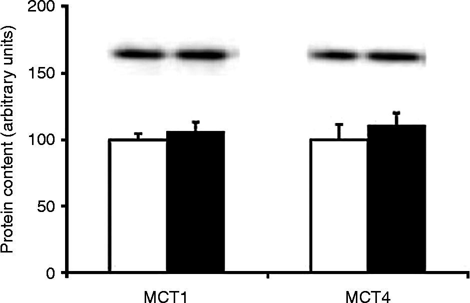
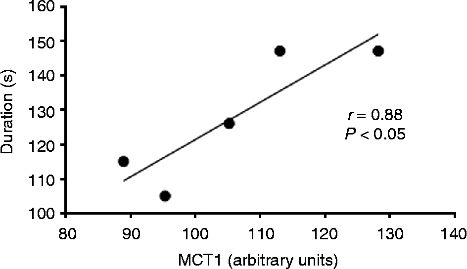
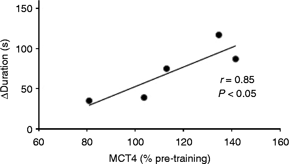
A single clear band with a molecular mass of 40–45 kDa for MCT1 and MCT4 was detected in the equine gluteus medius muscle. Protein content of MCT1 and MCT4 in the muscle was not significantly different before and after 6 weeks of high-intensity training period (Fig. 1). There was a statistically significant relationship between MCT1 protein expression and duration of the all-out test after the training (Fig. 2). Increase in the duration of exercise in the incremental all-out test showed a significant positive relationship with the change in MCT4 expression after the training (Fig. 3). No significant relationship was found between the duration of exercise in the all-out test and VO2max.
MCT1 and MCT4 protein content in the gluteus medius muscle before and after 6 weeks of high-intensity training. Values are means ± SE

Relationship between MCT1 in the gluteus medius muscle and the duration of exercise in an incremental all-out running test after 6 weeks of high-intensity training

Relationship between MCT4 in the gluteus medius muscle relative to the pre-training value and increase in the duration of exercise in an incremental all-out running test after 6 weeks of high-intensity training

Discussion
Thoroughbred horses are extraordinary athletes that can run at high speed (60–65 km h− 1) for more than a few minutes. They have a high maximal aerobic capacity, high mitochondrial volume in their skeletal muscles and high glycogen content in their muscles with a high proportion of fast fibresReference Rivero, Ruz, Marti-Korff, Estepa, Aguilera-Tejero and Werkman21. In Thoroughbreds, the gluteus medius muscle consists of about 90% fast fibres and 10% slow fibresReference Langsetmo and Poole22. A high proportion of fast-type fibres in skeletal muscles is one of the major characteristics of Thoroughbreds, with high production of lactate during exercise. However, almost half of the fast fibres in Thoroughbreds are type IIA and type IIAX fibresReference Yamano, Eto, Hiraga and Miyata14. Type IIA fibres have a capacity not only for the production of lactate but also for the oxidation of lactate, comparable to that of type I fibresReference Baldwin, Hooker and Herrick23. Thus, since Thoroughbreds have a high percentage of type IIA and IIAX muscle fibres, it is possible that they have a high capacity for both the production and oxidation of lactateReference Yamano, Eto, Hiraga and Miyata14, Reference Rivero, Ruz, Marti-Korff, Estepa, Aguilera-Tejero and Werkman21. In this study, concentrations of blood lactate at exhaustion were around 12 mmol l− 1, which was not so high as expected. However, according to Poso et al. Reference Poso, Essen-Gustavsson and Persson24, a blood lactate concentration of around 12 mmol l− 1 can be estimated based on a plasma lactate concentration of around 20 mmol l− 1, which is not so low but not very high as expected. In this study, we used treadmill running with a 10% incline because of safety reasons, i.e. the inadvisability on running a horse to exhaustion. Running with an incline of the treadmill should reduce the volume of muscle working during running compared with flat running, so it is possible that the treadline incline decreased blood lactate concentration at exhaustion.
Changes in lactate metabolism are associated with changes in the expression of MCT1 and MCT4Reference Brooks3, Reference Juel and Halestrap4. However, at present, little is known about the relationship between MCTs and exercise capacity in horses. In this study, while we could not find a significant increase in MCT1 and MCT4 after training, MCT1 was positively correlated with the duration of exercise in the incremental all-out test after training. Moreover, we found a significant relationship between change in MCT4 and increase in duration in the incremental all-out test during training. On the other hand, no significant relationship was found between the duration of exercise in the all-out test and VO2max after training. These results indicate that the protein content of MCT1 and MCT4 influences the capacity to sustain maximal running exercise rather than VO2max in horses. In humans, Thomas et al. Reference Thomas, Perrey, Lambert, Hugon, Mornet and Mercier25 showed that MCT1 content is related to blood lactate removal and fatigue indices after 1 min of supramaximal exercise, and that MCT1 is one of the major factors in determining fatigue in supramaximal exercise. Their study supports our result that MCT1 is related to the duration in the all-out test. In previous reports in humans and rats, MCT4 has often not been considered as a major mediator of the MCT-related effects of endurance training, because its protein level does not increase after endurance training, partly because of the difference in K m value for lactate transport of the transporters MCT4, 17–34 mmol l− 1 and MCT1, 4–6 mmol l− 1 Reference Bonen5, Reference Bonen, Heynen and Hatta6, Reference Dimmer, Friedrich, Lang, Deitmer and Bröer26. It is reported that muscle lactate concentration can reach over 30–40 mmol kg− 1 during maximal exercise in horsesReference Snow, Harris and Gash1. Therefore, it is possible that muscle MCT4 has more effect on lactate metabolism compared with that in humans and rats. Altogether, the results of this study suggest that not only MCT1 but also MCT4 could play important roles in horses during maximal exercise.
In summary, we have demonstrated the relationship between MCT1 and MCT4 protein content and exercise capacity in Thoroughbred horses. Although the overall changes in MCT protein expression after endurance training were small, MCT1 showed a significant relationship with the duration of maximal exercise, and changes in MCT4 showed a significant relationship with an increase in the duration of exercise in the incremental all-out test during training. Taken together, the results suggest that MCT1 and MCT4 in the skeletal muscles can play important roles in the effect of high-intensity training for sustaining maximal exercise in Thoroughbred horses.
Acknowledgement
This study was funded by Equine Research Institute, Japan Racing Association.