Coronary arteriovenous fistula is a rare anomaly that consists of abnormal communication between the coronary artery and one of the cardiac chambers or vessels adjacent to the heart.Reference Ata, Turk, Bicer, Yalcin, Ata and Yavuz 1 Coronary arteriovenous fistula is present in 0.002% of the general population and is visualised in nearly 0.25% of patients undergoing cardiac catheterisation.Reference Fernandes, Kadivar, Hallman, Reul, Ott and Cooley 2 These patients are frequently asymptomatic; however, they can present with symptoms of dyspnoea, chest pain, and arrhythmias necessitating therapy.Reference Von Arnim and Reuschel-Janetschek 3 – Reference Ozhan, Akdemir and Duran 5 As an alternative to surgery, percutaneous closure of the coronary arteriovenous fistula may be proposed in symptomatic patients.Reference Collins, Mehta, Benson and Horlick 6 , Reference Kilic, Akdemir, Bicer and Dogan 7 We describe a case of a successful percutaneous closure of a coronary arteriovenous fistula in a patient who experienced transient myocardial ischaemia during the procedure along with changes in electrocardiography.
Case report
A 38-year-old man was admitted to our hospital with chest pain and reduction in exercise tolerance since the past 6 months. Percutaneous coil occlusion was performed for a large coronary artery that was connected to a coronary sinus fistula 8 months before. The results of the physical examination as well as those of electrocardiography, chest radiography, laboratory tests, and echocardiographic examination were normal. His chest pain was typical, and ST depressions were observed during the treadmill exercise stress test. Coronary angiography showed the persistence of fistula and retained coils in the fistula artery. Fistula was present between the left circumflex artery and the coronary sinus, which was confirmed by multiple angiographic projections. Primarily, additional coil placement inside the arteriovenous fistula was decided as the mode of treatment. Vascular access was obtained through the right femoral artery using the Seldinger technique. An 8-French sheath was introduced, and 10.000 units of heparin were administered. An 8-French Judkins left-guiding catheter (Medtronic, Inc., Minneapolis, Minnesota) was used to gain access into the left coronary ostium. A microcatheter was cannulated into the fistula over a 0.014-inch guide wire (Echelon Microcatheter, Micro Therapeutics, Irvine, California, United States of America). The selected coils were electrolytically detachable. A microcoil system was used, consisting of a platinum embolic coil (microcoil) and attached to a device-positioning unit, the detachment control box, and the connecting cable.
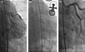
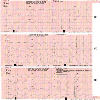
We placed the first coil inside the fistula artery successfully and tried to detach it; however, it did not detach. We tried four times with a long-awaiting electrical stimulus. Finally, a 2.5 × 18-millimetre graft stent was deployed under 20 atmospheric pressure. This resulted in the occlusion of the fistulous communication and good flow in the distal left circumflex artery (Fig 1a–c). A synchronised electrocardiogram was recorded simultaneously, which showed ST segment elevation in leads DII, DIII, and AVF. During the second coil detachment, electrocardiographic recordings showed ST segment elevation and bizarre changes in the inferior leads (Fig 2a–c). The patient remained haemodynamically stable during the procedure, but he described severe atypical chest discomfort at every long electrical detachment. Cardiac enzyme levels were in the normal range the day after the procedure. The patient was discharged after 2 days in hospital without any other adverse events. He remained asymptomatic for 3 years.
Angiography showing the coronary fistula (a), coils (b), and repair using a graft stent (c).

Normal electrocardiogram before the electrical current opening (a), ST segment elevation, and deep T-wave inversion during early (b) and later (c) application of electrical current.

Discussion
The majority of the coronary arteriovenous fistulae remain asymptomatic in the absence of compression signs of the aneurysmatic fistula, coronary steal, or intracardiac shunt. Some patients, however, report angina and dyspnoea, which are difficult to be attributed to any specific cause.Reference Ata, Turk, Bicer, Yalcin, Ata and Yavuz 1 , Reference Fernandes, Kadivar, Hallman, Reul, Ott and Cooley 2 , Reference Olearchyk, Runk, Alavi and Grosso 8 The usual cause of chest pain is supposed to be the coronary steal phenomenon, which could be a persistent or an episodic steal.Reference Ata, Turk, Bicer, Yalcin, Ata and Yavuz 1 , Reference Fernandes, Kadivar, Hallman, Reul, Ott and Cooley 2 , Reference Olearchyk, Runk, Alavi and Grosso 8 Some percutaneous coronary interventions are associated with angina and asymptomatic ischaemic episodes, called silent myocardial ischaemic episodes.Reference Von Arnim and Reuschel-Janetschek 3 – Reference Ozhan, Akdemir and Duran 5 , Reference Konidala and Gutterman 9 Silent myocardial ischaemic patients have generally reduced sensitivity to pain felt during ischaemic episodes. Healthy asymptomatic patients and patients with coronary artery disease are all at higher risk for subsequent cardiovascular morbidity if there is evidence of silent ischaemia.Reference Von Arnim and Reuschel-Janetschek 3 – Reference Ozhan, Akdemir and Duran 5 , Reference Konidala and Gutterman 9
This report describes a patient whose coronary arteriovenous fistula was treated successfully with percutaneous intervention. Although no complication occurred during the procedure, electrocardiography recorded episodes of ischaemia at the time of closure of the fistula artery. ST segment elevation occurred in inferior leads, and the patient experienced some atypical chest discomfort. Electrocardiographic changes during coil occlusion of the coronary arteriovenous fistula are unusual. Aetiology of the ST segment elevation and details of other bizarre electrocardiographic changes are not clear. However, electrical effects of coil detachment on electrocardiography, temporary coronary spasm because of fistula artery occlusion, and silent ischaemia may be causes of the electrocardiographic changes.
We did not find any articles in the database on ischaemic episodes during percutaneous coronary fistula interventions. In this report, we describe an electrocardiographic sign of silent ischaemia during electrical coronary coil detachment accompanying the patient's atypical chest discomfort. This electrocardiographic sign was possibly caused by a spasm or electrical artefact. Besides, at the time of the closure of the fistula vessel, electrocardiographic changes were possibly caused by reperfusion toxicity in the myocardial area, the perfusion of which reduced until that time because of the steal phenomenon.
Adverse effects because of reperfusion in the ischaemic myocardial area are frequently seen in acute myocardial infarction patients whose occluded infarct-related artery recovers as a result of reperfusion therapy. In the present case, electrical instability in the myocardial area, which was reperfused, caused reperfusion arrhythmias. In our patient, reperfusion in the myocardial area, which was the ischaemic cause of the steal phenomenon, might have been caused by motion abnormalities in this myocardium. The bizarre electrocardiographic changes and ST elevation were believed to be associated with a specific pattern of abnormal left ventricular motion or electrical current effects. In our patient, the fistula artery was closed previously and, subsequent to treatment, flow in this artery resumed.
Finally, we have concluded that the electrocardiographic changes in this case are due to the application of electrical current during coil detachment. Electrical activity during the detachment process may disturb the electrical system of the heart.
In conclusion, it should be noted that ischaemic episodes, either silent or asymptomatic, are as common after coronary interventions as in coronary fistula coil occlusion. Close monitoring of patients during the electrical coil detachment process is important. Long-duration electrical currents for coronary coil detachment may cause serious cardiac events.