Introduction
Tapeworms are the most important group of parasites infecting elasmobranch fishes (Haseli et al., Reference Haseli2012; Caira et al., Reference Caira, Caira and Jensen2017b). Of the 19 recognized orders of tapeworms, those parasitizing elasmobranch fishes include representatives from nine orders (1034 species in 202 genera) (Caira et al., Reference Caira, Caira and Jensen2017b), each with very distinct morphological differences, thus making this group the most speciose infecting elasmobranch fishes (Caira and Healy, Reference Caira, Healy, Carrier, Musick and Heithaus2004). As adults, these tapeworms reside in the spiral intestine and demonstrate a noticeable specificity to their definitive elasmobranch host (Williams, Reference Williams1966; Caira and Jensen, Reference Caira and Jensen2014). This specificity has been attributed to morphological adaptations of the attachment structures to the host gut mucosa, host physiological adaptations allowing or preventing establishment of parasites, and ecological features of the hosts, such as resource availability, substrate preference, diet and phylogenetics (Randhawa and Burt, Reference Randhawa and Burt2008, and references therein). Furthermore, congeners sharing hosts have been shown to exploit separate niches by exhibiting attachment-site preferences within the spiral intestine of rajid skates (Carvajal and Dailey, Reference Carvajal and Dailey1975; Randhawa, Reference Randhawa2012).
Globally, several studies have focused on the taxonomy, systematics, distribution, and host specificity of tapeworms infecting elasmobranchs (e.g. Euzet, Reference Euzet1959; Alexander, Reference Alexander1963; Williams, Reference Williams1966, Reference Williams1968, Reference Williams1969; Caira et al., Reference Caira, Jensen and Healy1999, Reference Caira2017a; Jensen, Reference Jensen2005; Tyler, Reference Tyler2006; Randhawa and Burt, Reference Randhawa and Burt2008; Ruhnke, Reference Ruhnke2011; Caira and Jensen, Reference Caira and Jensen2017). Many outcomes were the subject of passionate discussions, others spurred complementary or corrective research, especially regarding the advance of taxonomic and systematics research with the advent of molecular tools (e.g. Waeschenbach et al., Reference Waeschenbach2007; Healy et al., Reference Healy2009; Caira et al., Reference Caira2013, Reference Caira2014; Ruhnke et al., Reference Ruhnke, Caira and Cox2015). These have led to important progress, e.g. the description and classification of 1034 tapeworm species from elasmobranchs (Caira et al., Reference Caira, Caira and Jensen2017b). However, study effort has not been consistent geographically and the species inventory of the global biota for these groups of parasites is far from complete (e.g. Randhawa and Poulin, Reference Randhawa and Poulin2010). For instance, few studies have examined the parasite fauna of elasmobranch fishes off the Algerian coast (Tazerouti et al., Reference Tazerouti, Euzet and Kechemir-Issad2007, Reference Tazerouti, Kechemir-Issad and Euzet2009). This incomplete inventory is typical of most host–parasite checklists worldwide (Poulin et al., Reference Poulin2016).
The Mediterranean Sea is situated at the junction of three continents. Covering an area of c. 2.5 million km2 (Jereb et al., Reference Jereb2015), it is a hub of chondrichthyofauna diversity, and is home to between 15,000 and 20,000 marine species, including 85 species of elasmobranchs (Bradai et al., Reference Bradai, Saidi and Enajjar2012). Of these, 36 species of Rajidae inhabit these waters (Refés et al., Reference Refés, Semahi and Boulahdid2010). The combination of climatic and anthropogenic factors has favoured the migration and appearance of certain species of fish in the Mediterranean (Bianchi and Morri, Reference Bianchi and Morri1994). The Algerian Basin itself hosts a wide variety of Rajidae (Hemida et al., Reference Hemida, Sergoua and Seridji2007), which host a high diversity of parasites; the present study on parasites of rajid skates in this region has led to the description of the Echeneibothrium community and species richness/diversity in these skates. Further to a parasitological survey of tapeworms infecting rajid skates along the coast of Algeria, we focused on the tapeworm assemblage of Dipturus batis Linnaeus, 1758. This marine benthic skate is found essentially in coastal waters within the 100 m and 1000 m range (FAO, 2009). This species is extirpated from large parts of its range, including habitats within the Mediterranean Sea, North Sea, Irish Sea, and English Channel (Dulvy and Reynolds, Reference Dulvy and Reynolds2002). However it still occurs in some parts of the Mediterranean Sea (Griffiths et al., Reference Griffiths2010). It feeds demersally on a wide variety of prey, including other skates, dogfish, catsharks, anglerfish, gurnards, flatfishes, pilchards, herring, scads, lobsters, crabs and cephalopods (Wheeler, Reference Wheeler1969).
During this parasitological survey a new species of tapeworm, of consistent morphology with the genus Echeneibothrium, was recovered from the spiral valve of D. batis. The detailed study of the collected specimens shows that they have features sufficiently distinctive to represent a new species. The new species is described and illustrated in this paper. Furthermore, the host specificity of this rhinebothriid genus infecting skates in Algerian waters is discussed, along with comments on the attachment-site specificity of this new species, and its congeners, infecting D. batis.
Materials and methods
Host and parasite collection
From January to May 2014, 33 specimens of Dipturus batis were examined. Samples were collected from the east coast of Algeria in the Béjaïa Gulf (36°45′00″N, 5°04′00″E) and from Annaba (36°53′59″N, 7°46′00″E), using gill nets or small bottom trawls in conjunction with local fishermen. These skates were identified using FAO keys (Fischer et al., Reference Fischer, Bauchot and Schneider1987; FAO, 2009).
The fresh spiral intestine from each skate was removed with a mid-ventral longitudinal incision. Spiral intestines were injected with 8% formalin and kept in 5% formalin. Under stereomicroscope, the spiral intestines were opened through the turns of the spires to expose the surface of each chamber, rinsed with seawater. The mucosa and the resulting washings were examined for tapeworms, and the parasites were then removed and transferred to 70% ethanol for taxonomic study; some were fixed in 10% formalin for scanning electron microscopy (SEM). The attachment site was noted, whenever possible, to determine the distribution of the tapeworm parasites within the spiral valve for further studies.
Morphological methods
For morphological description, the worms were prepared as follows: rehydrated in distilled water, stained in acetic acid alum carmine, dehydrated through a graded ethanol series, cleared in clove oil, and mounted on glass slides in Canada balsam. Specimens for examination with SEM were rinsed in distilled water, post-fixed in 1% osmium tetroxide for 2 h, dehydrated through a graded ethanol series, critical point dried (Balzers CPD020 critical point dryer), mounted on aluminium stubs with double-sided carbon adhesive tape and sputter coated with gold for 2 minutes (Edwards S150A sputter coating unit), then examined in a JEOL 6400 scanning electron microscope at 15 kV accelerating voltage using Gatan Digital Micrograph software.
Identification and all measurements were carried out using light microscopy. Measurements are presented as the range, with the mean in parentheses; number of worms examined (n). All measurements are in micrometers except total length, which is in millimetres. Drawings were made with the aid of a drawing tube, scanned and redrawn by computer with Adobe Illustrator CS6.
Morphological characterization of intestinal villi and tapeworm attachment structures
The spiral intestines of the infected D. batis examined herein were measured and then opened following the turn of the spire. Each whorl was exposed separately. Under stereomicroscope, the villi of chambers with higher parasite abundance (whorl 3) and those of the other chambers were removed carefully, and processed for histology. To determine whether the attachment structures of the scolex, especially the bothridia (which are continuously involved in attachment), are compatible with the size of the villi associated with the gut mucosa, we measured both the bothridia of each Echeneibothrium species found in the spiral valves of D. batis and villi from the different whorls. A series of Student t-tests were performed to compare statistically the size (length, width) of the attachment organs (bothridia) of the different species of Echeneibothrium and the size (length, width) of villi constituting the ecological niche of these tapeworms.
Results
Of the 33 D. batis specimens examined (total length range: 25–106 cm), nine were male (25–76 cm) and 24 were female (45–106 cm). Three of 33 (9%) specimens were infected with the new species described herein (all female, of 84, 91 and 106 cm total length). Other tapeworm species recovered from D. batis during this survey include Echeneibothrium dubium van Beneden, 1861, Echeneibothrium demeusiae Euzet, 1959, and Echeneibothrium variabile van Beneden, 1850. The new species is described below:
Echeneibothrium algeriensis n. sp.
Description (figs 1 & 2)
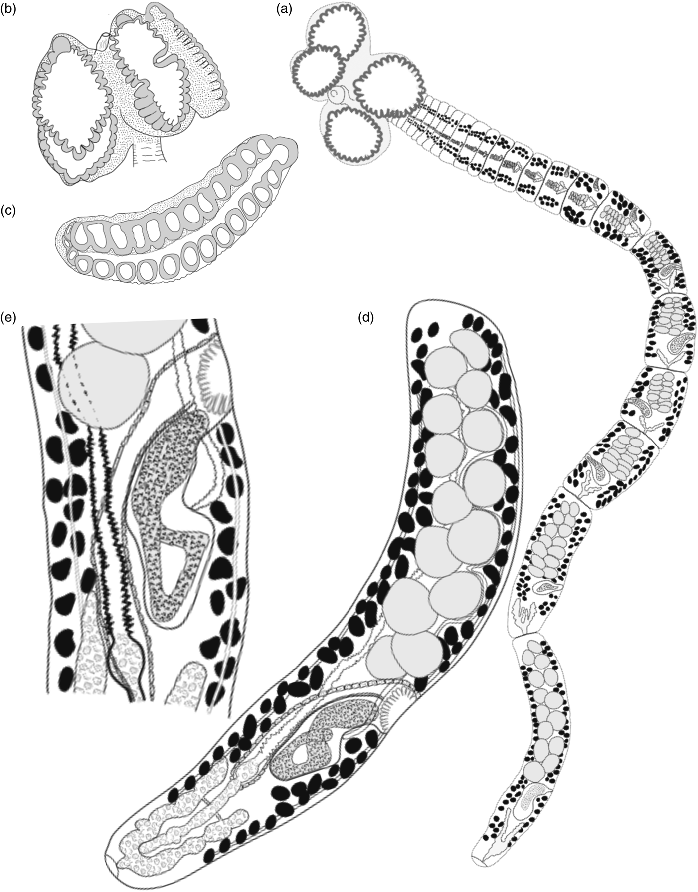
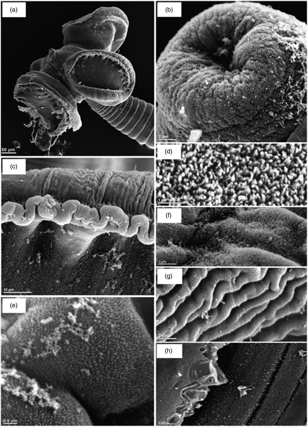
Measurements based on 27 mature specimens, two immature specimens (light microscopy), and six scolices (SEM). Adult worms (fig. 1a) slightly craspedote, euapolytic. Strobila 2.0–5.7 mm (3.5 mm; n = 25) long; greatest width 170–200 (180; n = 27); 17–44 (33; n = 27) proglottides per worm. Scolex proper 260–538 (378; n = 25) long, 417–648 (499; n = 25) wide. Scolex with four pyriform or round bothridia, slightly pedicellate (fig. 1b), 224–538 (308; n = 27) long, 115–256 (236; n = 27) wide. Slender extensible myzorhynchus 69–121 (96; n = 19) long, 45–109 (76; n = 19) wide, wider at base than the apical ending; fully evaginated myzorhynchus 112–121 (118; n = 3) long, 74–109 (91; n = 3) wide. Proximal bothridia and proximal bothridial margins covered with acicular filitriches (fig. 2e–g), interrupted in distal half of proximal bothridial surface by capilliform filitriches (fig. 2f). Myzorhynchus surface and apical organ covered entirely with acicular filitriches (fig. 2b). Distal bothridial surfaces covered with small capilliform filitriches (fig. 2c–d). Strobila proper covered with acicular filitriches (fig. 2h). Bothridia bordered by (28–30) marginal loculi (fig. 1c); arrangement: anterior loculus 30–53 (49; n = 25) wide; 26–28 facial loculi arranged in 13–14 opposite pairs, 17–44 (28; n = 22) wide; one posterior loculus, lined by a thick membrane; septa thin. Cephalic peduncle 56–114 (84; n = 22) long, 52–103 (76; n = 22) wide. Mature proglottides initially as long as wide, becoming longer than wide posteriorly. Terminal proglottis 284–1056 (751; n = 25) long, 130–347 (190; n = 25) wide (fig. 1d). Cirrus-sac subspherical in mature proglottides, pyriform in gravid proglottides, in medial-third field immediately anterior to ovary. Cirrus-sac (fig. 1e) in mature proglottides 52–126 (82; n = 15) long, 41–69 (47; n = 15) wide. Cirrus armed with small spinitriches. Vas deferens (fig. 1d) dorsal, slightly coiled, extending laterally, anterior to cirrus-sac, bypasses the testes into anterior part of proglottis, joining the cirrus sac between the anterior and middle margins. Genital pore, in posterior half of proglottis; irregularly alternating. Testes globular, 13–22 (15; n = 23) in number per proglottis; 15–56 (32; n = 17) long, 10–22 (14; n = 17) wide; arranged in two columns, occupying both poral and anti-poral field extending from anterior margin of proglottis to anterior margin of cirrus-sac, absent in post vaginal field. Ovary U–shaped posterior; bilobed in facial view; tetralobed in cross section; with discreet isthmus; lobes of approximately equal size. Mehlis’ gland localized posterior to ovarian lobes. Vagina (fig. 1e) straight or slightly sinuous, thin walled, large at its base forming a seminal receptacle, extending medially from Mehlis’ gland, bypassing the cirrus-sac and opening into the genital atrium anterior to cirrus-sac. Genital atrium common to the male and female reproductive organs with obvious muscular sphincter. Uterus, ventral, sacciform, tubular, mostly clouded by the testes, extending along the proglottis from posterior ovarian lobes to the anterior part of proglottis. Uterine duct discreet. Vitelline follicles (fig. 1d) well developed; subspherical, 10–24 (18; n = 15) by 7–14 (10; n = 15); forming two marginal rows extending throughout length of proglottis, continous through the cirrus-sac. Vitelline ducts, extending laterally, along the proglottis. Excretory ducts conspicuous.
Drawings of Echeneibothrium algeriensis n. sp. (a) Complete fixed mature specimen; (b) scolex detail with retracted myzorhynchus; (c) bothridia (side view); (d) terminal mature proglottis; (e) close-up of the reproductive structures. Scale bars: a. 157 μm; b. 100 μm; c. 59 μm; d. 51 μm; e. 37μm.

Scanning electron micrographs of Echeneibothrium algeriensis n. sp. (a) Scolex; (b) acicular filitriches on myzorhynchus; (c) detail of bothridial margin; (d) small capilliform filitriches on distal bothridial surfaces; (e) acicular filitriches on proximal bothridial margins; (f) capilliform filitriches on distal half of proximal bothridial surface; (g) acicular filitriches on proximal surface of bothridium; (h) acicular filitriches on strobila proper.

Taxonomic summary
Type host. Dipturus batis (Linnaeus, 1758) (Chondrichthyes: Rajiformes: Rajidae); blue skate.
Site of infection. Spiral intestine, in anterior half.
Type locality. Annaba (36°53′59″N, 7°46′00″E), east coast of Algeria, Mediterranean Sea.
Additional locality. Béjaïa Gulf (36°45′00″N, 5°04′00″E), east coast of Algeria, Mediterranean Sea.
Prevalence of infection. 9% of examined hosts (3 of 33 examined).
Mean intensity. 52 per infected host (156 parasites in three hosts; range = 7–140).
Mean abundance. 4.72 individuals per host examined.
Material. The holotype (NBM-010663) and four paratypes (NBM-010664–NBM-010667) are deposited in the New Brunswick Museum, Saint John, New Brunswick, Canada.
Etymology. The species was named according to the country where the host was collected.
Remarks
Specimens of E. algeriensis n. sp. recovered from D. batis off Annaba share the same range of measurements and all morphological characteristics with those provided from Béjaïa Coast.
The species described herein shares a set of morphological criteria consistent with the diagnosis for Rhinebothriidea established by Healy et al. (Reference Healy2009) and the genus Echeneibothrium (see Euzet, Reference Euzet, Khalil, Jones and Bray1994). Although E. algeriensis n. sp. described above shows similarities to the other species of the genus in general morphology, especially scolex morphology, i.e. scolex bearing bothridia divided by septa into multiple loculi and the presence of an apical modification (myzorhynchus), it can be distinguished from other species of the genus.
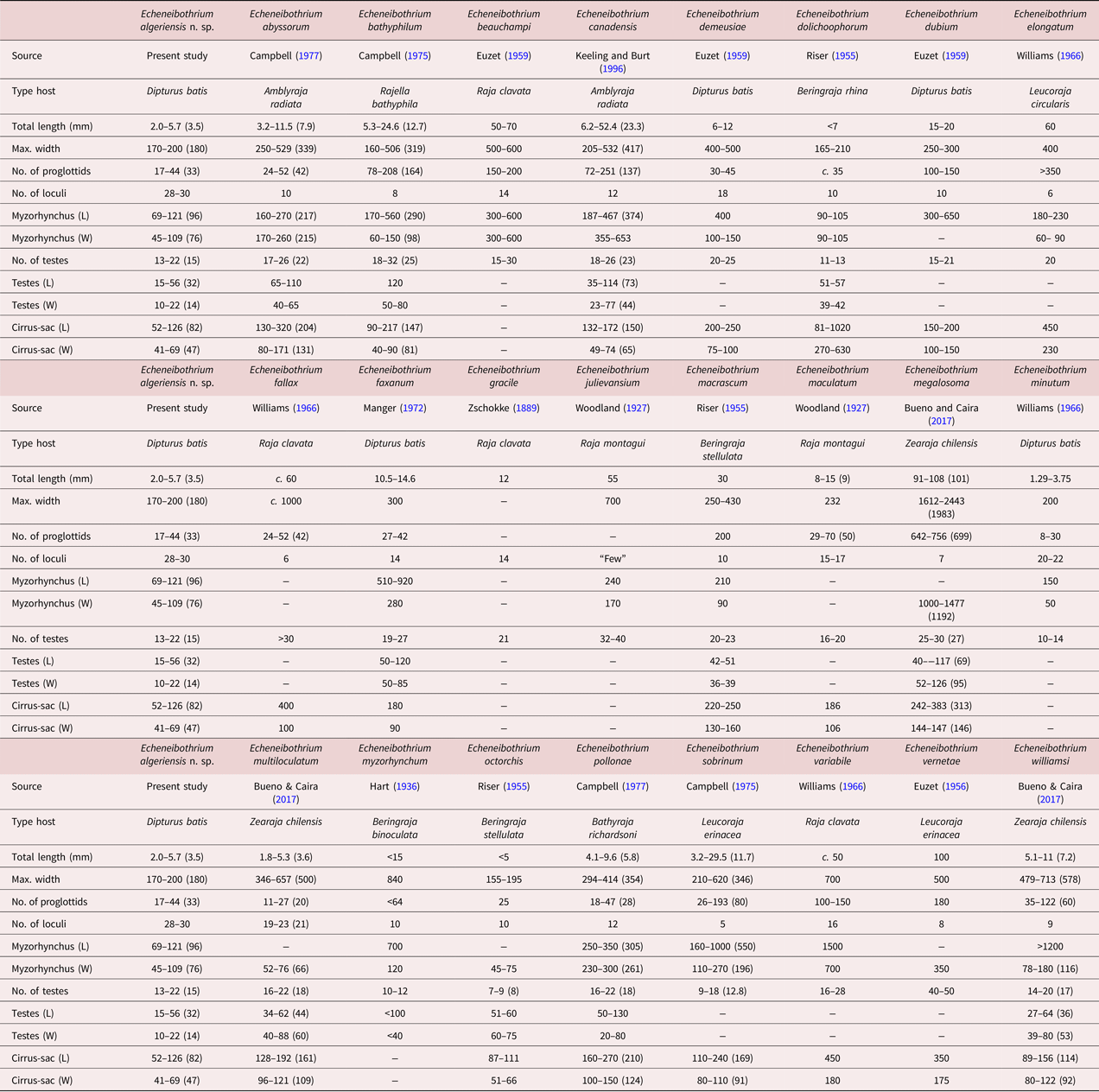
Echeneibothrium algeriensis n. sp. is easily distinguishable from other species of the genus that were recovered from D. batis during this survey. Echeneibothrium algeriensis n. sp. is most similar to E. demeusiae in terms of size (2.0–5.7 mm and 6–12 mm, respectively) and overall morphology (table 1). However, E. algeriensis n. sp. possesses more loculi per bothridium than E. demeusiae (28–30 vs 18), a shorter myzorhynchus (69–121 μm vs 400 μm), smaller bothridia (224–538 × 115–256 μm vs 600–800 × 250–300 μm) and a shorter cirrus-sac (52–126 μm vs 200–250 μm) (table 1). Echeneibothrium dubium and E. variabile possess fewer loculi per bothridium (10 and 16, respectively), a longer myzorhynchus (300–650 μm and up to 1.5 mm, respectively), and are generally larger (15–20 mm and c. 50 mm, respectively) compared to E. algeriensis n. sp. (28–30 loculi, 69–121 μm, and 2.0–5.7 mm, respectively) (table 1). Another species described from D. batis, but not recovered during this survey, Echeneibothrium minutum Williams, 1966, is akin to E. algeriensis n. sp. in size (1.29–3.75 mm for the former and 2.0–5.7 mm for the latter). However, E. minutum possesses pronounced pedicels that carry bothridia, fewer loculi per bothridium (20–22), fewer testes per proglottis (10–14), and a longer myzorhynchus (150 μm) compared to E. algeriensis n. sp. (28–30 loculi, 13–22 testes, and 69–121 μm, respectively) (table 1). Therefore, the new species described herein can clearly be distinguished from E. minutum.
Morphological comparison of Echeneibothrium algeriensis n. sp. with the other 24 valid species of the genus (see Ruhnke et al., Reference Ruhnke, Reyda, Marques, Caira and Jensen2017). All measurements are in micrometers unless stated otherwise.

L, length; W, width
Echeneibothrium algeriensis n. sp. is particularly close to Echeneibothrium multiloculatum Carvajal & Daily, 1975 in its general morphology. For instance, based on the redescription of E. multiloculatum ex Zearaja chilensis from the South Pacific Ocean (Chile), Bueno and Caira (Reference Bueno and Caira2017) provided a detailed description akin in its general morphology to E. algeriensis n. sp. Echeneibothrium multiloculatum is similar in length (1.8–5.3 mm), size of bothridia (274–447 × 172 – 298 μm), width of the myzorhynchus (52–76 μm), number of testes per proglottis (16–22), and length of testes (34–62 μm) to E. algeriensis n. sp. (see table 1). They also manifest other similarities in their scolices. In fact, the loculi of these two species are limited to the bothridial margins, leaving visible a clear central area. However, the main difference is in the number of bothridial loculi: E. algeriensis n. sp. loculi are arranged into an apical sucker, 26–28 marginal loculi disposed into 13 or 14 opposite pairs, and one posterior loculus, whereas E. multiloculatum has a similar arrangement of loculi, but only 9–11 pairs of marginal loculi. The proximal and distal bothridial surface of E. multiloculatum is covered by gladiate spinitriches interspaced by capilliform filitriches; the scolopate spinitriches cover the bothridial rim, whereas the proximal bothridial surface of E. algeriensis n. sp. is covered by acicular and capilliform filitriches in the distal half of proximal bothridial surface and the distal bothridial surfaces are covered with small capilliform filitriches only. The acicular filitriches cover both the bothridial rim and the global surface of the myzorhynchus. Another major difference is the size of the cirrus-sac, which is longer in E. multiloculatum (128–192 μm) than in E. algeriensis n. sp (52–126 μm).
Of the remaining Echeneibothrium species, E. algeriensis n. sp. is most similar in length (2.0–5.7 mm) to Echeneibothrium abyssorum Campbell, 1977 (3.2–11.5 mm), Echeneibothrium dolichoophorum Riser, 1955 (< 7 mm), Echeneibothrium octorchis Riser, 1955 (< 5 mm), Echeneibothrium pollonae Campbell, 1977 (4.1–9.6 mm), and Echeneibothrium williamsi Carvajal & Dailey, 1975 (5.1–11.0 mm) (table 1), and is significantly smaller than all other valid Echeneibothrium species (table 1). However, E. algeriensis n. sp. possesses more loculi per bothridium (28–30) than all currently recognized Echeneibothrium species (see table 1), with E. multiloculatum being the most similar (19–23; see above). Additionally, E. algeriensis n. sp. has more testes per proglottis than E. dolichoophorum, Echeneibothrium myzorhynchum Hart, 1936 and E. octorchis, and significantly fewer than E. abyssorum, Echeneibothrium bathyphilum Campbell, 1975, Echeneibothrium canadensis Keeling & Burt, 1996, Echeneibothrium fallax (Woodland, 1927), Echeneibothrium faxanum Manger, 1972, Echeneibothrium julievansium Woodland, 1927, Echeneibothrium megalosoma Carvajal & Dailey, 1975, and Echeneibothrium vernetae Euzet, 1956 (see table 1). The cirrus-sac of E. algeriensis n. sp. is smaller (52–126 × 41–69 μm) than that of all other species for which this structure is described, except E. octorchis and E. williamsi (see table 1).
Echeneibothrium algeriensis n. sp. differs from other undescribed species of the genus. It differs from all species of Echeneibothrium listed by Williams (Reference Williams1966); it possesses more loculi per bothridium than Echeneibothrium sp. ex Amblyraja radiata ( Donovan, 1808) (28–30 vs 8) and is much shorter than Echeneibothrium sp. ex Raja montagui (2.0–5.7 vs 55 mm), and possesses fewer testes per proglottis (13–22 vs 30) and more loculi per bothridium (28–30 vs 8). Additionally, it possesses more loculi per bothridium than Echeneibothrium sp. ex Zearaja nasuta, (originally described as Phormobothrium affine (Alexander, 1963)) and has fewer testes per proglottis than Echeneibothrium sp. ex Okamejei kenojei (originally identified as Raja porosa) (13–22 vs 40) and more loculi per bothridium (28–30 vs 4).
Morphological description of intestinal villi and tapeworm attachment structures
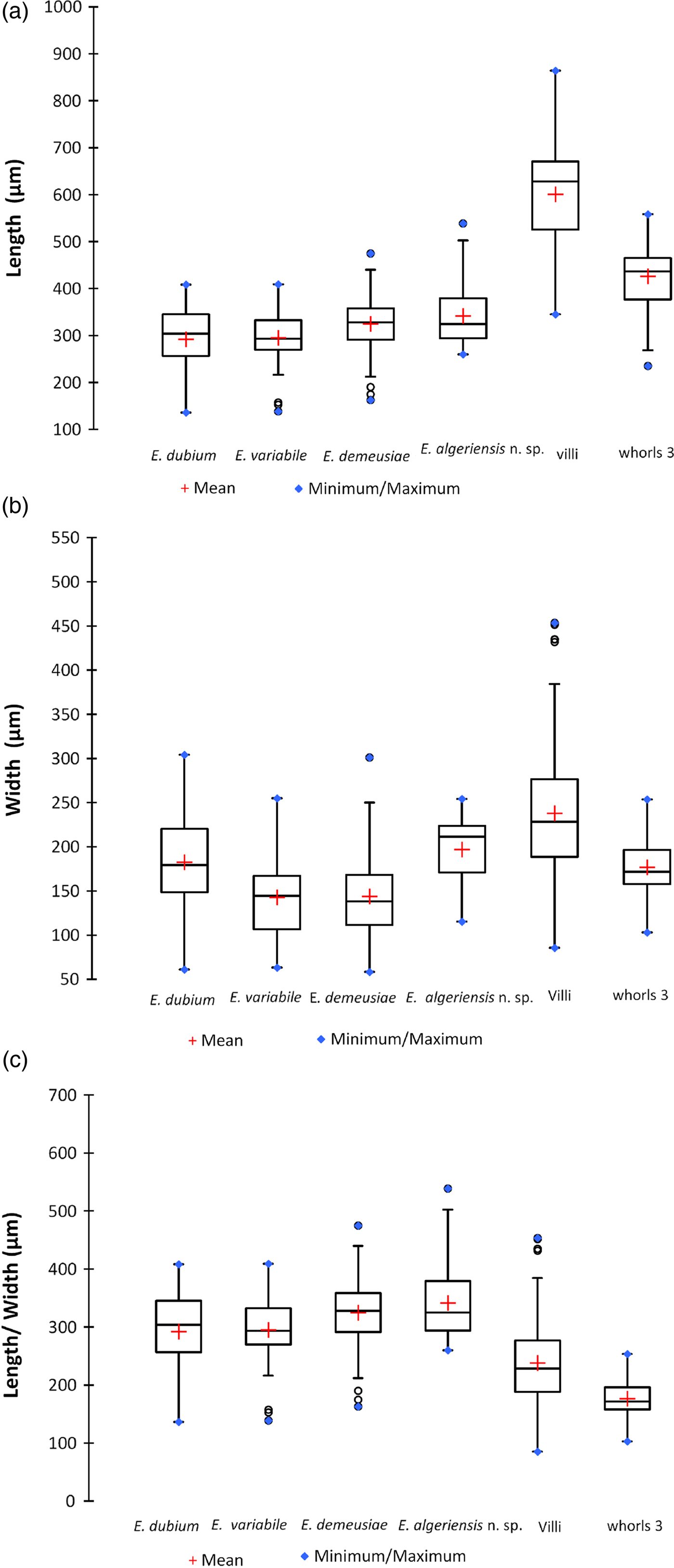
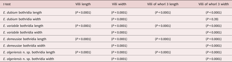
The spiral valves of infected D. batis (total length 84, 91 and 106 cm) measured 16, 17 and 21 cm in length, respectively. The inner surface of the spiral valves consists of 11 whorls; the two faces of each whorl and the inner face of the spiral valve's walls are lined with villi (not ridges like in those observed in genus Raja), fixed at their base to the intestinal mucosa, appearing as finger-like projections. Villi of the first whorls are relatively smaller and distant compared to those in median chambers, where they transform to become thick and dense; at the posterior end of the intestine, villi atrophy and tend to separate and decrease in density until none are found at the posterior extremity. Figure 3a and tables 2 & 3 show the means and variability of individual villus lengths in whorls with high tapeworm abundances (whorl 3), those occupied less frequently by the parasites, and length of bothridia for each of the Echeneibothrium species recovered from D. batis. Using a t-test approach, the analyses demonstrated that the two types of villi (from whorl 3 and other whorls) were significantly longer than the bothridia of each species of Echeneibothrium (P < 0.0001). Figure 3b and tables 2 & 3 show the means and variability of villi widths in whorls with high tapeworm abundances (whorl 3), those occupied less frequently by the parasites, and the widths of bothridia for each Echeneibothrium species found in D. batis. The t-tests indicate that there is an insignificant difference between the width of the villi (whorl 3) and the width of bothridia of E. dubium (P = 0.39), indicating that these worms should have the ability to attach to the mucosa of D. batis. The same could be said for E. algeriensis n. sp., in which the bothridium is slightly larger than the villi (P < 0.0001). However, the bothridia of E. variabile and E. demeusiae are generally narrower than the villi. Furthermore, the t-tests show a significant difference between the various widths of the bothridia and all the villi (whorl 3 and other villi). Villi measurements are summarized in table 2 and fig. 3. Table 3 summarizes the results from the t-tests.
Graphical representations of size distributions: (a) bothridial length with villiar length; (b) bothridial width with villiar width; (c) bothridial length with villiar width.

Measurements of all variables included in t-test analyses, with range, mean and coefficient of variation.

SD, standard deviation; CoV, coefficient of variation
Comparison of bothridial measurements from E. dubium, E. variabile, E. demeusiae and E. algeriensis n. sp. with villiar measurements from D. batis studied herein (t-test). Statistical significance set at P < 0.05.

Discussion
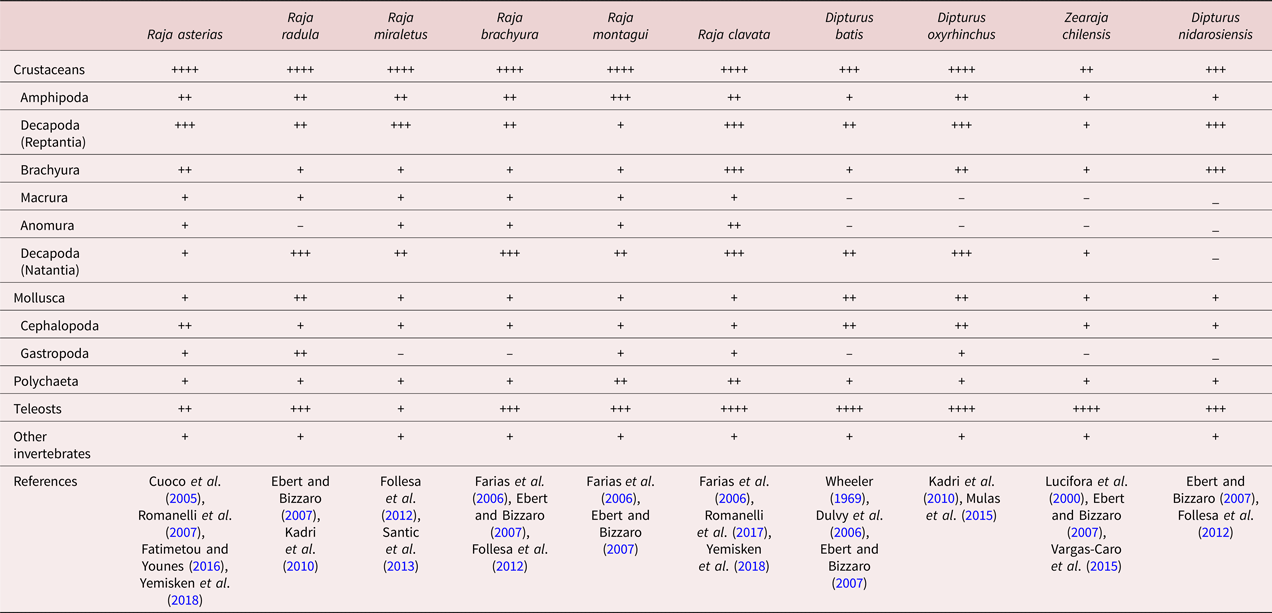
Diet is the port of entry for trophically transmitted parasites. Yet, once in, other factors will influence the establishment of these parasites. For instance, E. vernetae infects and reaches maturity in both congeneric rajid skates Leucoraja erinacea Mitchill, 1825 and Leucoraja ocellata Mitchill, 1815 (Randhawa et al., Reference Randhawa, Saunders and Burt2007). However, only immature specimens of this species have been recovered from the sympatric skate A. radiata in the north-western Atlantic Ocean, suggesting that the parasite is transmitted via a shared prey species common to all three skate species, yet other mechanisms, such as physiological compatibility, are preventing the establishment of this species in A. radiata (Randhawa et al., Reference Randhawa, Saunders and Burt2007). Based not on evidence but on our understanding of the transmission of these parasites, their presence in these respective hosts must be linked to their respective diets. For instance, adult specimens of the following Echeneibothrium species have been recovered from these respective hosts during recent surveys: (1) Echeneibothrium beauchampi Euzet, 1959 ex Raja asterias Delaroche, 1809 and Raja miraletus Linnaeus, 1758; (2) E. dubium ex R. asterias, D. batis and D. oxyrhinchus; (3) Echeneibothrium maculatum Woodland, Reference Woodland1927 and (4) E. fallax ex Raja clavata Linnaeus, 1758; and (5) E. demeusiae and (6) E. variabile ex D. batis and D. oxyrhinchus (Benmeslem and Tazerouti, pers. obs.). The composition of the diet of the various skate species probably influences the host distribution of these parasites. However, diet is influenced not only by the availability of prey but also by food preference demonstrated by some predators in the presence of a variety of prey; some rays are categorically selective in their diet (Braccini and Perez, Reference Braccini and Perez2005). A comparative dietary study of various species of Rajidae (table 4) indicates that overlap in prey species exists primarily for crustaceans, teleosts and cephalopods (Vannucci et al., Reference Vannucci2006; table 4 and references therein). Focusing on specific rajid species pairs sharing species of Echeneibothrium, both R. asterias and R. miraletus share E. beauchampi and have the most similar diet among Raja species pairs included in table 4, suggesting transmission via shared prey items. Similarly, both D. batis and D. oxyrhinchus have the most similar diets among Dipturus species pairs (table 4) and share E. demeusiae and E. variabile (see above), also suggesting that these two species of Echeneibothrium are transmitted via prey items common to the diets of both skate species. Additionally, E. dubium infects three skate species (see above) that have similar feeding habits in a subset of their diet, i.e. crustaceans and cephalopods, while differing significantly in the proportion of their diet comprising teleost fishes (table 4), suggesting that this tapeworm is probably acquired by skates via ingestion of infected crustacean hosts. The comparison of feeding habits (table 4) doesn't explain unequivocally the inter-specific variations in tapeworm assemblages in these skate hosts. Consistent with Randhawa et al. (Reference Randhawa2008), we conclude that diet is not the only filter determining tapeworm assemblages in rajid skates; the specificity of different tapeworms towards the variety of encountered hosts is due to other ecological (e.g. substrate preference) or host-related factors (e.g. morphological or immunological compatibility).
Comparison of feeding habits of some species of the genera Raja and Dipturus.

++++ Predominant prey; +++ frequently consumed; ++ frequently consumed, but less than (+++); + rarely/occasionally consumed; – not found in the stomach contents
Echeneibothrium species exhibit a high degree of specificity towards their respective hosts (Williams, Reference Williams1966). Obligate parasites must cross two filters (encounter filter and compatibility filter) in order to establish in their host (Euzet and Combes, Reference Euzet and Combes1980). This requires both parasites and hosts being present in the same biotope. With respect to the encounter filter, if spatial, temporal and behavioural conditions are met, then this filter is open (Combes et al., Reference Combes, Renaud and Le Brun1991). For the compatibility filter, if the parasite can find adequate resources and is compatible with its hosts, then it is able to persist in this host (Euzet and Combes, Reference Euzet and Combes1980). Species of the genus Echeneibothrium developed strategies to adapt in order to co-exist in their host organism. This begins by settling in their microhabitat; indeed, the attachment of the parasite to the intestinal mucosa is a major requirement for the establishment of the tapeworm–host relationship, and therefore morphological compatibility between parasite attachment structures and topography of the host intestinal mucosa should not be overlooked.
It is well established that host and attachment-site specificity in skates are determined, to a certain extent, by the close fit between a tapeworm's scolex and the host intestinal mucosa (Williams, Reference Williams1960, Reference Williams1966, Reference Williams1968; Randhawa and Burt, Reference Randhawa and Burt2008 and references therein). The adaptation of the scolex to the host mucosa allows tapeworms to remain attached despite physiological processes occurring in the digestive tract, such as peristaltic movements, which could mechanically expel them from their microhabitat. Species of Echeneibothrium possess robust attachment structures and well-established scolex morphology adapted to a mode of attachment compatible with mucosa characterized by villi, seemingly accommodated to the structure of their host mucosa. This muscular nature of the attachment in Echeneibothrium (Euzet, Reference Euzet1959; Williams, Reference Williams1966; Randhawa and Burt, Reference Randhawa and Burt2008) is characterized by the highly polymorphic and multiloculated bothridia wrapping around individual intestinal villi, by the myzorhynchus engulfing one to several villi, or by using bothridia and myzorhynchus to securely attach (Euzet, Reference Euzet1959; Williams, Reference Williams1966).
The studies conducted by Williams (Reference Williams1960, Reference Williams1968) attest that the differences in topography of the gut mucosa determine the specificity of tetraphyllidean (sensu lato) cestodes. Further to our observations, we conclude that the intestinal structure of D. batis facilitates the easy attachment of Echeneibothrium to villi, not only via each bothridium grabbing a villus but also assisted by the myzorhynchus. Villi are long enough to let the bothridium attach using all its loculi, thus setting up the fastening of the parasite (tables 2 & 3; fig 3). Consistent with Randhawa and Burt (Reference Randhawa and Burt2008), we suggest that compatibility between the tapeworm's attachment structures and the size of the villi determines the suitability of sites of attachment, as does the use of spaces that exist between the villi, which allow the parasites to slide their strobila between them, providing them with good support.
Observations of Echeneibothrium indicate that these species have pedunculated, highly polymorphic, flexible and muscular bothridia (Williams, Reference Williams1966) and may attach to the mucosa of the host's gut by contracting or expanding their bothridia and bending them according to the relief of their attachment site in an effort to expose all their loculi to the mucosal surface. Unsurprisingly, significant differences were observed between lengths of the bothridia and widths of the villi (fig. 3c; tables 2 & 3) (P < 0.0001), with bothridial lengths exceeding villiar widths. This result suggests that these parasites may use their bothridia (length-wise) to surround and squeeze individual villi. Hence, we deduce that not only is the size of these villi important for adequate attachment of the parasites, but so is their shape. For instance, the cylindrical shape of the villi coheres perfectly with the attachment mode of Echeneibothrium (Williams, Reference Williams1966).
Results in this study further reinforce that no single factor determines host specificity in the skate–Echeneibothrium spp. host–parasite relationship. For instance, diet alone (ecological factor) does not suitably explain why skate species with similar diets are infected by different species of Echeneibothrium, nor do shape and size of villi in the spiral intestine (morphological factor). These results are consistent with other studies focusing on the same model system that suggested that: (1) ecological factors other than diet, such as substrate preferences and associated prey biota, could explain differences in echeneibothriid tapeworm fauna (Randhawa et al., Reference Randhawa2008); (2) diet is a point of entry for parasites into the host, but establishment is dictated by other determinants, such as host compatibility (Randhawa et al., Reference Randhawa, Saunders and Burt2007; Randhawa and Burt, Reference Randhawa and Burt2008); and (3) agreement between morphology of attachment structures, i.e. bothridia, and villi, is more important in attachment-site specificity when examining the parasite fauna of closely related hosts (Randhawa and Burt, Reference Randhawa and Burt2008). Furthermore, it is possible that diet studies have simply not described diets of batoid skates to scales at which differences are perceivable. For instance, many studies describe diet by identifying prey to order or family (see table 4 and references therein), whereas perhaps specificity in interactions between echeneibothriid worms and their intermediate host(s) is discernable only at the level of prey genus or species. In short, this study reiterates two main aspects: (1) no single factor should be examined in isolation when examining host specificity of parasites; and (2) the description of a new tapeworm species from a relatively well-studied host, but from a poorly sampled geographical region, reinforces the need to examine parasite fauna from host species across their entire range in order to gain a good understanding of the biodiversity these hosts harbour and their ecology.
Acknowledgements
The kind and generous assistance of fishermen with sample collection at the following Algerian ports is acknowledged: Béni Saf, Bouharoun, Béjaïa and Annaba. We thank N. Trieu of the Microscopy and Microanalysis Facility at the University of New Brunswick (Fredericton, New Brunswick, Canada) for the sample preparation and acquisition of scanning electron micrographs.
Financial support
This research received no specific grant from any funding agency, commercial or not-for-profit sectors.
Conflict of interest
None.