INTRODUCTION
Compounding is a productive process that allows speakers of most languages to create complex words by combining two or more simple lexical units. Compound words can be semantically transparent, when the meaning of the word can be easily inferred from the meaning of its constituents (e.g., bedroom), or semantically opaque, when no semantic relationship can be found between the whole word and its single elements (e.g., eggplant).
Since the 1980s, the main issue has concerned how multimorphemic words, such as compounds, are mentally represented. Two opposing theories have been proposed. Full-listing theories suppose that morphologically complex words are stored and retrieved as whole words, just like morphologically simple words (see, e.g., Butterworth, Reference Butterworth and Butterworth1983). However, this hypothesis does not consider the principle of cognitive economy, which claims that the more productive a word formation process (such as in the agglutinated languages like Turkish), the more likely it will generate a potentially infinite number of complex words.
Conversely, full-parsing theories (e.g., Taft & Forster, Reference Taft and Forster1975) argue that complex words are processed through the full decomposition of their single constituents. Based on this explanation, however, it is impossible to understand how semantically opaque compounds (such as the Italian word corrimano -handrail- lit. run-hand) are processed.
More recently, an alternative hypothesis, the so-called dual route model, was proposed. This model can be considered a sort of compromise between the full-listing and full-parsing accounts. Dual route theories assume that complex words can be stored and retrieved whole, through a single-lemma-single-morpheme structure, or by decomposition into their constituents and processed through a single-lemma-multiple-morpheme route. The former would apply to very frequent and/or opaque compounds (i.e., hotdog or passport) and the latter to less frequent and/or transparent compounds (i.e., blackboard or sunshine) (Levelt, Roelofs, & Meyer, Reference Levelt, Roelofs and Meyer1999). Evidence for decomposition comes from psycholinguistic studies in healthy subjects and from reports of patients with language deficits. Several studies using priming techniques in healthy subjects have showed that the recognition of compounds is facilitated if it is preceded by presentation of either the first or the second element, thus demonstrating the parallel activation of both constituents (Jarema, Busson, Nikolova, Tsapkini, & Libben, Reference Jarema, Busson, Nikolova, Tsapkini and Libben1999; Libben, Gibson, Yoon, & Sandra, Reference Libben, Gibson, Yoon and Sandra2003).
Further evidence in favor of the decompositional hypothesis comes from studies investigating the processing of compounds in aphasic patients. In a group study, Hittmair-Delazer Andree, Semenza, DeBleser, and Benke (Reference Hittmair-Delazer, Andree, Semenza, DeBleser and Benke1994) found that when patients performed a compound naming task they preferentially produced semantic paraphasias that closely resembled the decomposed structure of the stimuli. Errors consisted of existing and nonexisting compounds whose single constituents had a close semantic relationship with the targets. Semenza, Luzzatti, and Carabelli (Reference Semenza, Luzzatti and Carabelli1997) found that Broca’s agrammatic aphasics, unlike Wernicke’s patients, tended to omit the verb component in verb-noun (VN) compounds even though in Italian they belong grammatically to the noun category. Moreover, this pattern was not due to a positional effect (such as impaired access to the first component) because in noun-noun (NN) compounds errors were equally distributed on the first and the second constituent. Similarly, Mondini, Luzzatti, Zonca, Pistarini, and Semenza (Reference Mondini, Luzzatti, Zonca, Pistarini and Semenza2004) confirmed that patients with disproportionate verb naming deficits were also more impaired on VN compounds than NN compounds. As patients with Broca’s aphasia generally omit more verbs than nouns when processing simple words, the authors concluded that the omission of the verb component in VN compounds should be attributed to their decomposition into separate noun and verb forms.
More recently, Nasti and Marangolo (Nasti & Marangolo, Reference Nasti and Marangolo2005) described a patient who frequently omitted either the first or the second element in a compound word reading task and less frequently in naming and writing to dictation tasks. Moreover, as in previous studies the patient’s errors always respected the decomposed structure of the stimuli. Furthermore, coherently with his residual agrammatism he showed a deficit in retrieval of the verb component in VN compounds.
In a sample of Italian agrammatic patients, Mondini, Luzzatti, Saletta, Allamano, & Semenza (Reference Mondini, Luzzatti, Saletta, Allamano and Semenza2005) explored the processing of prepositional compounds (e.g., mulino a vento - windmill) and found that in naming, omission was the most frequent type of error; in repetition, reading, writing, and completion tasks errors were mostly substitutions of the target preposition. This trend was observed even with fully lexicalized compound forms, namely, those forms in which the linking preposition is syntactically and semantically opaque.
Taken together these results seem to be consistent with a dual-route interpretation of lexical access, which hypothesizes decomposition even in the case of opaque compounds (Badecker, Reference Badecker2001; Mondini et al., Reference Mondini, Luzzatti, Saletta, Allamano and Semenza2005). In fact, analysis revealed that the errors were almost always substitutions or omissions of one component only. Moreover, Italian aphasic patients with a verb-relative-to-noun deficit systematically drop the verb component in VN compounds [i.e., aspirapolvere (vacuum cleaner)]. This finding has been considered the strongest evidence of decomposition.
Despite great efforts to investigate the cognitive mechanisms involved in processing complex words, we still know little about the neural correlates of compound processing. The only report in the literature is a recent study by El Yagoubi and colleagues (El Yagoubi, Chiarelli, Mondini, Perrone, Danieli, & Semenza, Reference El Yagoubi, Chiarelli, Mondini, Perrone, Danieli and Semenza2008) in which NN compound processing was investigated by means of an event-related potential (ERP) technique. Normal Italian-speaking subjects were asked to perform a lexical decision task in which items were composed of noun-noun compounds [e.g., capobanda (band leader)], noncompounds with an embedded word [e.g., coccodrillo (crocodile), where cocco means “coconut”] and nonwords generated either by reversing the positions of the two constituents of the compound [e.g., for capobanda (leader) the corresponding nonword was bandacapo] or by reversing the positions of the word embedded in the noncompounds (e.g., for coccodrillo the nonword was drillococco).
Two main results were found. The first was a larger N400 lexicality effect for noncompound than for compound words. The authors argued that this effect was likely due to the presence of nonwords created by inverting the component word. Because the nonwords were derived from compound words that contained two real words, participants may have accessed the meaning of the two constituents by means of a decomposition process. Second, results showed a more negative peak in the left anterior negativity component (LAN, which is thought to be related to morphosyntactic processing) for compounds than for noncompounds, which was attributed to the formation of morphosyntactic constituents. Taken together these results are compatible with data from behavioral studies suggesting a decomposed representation of compound constituents.
Although these data provide remarkable insights into the temporal dynamics of compound words processing, they are less informative at the level of spatial localization.
The goal of the present study was to explore the brain areas involved in compound word processing. Previous behavioral findings in normal subjects (e.g., Jarema et al., Reference Jarema, Busson, Nikolova, Tsapkini and Libben1999; Libben et al., Reference Libben, Gibson, Yoon and Sandra2003) as well as in aphasic patients (e.g., Hittmair-Delazer et al., Reference Hittmair-Delazer, Andree, Semenza, DeBleser and Benke1994; Mondini et al., Reference Mondini, Luzzatti, Zonca, Pistarini and Semenza2004; Nasti & Marangolo, Reference Nasti and Marangolo2005; Semenza et al., Reference Semenza, Luzzatti and Carabelli1997) seem to suggest that the single elements of compound words are processed separately. Nevertheless, it is still unclear whether or not this hypothetical cognitive independence indicates the existence of distinct neural mechanisms devoted to processing the different constituents of compounds.
Currently, there is substantial evidence that the brain systems required for retrieving verbs and nouns are at least partially segregated. Indeed, some studies have suggested that areas lying outside the frontal regions (i.e., temporo-parietal cortices, Aggujaro, Crepaldi, Pistarini, Taricco, & Luzzatti, Reference Aggujaro, Crepaldi, Pistarini, Taricco and Luzzatti2006; Luzzatti, Aggujaro, & Crepaldi, Reference Luzzatti, Aggujaro and Crepaldi2006; Piras & Marangolo, Reference Piras and Marangolo2007) might be responsible for processing verbs. Other studies have found that the frontal areas play a key role in processing verbs (Daniele, Giusstolisi, Silveri, Colosimo, & Gainotti, Reference Daniele, Giustolisi, Silveri, Colosimo and Gainotti1994; Shapiro, Moo, & Caramazza, Reference Shapiro, Moo and Caramazza2006; Tranel, Martin, Damasio, Grabowski, & Hichwa, Reference Tranel, Martin, Damasio, Grabowski and Hichwa2005). It has also been suggested that the inferior frontal gyrus (due to its location near the motor areas) supports the production of action names for which the availability of motor features is important (Parsons et al., Reference Parsons, Fox, Downs, Glass, Hirsch and Martin1995). With regard to noun processing, as nouns usually refer to concrete entities (i.e., objects, tools, animals) that involve the processing of perceptual features, previous lesion studies have frequently reported activation of the temporal lobe (Daniele et al., Reference Daniele, Giustolisi, Silveri, Colosimo and Gainotti1994; Luzzatti et al., Reference Luzzatti, Aggujaro and Crepaldi2006; Tranel et al., Reference Tranel, Martin, Damasio, Grabowski and Hichwa2005).
The rationale behind our investigation was that if compounds are processed through a whole word representation both NN and VN, as nominal compounds (they all belong to the grammatical category of nouns), should recruit similar cerebral regions.
On the contrary, if the two elements of the compound are processed by separate brain regions involvement of different damaged areas might be found only in the case of VN compounds.
The present work was aimed at addressing this issue in a group of 20 patients with left brain damage using a recently devised lesion-behavior methodology, VLSM (Bates et al., Reference Bates, Wilson, Saygin, Dick, Sereno and Knight2003). VLSM uses fully continuous information both at the behavioral (no arbitrary cutoffs are stipulated) and the neuroanatomical level (all patients are included regardless of lesion location). Statistical analyses on the relationship between tissue damage and observed behavior are carried out on a voxel-by-voxel basis, as in functional imaging, and the results are plotted as color maps depicting the degree of behavioral involvement of each voxel. Here, VLSM was used to identify lesions associated with naming difficulties in four classes of stimuli: simple nouns, simple verbs, noun-noun compounds (NN), and verb-noun (VN) compounds.
MATERIALS AND METHODS
Subjects
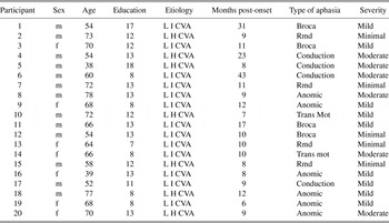
Twenty participants (13 males, 7 females) who had suffered a single left hemisphere cerebrovascular accident (CVA) were included in the study. Subjects were recruited and tested at the Center for Neuropsychological Diagnosis and Rehabilitation of the Fondazione I.R.C.C.S. Santa Lucia in Rome, Italy. Inclusion criteria were native Italian language proficiency, premorbid right-handedness, a single left CVA at least 6 months before the investigation, suitability for MRI scanning and no previous neurological, psychiatric, or substance abuse history. Mean age of the patients was 62 years (SD 11), mean time poststroke was 13 months (SD 9) and mean education level was 12 years (SD 3) (see Table 1). The data analyzed in the current study were collected in accordance with the Declaration of Helsinki and the Institutional Review Board of the Fondazione Santa Lucia. Before participation, all patients signed informed consent forms.
Clinical and sociodemographic data of the aphasic group

Note
LHCVA = left hemorrhagic cerebrovascular accident; LICVA = left ischemic cerebrovascular accident; Trans mot = transcortical motor; Rmd = residual minimal disorders.
Clinical Assessment of Aphasia
Aphasic disorders were assessed using the Italian version of the Western Aphasia Battery, which includes different subtests for the different language modalities, that is, naming, repetition, reading, and writing. The aim of the battery is to classify the patient in one of the major syndromes according to the standard aphasia taxonomy (Broca’s, Wernicke’s, Transcortical, Conduction, and Anomic aphasia) on the basis of his/her score on each language task (Kertesz, 1982). The aphasic symptoms shown by each participant varied from severe to residual minimal disorders. Thus, they covered a wide range of linguistic impairments (see Table 1). None of the patients had articulatory difficulties that could have confounded the error analysis of the picture-naming task.
Experimental Stimuli and Tasks
To investigate compound-word-naming performance, a total of 90 stimuli that could be depicted were divided into (i) 30 simple nouns; (ii) 30 simple verbs; (iii) 12 NN compounds (e.g., mappamondo -globe-); and (iv) 15 VN compounds (e.g., apribottiglie -bottle opener) (Appendix). Stimuli frequencies were obtained from the CoLFIS web source (Laudanna, Thornton, Brown, Burani, & Marconi, Reference Laudanna, Thornton, Brown, Burani and Marconi1995; http://www.istc.cnr.it/material/database/colfis/), which is based on a large corpus of 3,798,275 lexical units gathered from Italian newspapers, magazines and books. Stimuli were matched across categories for frequency [means and standard deviations in parentheses for simple nouns: 9 (9); simple verbs: 8.7 (10); NN compounds: 6.3 (8.2); VN compounds: 8.6 (12.6)] and length [simple nouns: 8.2 (1.5); simple verbs: 8.7 (1.2); NN compounds: 8.8 (1); VN compounds: 9 (1.3)]. Ten age-and educational level matched normal controls were asked to rate the imageability of each noun, verb, NN and VN compound on a 7-point scale (1 = low imageability; 7 = high imageability). Statistical tests revealed no significant difference among the four groups of stimuli, which were all judged as highly imageable. The pictures were randomly presented to each patient in three separate sessions. Subjects were asked to name each picture without a time limit.
MRI Acquisition
Images were acquired using a 1.5 Tesla (T) Siemens Vision Magnetom MR system (Siemens Medical Systems, Erlangen, Germany).
To obtain a precise brain definition and to distinguish between the gliotic and necrotic parts of the vascular lesions and healthy tissue, four different anatomical sequences were acquired: (1) a T-2 weighted turbo spin echo image [repetition time (TR) = 3800 ms; echo time (TE) = 90 ms; field of view (FoV) = 173 × 230]; (2) a proton density image (TR = 3800 ms; TE = 20 ms; FoV = 173 × 230); (3) a fluid attenuated inversion recovery image (TR = 9999 ms; TE = 105 ms; FoV = 188 × 250); (4) a dedicated high-resolution (1 × 1 × 1 mm) T1-weighted image of the whole brain, using a 3-D Magnetization Prepared Rapid Gradient Echo (MPRAGE) sequence (TR = 11.4 ms; TE = 4.4 ms; flip angle = 10°, 256 × 256 matrix, 1 × 1 mm in-plane resolution, 220 contiguous 1-mm coronal slices).
Lesion Analysis and VLSM
Lesions were drawn manually slice-by-slice on the digital MPRAGE images using free MRIcro software (Rorden & Brett, Reference Rorden and Brett2000) and were saved as Regions of Interest (ROIs). During this process, the other clinical images acquired were visually co-inspected to obtain a detailed lesion image, which was corroborated by two clinical neuroradiologists who were blind to the aims of the study and the patients’ behavior. The gliotic tissue was considered as part of the lesion.
Normalization of each patient’s MRI to a common spatial framework was performed using SPM99 software (Wellcome Department of Cognitive Neurology, London, UK), implemented in MATLAB (The MathWorks Inc., Natick, MA, USA), through an automatic nonlinear stereotaxic normalization procedure (Friston, Ashburner, Poline, Frith, Heather, & Frackowiak, Reference Friston, Ashburner, Poline, Frith, Heather and Frackowiak1995). Estimation of the normalization parameters was restricted to healthy tissue (Brett, Leff, Rorden, & Ashburner, Reference Brett, Leff, Rorden and Ashburner2001). Distorted lateral ventricles were excluded from the computation.
Each patient’s lesion image was entered in the VLSM analysis together with four behavioural scores: (i) number of correct responses in the noun-naming task; (ii) number of correct responses in the verb-naming task; (iii) number of correct responses in the NN-naming task; and (iv) number of correct responses in the VV-naming task. The VLSM method is as follows: at each voxel, patients are divided into two groups according to whether or not the voxel is lesioned. These groups are then compared (e.g., with a t test) and the resultant t values are overlaid on the single-subject MNI brain as color maps showing the degree of behavioural involvement of each voxel in the single tasks. The VLSM algorithms programmed in MATLAB (The Mathworks, Natick, MA) are available online at http://crl.ucsd.edu/vlsm. In the present study, a customized version of VLSM was used. Therefore, we were also able to submit lesion images to an automated anatomical labeling procedure based on macroscopic anatomical parcellation of the MNI single-subject brain (Tzourio-Mazoyer et al., Reference Tzourio-Mazoyer, Landeau, Papathanassiou, Crivello, Etard and Delcroix2002), which includes all main gyri of the cerebral cortex. Thus, for each patient we were able to compute the total volume of the lesion and the percentage of each damaged brain region.
Statistically, multiple comparisons were controlled by performing a correction so that the false discovery rate (FDR) was set at p = .05. Time postonset and lesion volume were used as covariates to ensure that these factors would not affect our results. Moreover, to maintain a reasonable level of statistical power, statistical analyses were restricted to those voxels where at least 5 patients were lesioned. Finally, to highlight differences and commonalities across tasks, the single t-maps were compared by conjunction and correlation analyses. For conjunction analyses, VLSM computes a map showing the minimum value of two images for each voxel. For correlation analyses, VLSM calculates the similarity between two maps by computing a correlation between the t values and generating a map on which each voxel is colored according to the t difference between the two maps.
RESULTS
Behavioral Results
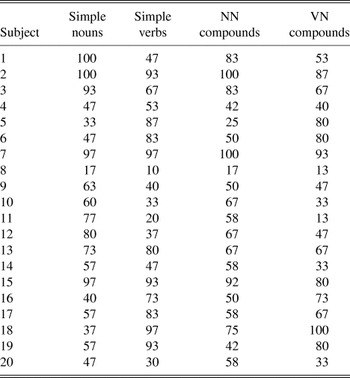
Percentages of correct responses in the four tasks are reported in Table 2. To assess differences among tasks, a repeated measures analysis of variance (ANOVA) with type of stimulus (i.e., noun, verb, NN, and VN compound) as within-subjects variable was computed. It revealed no significant differences in the percentage of correct responses (F(3,57) = .025) among noun (mean = 64%; SD = 24.7), verb (mean = 63.2%; SD = 28.3), NN (mean = 62.1%; SD = 22.5), and VN (mean = 59.3%; SD = 25.8) picture naming. Pearson correlation tests were computed (i.e., noun-verb; noun-NN compound; noun-VN compound; verb-VN compound; verb-NN compound; VN compound-NN compound). Results showed a highly significant correlation only for the verb-VN compound (r = 0.84; p < .001) and the noun-NN compound (r = 0.86; p < .001).
Percentages of correct responses in the four naming tasks

As in previous studies (Nasti & Marangolo, Reference Nasti and Marangolo2005), errors were classified for noun and verb naming as: omissions (no response), semantic paraphasias [elefante (elephant) → rinoceronte (rhino)] and phonological paraphasias [poliziotto (policeman) → piziotto].
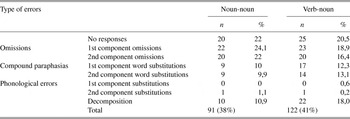
For the compound-naming task, they were categorized as: omissions (no response); omission of the first component [e.g., apribottiglie (bottle opener) lit. open bottles →…bottiglie (…bottles)]; omission of the second component [e.g., apribottiglie → apri… (open…)]; compound paraphasias in which word substitutions could affect either the first [e.g., apribottiglie (corkscrew) → portabottiglie [bottle holder], lit. carry bottles`) or the second element of the compound (e.g., mappamondo [globe], lit. map world → mappaterra [map earth]; phonological paraphasias in which phonological errors could be either in the first (e.g., apribottiglie → acribottiglie) or the second element of the compound (e.g., apribottiglie → apribottoglie); decomposition [when the compound was broken up into its constituents, such as in asciugamani (towel), lit. dry hands → asciugare le mani (to dry the hands)]. Results are summarized in Table 3.
Number, percentage, and type of errors in compound naming tasks

First, we were interested in assessing differences between the percentage of whole word (i.e., no responses) and decompositional errors (i.e., 1st component omission, 2nd component omission, 1st component substitution, 2nd component substitution, 1st or 2nd component phonological errors, compound decomposition) in NN- and VN-compound-naming tasks. To this aim, we computed a 2 × 2 repeated measures ANOVA with type of compound (i.e., NN and VN) and type of error (i.e., whole word vs. decompositional) as the within-subjects variables. Results revealed a significant effect only for type of error (F(1,19) = 122,11; p < .0001), which indicated that patients made significantly more decompositional errors (p < .0001; post hoc Scheffè analysis).
To determine whether a positional effect of the decompositional errors influenced NN and VN naming, a second 2 × 2 repeated measures ANOVA was computed with type of compound (i.e., NN and VN) and error position (i.e., 1st and 2nd position) as the within-subjects variables. Results showed no significant effects. Furthermore, no statistical differences emerged when we investigated whether the different aphasia categories affected processing of the first or second component of the compounds.
Voxel-Based Lesion-Symptom Mapping Results
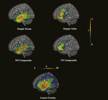
We used VLSM to identify brain regions associated with performance on the four naming tasks. To this aim, we computed single t-maps for each word category (i.e., noun, verb, NN, and VN compounds) (see Figure 1).
First two rows: Lateral volume rendering of VLSM maps computed for the four naming tasks: simple nouns, simple verbs, noun-noun (NN) compounds, and verb-noun (VN) compounds. High t scores (yellow-to-red) indicate that lesions in the corresponding voxels affected behavior significantly. Low t scores (blue) indicate left hemispheric regions whose lesions had relatively little impact on behavior. All voxels above t = 2.97 were significant at p = .05 (FDR correction). Last Row: Overlay of patients’ lesions. Maximum overlap is highlighted in red.

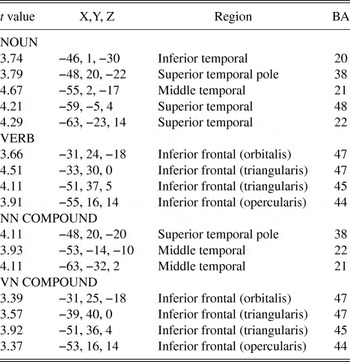
Noun naming was primarily affected by lesions located in the temporal areas. Specifically, the highest t values (see Table 4) were found in the inferior, middle and superior temporal areas (BAs 20, 21, 22, 38, and 48). Similarly, lesions involving the temporal areas significantly affected the NN-compound-naming task (BAs 21, 22, and 38). On the contrary, verb naming recruited regions located in the inferior frontal areas (pars orbitalis, triangularis, and opercularis, BAs 44, 45, and 47). Of interest, VN-compound naming was also associated with infero-frontal regions (BAs 44, 45, and 47) (see Table 4).
Highest t values for VLSM maps in the four naming tasks

Note
Approximate MNI coordinates, region labeling and Brodmann’s areas are given. NN Compound = Noun-noun compound : VN Compound = Verb-noun compound.
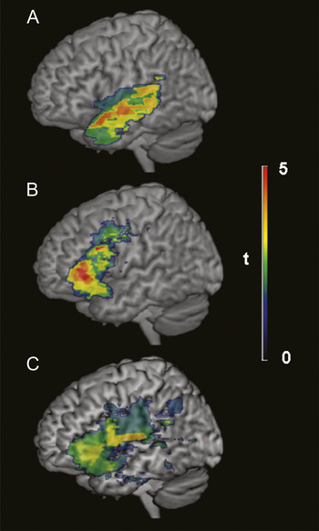
To further highlight commonalities across tasks, VLSM conjunction maps were computed for N and NN compounds and for V and VN compounds which confirmed the previous results (Bates et al., Reference Bates, Wilson, Saygin, Dick, Sereno and Knight2003) (see Figure 2).
Lateral volume rendering of VLSM conjunction maps computed for simple nouns and noun-noun (NN) compounds (A) and for simple verbs and verb-noun (VN) compounds (B). Yellow-to-red colors indicate left hemispheric regions where the maximum overlap between tasks was found. In C, VLSM map for VN including percentage of patient’s lesion affecting the inferior frontal areas as covariate.

Therefore, its seems likely that while, as expected, NN compounds were processed by the same anatomical regions as simple nouns, VN compounds, although they belong to the noun category, recruited only those frontal regions recruited by simple verbs.
Because, as previously reported, frontally damaged patients sometimes make errors that systematically affect the first component in the VN-naming task, we asked ourselves whether the above result could have been partly due to the impact of inferior frontal lesions on VN-compound naming. We also wished to determine whether there was a correlation between the extent of damage in frontal areas and the type of decomposition errors found for VN compounds.
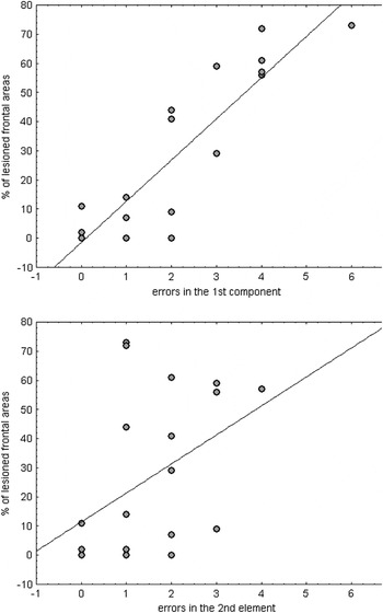
To this aim, we computed a VLSM map for the VN-compound task. The former included the proportion of the patient’s lesion that affected the inferior frontal areas as covariate. Results revealed that even though frontal regions were still significantly affected, other regions, specifically the superior temporal areas, were also involved (see Figure 2). Moreover, two separate correlation analyses between the percentage of lesioned frontal areas and i) the number of first component errors and ii) the number of second component errors revealed a highly significant correlation only for the first component errors (r = .89; p < .001) (see Figure 3).
Scatterplots showing the impact of a lesion in the left inferior frontal areas (y axis) in modulating the number of errors affecting the first (x axis on the top) or the second (x axis on the bottom) component of verb-noun (VN) compounds. Regression lines are reported in black.

DISCUSSION
The present study was aimed at investigating the brain areas involved in processing compound words. Previous behavioural and neuroimaging studies in normal subjects (e.g., El Yagoubi et al., Reference El Yagoubi, Chiarelli, Mondini, Perrone, Danieli and Semenza2008; Jarema et al., Reference Jarema, Busson, Nikolova, Tsapkini and Libben1999; Libben et al., Reference Libben, Gibson, Yoon and Sandra2003) and in aphasic patients (e.g., Hittmair-Delazer et al., Reference Hittmair-Delazer, Andree, Semenza, DeBleser and Benke1994; Koester, Gunter, & Wagner, Reference Koester, Gunter and Wagner2007; Mondini et al., Reference Mondini, Luzzatti, Zonca, Pistarini and Semenza2004; Nasti & Marangolo, Reference Nasti and Marangolo2005; Semenza et al., Reference Semenza, Luzzatti and Carabelli1997) seem to suggest that complex words undergo a decompositional process. In the present study, we analyzed the MRI data of 20 subjects with left brain damage together with their performances on two single-word (nouns and verbs) and two compound-word (NN and VN) naming tasks using VLSM analysis (Bates et al., Reference Bates, Wilson, Saygin, Dick, Sereno and Knight2003). With this approach, we were able to explore the brain areas that had the greatest effect on the four tasks using fully continuous behavioral and lesion information. Stimuli consisted of simple nouns, simple verbs, noun-noun (NN), and verb-noun (VN) nominal compounds. We reasoned that if compounds are processed through a whole-word representation, both NN and VN compounds, which are grammatically like nouns, should be represented in the same anatomical regions. Conversely, if decomposition of its constituent morphemes occurs, we should find different areas involved in processing the different constituents of VN.
Two main results are worth considering. First, N and NN compounds primarily recruited temporal areas. Several previous lesion studies have demonstrated that the processing of simple nouns affects different regions mainly located in the temporal areas (e.g., Daniele et al., Reference Daniele, Giustolisi, Silveri, Colosimo and Gainotti1994; Piras & Marangolo, Reference Piras and Marangolo2007; Tranel et al., Reference Tranel, Martin, Damasio, Grabowski and Hichwa2005). Nouns usually refer to concrete entities (i.e., objects, tools, animals) intrinsically denoted by a rich set of perceptual attributes. Therefore, it is likely that the temporal lobe will be implicated when the processing of a stimulus requires detailed analysis of its perceptual features. In line with these results, it was predictable that the areas involved in NN-compound naming would be the same as those recruited for simple nouns because they both belong to the same grammatical category and therefore would involve the temporal regions. Nevertheless, although the qualitative analysis of errors suggests the presence of a decompositional effect in NN processing and a strong correlation between nouns and NN, because both elements of the compound are nouns, these results do not allow us to make any prediction about the type of process that takes place at the neural level.
This observation brings us to our second result. It was predictable that verbs would be subserved by the frontal regions. Indeed, various studies have suggested that the link between the prefrontal cortex and verbs might be partly due to the activation of action representation (Cappa & Perani, Reference Cappa and Perani2002). The intriguing result of this study is the strong similarity that emerged in the first analysis between the areas involved in V and VN naming, suggesting greater involvement of the same frontal regions. This result is hard to reconcile with the whole-word-representation account. In fact, it indicates that in VN naming there was a bias in favor of the verb component, suggesting morphological decomposable processing. This finding appears to contrast with the qualitative analysis of errors, which shows that overall the patients had an equal distribution of errors for both elements of the VN compound. However, differences between subjects in processing the first and/or the second constituents of the VN compound could have been responsible for the observed results (Nasti & Marangolo, Reference Nasti and Marangolo2005; Semenza et al., Reference Semenza, Luzzatti and Carabelli1997). In other words, it is reasonable to assume that patients with anterior lesions will have greater difficulty in processing the verb component than those with different lesions, and this might have led to greater involvement of the frontal regions. This hypothesis was confirmed when patients’ percentage of lesion in the inferior frontal region was treated as covariate. Although the inferior frontal region was still involved, significant foci of interest were found in the superior temporal region. According to the morphological decomposition view, the two constituents of the VN compound were processed separately in different fronto-temporal regions.
Moreover, in line with previous behavioral reports on aphasic patients, there was a high correlation between the number of verb component errors and the percentage of lesioned frontal area. As stated in the introduction, frontal aphasics frequently drop the verb component in VN compounds; similarly, our anterior patients made more errors on the verb component in the VN-compound-naming task (Mondini et al., Reference Mondini, Luzzatti, Zonca, Pistarini and Semenza2004; Nasti & Marangolo, Reference Nasti and Marangolo2005; Semenza et al., Reference Semenza, Luzzatti and Carabelli1997).
In conclusion, taken together our data seem to support a decomposable representation of compound constituents at the neural level. Although no other studies thus far have directly investigated the neural correlates of compound processing, we believe that our data complement the existing neuropsychological literature by suggesting that the already reported morphological decomposition between the two compound constituents is supported by distinct cortical networks.
ACKNOWLEDGMENT
This work was partly funded by the National Institute of Deafness and Communication Disorders (NIH/NIDCD 2 R01 DC00216), USA.
APPENDIX
List of the experimental stimuli used (for noun-noun and verb-noun compounds, the literal translation is in parentheses).