Introduction
As more patients are surviving head and neck cancer with non-surgical treatments, the population living with the effects of radiation treatment presents an increasing caseload to surgeons. Reference Thorn, Hansen, Specht and Bastholt1 Following radiotherapy for head and neck cancer, a cohort of patients present with osteoradionecrosis (ORN). The reported incidence of ORN varies greatly between 5 to 38% and presents between 6 and 36 months post-irradiation. Reference Thorn, Hansen, Specht and Bastholt1–Reference Bettoni, Olivetto and Duisit3 Onset is initiated by trauma or infection (from tooth extraction, surgery, biopsy, denture irritation, accidents or infection) to the local area, and the mandible is a particularly high risk and problematic site. Other risk factors include age, sex, medical comorbidities, primary tumour site and stage, tumour location and extent, dental status/hygiene, smoking and alcohol Reference Store and Boysen2,Reference Mendenhall4,Reference Wang, Hu and Eisbruch5 , as well as dose received by the mandible. Reference Aarup-Kristensen, Hansen, Forner, Brink, Erikson and Johansen6
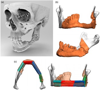
ORN is a debilitating side effect of radiotherapy that is challenging to treat, and while a definitive consensus on the management of ORN is lacking, the current standard of care for this patient cohort is symptom management. Recognised non-surgical has historically included hyperbaric oxygen therapy and more recently a combination of pentoxifylline, clodronate and vitamin E and long-term antimicrobial therapy. Reference Fan, Kim, Cho, Eo, Lee and Woo7 Response to medical management is variable and unpredictable. In refractory cases, where symptoms become unmanageable, the gold standard of care is mandibular resection and reconstruction such as a fibula osseo-fascio-cutaneous free flap. Reference Hirsch, Bell, Dierks, Potter and Potter8 Radiological assessment of high-resolution CT scans of the maxillofacial skeleton and fibula guides the resection and reconstruction (Figure 1). Locally, this is performed using a patient-specific implant system designed in conjunction with manufacturers specialising in implants and implant systems for craniomaxillofacial surgery (KLS Martin GmbH & Co, Tuttlingen, Germany). Additive manufacturing (3D printing) techniques are then used to produce patient-specific cutting guides and titanium alloy fixation devices to ensure an accurate execution of the surgical plan (Figure 1c). This computer-based virtual surgical planning approach has become the gold standard of care in mandible reconstruction for both malignant and benign pathologies. This process infers significant benefits over the traditional ‘free-hand’, ‘on-table’ approach in terms of accuracy, reducing surgical time and post-operative complications. Reference Levine, Patel, Saadeh and Hirsch9,Reference Ma, Van Dessel and Shujaat10 Unfortunately, this approach has not been universally adopted for osteoradionecrosis surgery as surgeons struggle to commit to a surgical plan without the flexibility to alter it based on the immediate intra-operative findings. Placement of a resection through areas of mandible exposed to high radiotherapy dose increases the risk of surgical failure. The additional information provided by merging radiotherapy dosimetry alongside the pre-surgical planning CT enables a more informed decision regarding the extent of the surgical excision. Areas of high dose can be avoided and resection margins placed in areas of bone with greater re-modelling potential, reducing the risk of further ORN and surgical failure, minimising the patient distress and healthcare expenditure that ensues.
Virtual surgical planning for resection of the mandible. (a) High definition CT is used to create a rendered image of the maxillofacial skeleton. (b) Radiological and clinical assessments guide the surgical planning. (c) Bone sections (in teal, red blue and green) taken from the patient’s fibula, guided by 3D-printed cutting guides form the reconstructed section replacing the resected mandible.

The exact relationship between radiotherapy dose and ORN in the mandible is thought to be complex and as yet still not perfectly defined, although some evidence suggests a trend between doses greater than 50Gy and the onset of ORN exists, with areas exposed to doses lower than 50Gy reported to be at significantly lower risk of ORN. Reference Thorn, Hansen, Specht and Bastholt1,Reference Aarup-Kristensen, Hansen, Forner, Brink, Erikson and Johansen6,Reference Fan, Kim, Cho, Eo, Lee and Woo7,Reference Tsai, Hofstede and Sturgis11
We present our experience of a novel approach to pre-operative planning for these complex cases. By incorporating radiotherapy dose registered and deformed onto a high-resolution diagnostic CT, volumes representing high risk of ORN can be incorporated into the 3D surgical design process. Importantly, areas that received low doses of radiotherapy can also be visualised to ensure that osteotomy sites and osteosynthesis plates are located in areas of mandible that received low doses of radiotherapy. To date, this is the first reported implementation of the 3D dose guided surgery (DGS) technique.
Materials and Methods
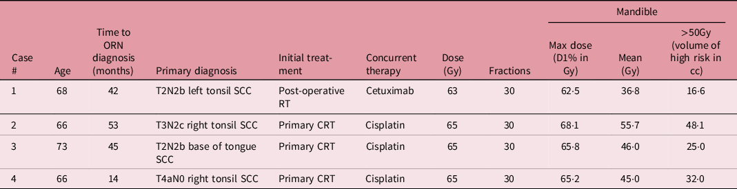
Four patients have undergone DGS for mandibular ORN between 2019 and 2021; the patients originally presented with head and neck cancer and were treated with radical radiotherapy (Table 1). The workflow was developed using a combination of the radiotherapy treatment planning system RayStation (RaySearch AB, Stockholm, Sweden) and the 3D surgical planning software IPS (KLS Martin GmbH & Co, Tuttlingen, Germany).
Patient baseline demographics with salient radiotherapy prescription and dosimetric parameters

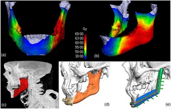
Patients presenting with symptoms of mandibular ORN are given a high-resolution CT covering the maxillofacial skeleton and fibula. In traditional 3D surgery, this high definition CT is rendered to aid visualisation osteoradionecrotic regions (Figure 1). In DGS, this diagnostic CT is deformably registered with the radiotherapy treatment planning CT. Using the resultant deformation matrix, the planned dose distribution is then propagated to the diagnostic CT creating the initial visualisation of planned radiotherapy dose aiding in the initial surgical design discussions. Outlining the mandible on the diagnostic CT in the treatment planning system then allows planned dose to be easily visualised on the structure’s surface (Figure 2a and b).
Stages of the dose guided surgery process: delivered radiotherapy dose is deformed onto the mandible surface as defined by the diagnostic CT scan (a) and (b). The isodose line representing high dose and high risk is segmented, displayed in red (c) to guide the surgical design (d) and (e).

The planned dose levels are then converted into 3D structures (STereoLithographic file or stl files) compatible with the 3D design application used by the surgical design company (Figure 2c). These structures, representing the planned radiotherapy dose to the mandible, guide the 3D surgical design by superimposing the regions onto the maxillofacial skeleton (Figure 2d) enabling the surgeon, in conjunction with the surgical design company, to remotely plan the surgery, locating fixation devices in regions of low radiotherapy dose and low risk, and resecting volumes at highest risk of ORN (Figure 2e).
Results
Visualising actual planned radiotherapy dose assists surgical planning by enabling the visualisation of areas of mandible that received high dose and at high risk of ORN to be visualised guiding resection design.
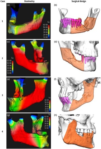
All patients were treated in the same number of fractions (30); therefore, physical dose was used in the estimation of risk. Displaying areas that received a range of physical radiotherapy doses also helps in guiding the location of fixation devices into areas of bone that received <40 Gy and at lower risk of ORN. The uncertainty associated with chemotherapy enhancement to normal tissue radiation was ignored since it is difficult to quantify and a poorly understood affect. This approach as a decision support tool increases the likelihood that areas of the mandible receiving high-dose radiotherapy, and at increased risk of osteoradionecrosis, are included in the resected volume, thereby minimising the risk of failure. The dosimetry and surgical design displaying the resected volume for all four cases are displayed in Figure 3. Resected volumes may extend beyond the high-dose/high-risk volumes since areas that present clinically or radiologically will be resected, and cut planes may also be surgical design purposes (case 3—not practical to leave a small portion of mandibular condyle remaining although it received a low dose). There are also situations when high dose remains and is managed conservatively.
Dosimetry and surgical designs for the 4 cases performed utilising dose guided surgery. Visualising and exporting the areas of mandible that received high dose and at increased risk of mandible guides the resection and ensure fixation devices can be located in areas of low risk having received low dose.

Visualising the dose and aiding the design process not only increases the chance of including high volumes but also embeds an efficiency, accuracy and objectivity in the process, reducing variations associated with subjective clinical and radiological assessments. In this small cohort of patients with short follow-up (6–13months), post-operative imaging suggests good prognosis, and all have evidence of bony healing with no suggestion of malunion (Table 2).
Cohort follow-up details

Discussion
Visualising the planned dose assists surgical planning by informing the surgeon about the volumes of mandible that received high-dose, guiding resection. Despite the lack of strict relationships between radiotherapy dose planned and development of ORN, viewing areas at risk of developing ORN in conjunction with other diagnostic information assists surgical planning.
From a technical perspective, we have developed a robust, reliable and accurate process that can be applied to all patients where mandibular resection and reconstruction are indicated due to osteoradionecrosis. As a novel technique, there have been no large-scale randomised controlled trials to evidence it. This approach does have tangible advantages; information known to be associated with the risk of ORN can be taken into account when planning a resection even when clinically and radiologically absent. Increasing the likelihood of mandible having receiving high dose, at increased risk of osteoradionecrosis, is included in the resected volume and fixation devices located in regions of low dose. There have been other reports of visualising the 3D clouds of radiotherapy dose to guide resection, Reference Costa, Costa and Netto12,Reference Kraeima, Steenbakkers, Spijkervet, Roodenburg and Witjes13 and our approach extends beyond visualisation and allows volumes of high dose to be incorporated directly into the decision-making and surgical planning process.
This study disseminates early yet positive results from a small cohort of patients with limited follow-up. However, DGS has changed local practice, representing a perceived enhancement in the standard of care for patient presenting with mandibular ORN.
Work formally quantifying the change in practice through a full retrospective analysis of volumes resected using the radiological approach compared with DGS is ongoing. The failure rates for these 2 cohorts of patients along with the dose distributions will supplement the discussion on what doses correlate with osteoradionecrosis and hopefully help generate more meaningful relationships.
DSG is not limited to mandibular resections. This approach is permeating into other disciplines guiding decision-making for patients undergoing surgery following radiotherapy, locating cochlear implants, performing maxilla reconstructions, sitting dental implants and assisting in identifying areas of good bone on which to locate fixation devices when stabilising the pelvis.
Conclusion
To our knowledge, this is the first demonstration of planned radiotherapy dose representing volumes at low- and high-risk osteoradionecrosis being registered, deformed to current anatomy and employed directly in the 3D surgical design process for ORN of the mandible. DGS employed in the management of patients presenting with osteonecrotic disease of the mandible is likely to have benefits in terms of minimising failed surgery or incorrectly located fixation devices. This approach appears promising; the authors anticipate DGS becoming the standard of care for refractory cases of osteoradionecrosis, where symptoms are medically unmanageable.
Declaration of Interests
None.
Ethics Statement
Ethical approval was not required for this study. Written informed consent was obtained from patients for the publication of images.