INTRODUCTION
Haemonchus contortus is a gastrointestinal parasitic nematode that infects sheep and goats, causing significant economic loss to the industry worldwide. The nematode occurs in 2 developmental stages in its life cycle: free-living and parasitic stages. It is infective in its L3 stage in an appropriate host. The worms have to adapt to the temperature shift and low-oxygen pressure as well as the immune responses in the infected host. Therefore, they must produce numerous molecules as survival strategies against these adverse changes (Bruschi, Reference Bruschi2002; Dzik, Reference Dzik2006). Better understanding of the roles of these molecules would help elucidate survival strategies of the parasites in the host and identify possible targets for vaccine development.
It has been suggested that the infective-L3 larvae of parasitic nematodes are analogous to dauer larvae of Caenorhabditis elegans, the dormant survival stage that are also resistant to harsh conditions. In C. elegans, dauer larval development is regulated through the TGF-β pathway (Birnby et al. Reference Birnby, Link, Vowels, Tian, Colacurcio and Thomas2000), cGMP pathway (Ren et al. Reference Ren, Lim, Johnsen, Albert, Pilgrim and Riddle1996) and insulin-like signalling pathway (Kimura et al. Reference Kimura, Tissenbaum, Liu and Ruvkun1997). Interestingly, many genes of these pathways have also been identified in parasitic nematodes. Gomez-Escobar et al. (Reference Gomez-Escobar, Gregory and Maizels2000) discovered the first nematode, likely a daf-7 (the TGF-β-like ligand) homologue, in the filarial nematode Brugia malayi. Brand et al. (Reference Brand, Varghese, Majewski and Hawdon2005) then demonstrated that the likely daf-7 homologue was expressed in the infective L3s of Ancylostoma caninum. In addition, Massey et al. (Reference Massey, Bhopale, Li, Castelletto and Lok2006) showed that Strongyloides stercoralis FKTF-1b, the fork head transcription factor, could almost fully rescue loss-of-function to a daf-16 mutant of C. elegans. A more recent study by Hu et al. (Reference Hu, Lok, Ranjit, Massey, Sternberg and Gasser2010) shows that a daf-16.2 from H. contortus could also restore daf-16 function to a C. elegans strain. These studies suggest that parasitic nematodes exploit similar regulatory mechanisms to C. elegans to ensure their survival. In the dauer stage of C. elegans, expression levels of Hsp70 and Hsp90 are up-regulated (Cherkasova et al. Reference Cherkasova, Ayyadevara, Egilmez and Reis2000), and Hsp70 has been shown to be part of the network of proteins that regulate longevity (Jones et al. Reference Jones, Riddle, Pouzyrev, Velculescu, Hillier, Eddy, Stricklin, Baillie, Waterston and Marra2001). Interestingly, H. contortus Hsp90 has been found to partially rescue the daf-21 mutant of C. elegans (Gillan et al. Reference Gillan, Maitland, McCormack, Nik Him and Devaney2009), suggesting similar functions of HSPs in parasitic nematodes and C. elegans.
Heat shock proteins (HSPs) belong to the family of chaperone proteins whose expression is induced by environment changes and different forms of stress (Li and Srivastava, Reference Li and Srivastava2004; Wieten et al. Reference Wieten, Broere, van der Zee, Koerkamp, Wagenaar and van Eden2007). These proteins are highly conserved and have been grouped into different families based on their molecular weights, such as HSP100, HSP90, HSP70, HSP60 and the small HSP12–40 family (Lindquist, Reference Lindquist1986). Recently, HSPs have been found to play important roles in antigen presentation, activation of lymphocytes, macrophages and dendritic cells (APCs) (Li et al. Reference Li, Menoret and Srivastava2002; Wallin et al. Reference Wallin, Lundqvist, More, von Bonin, Kiessling and Ljunggren2002; Tsan and Gao, Reference Tsan and Gao2004; Calderwood et al. Reference Calderwood, Mambula, Gray and Theriault2007; Pockley et al. Reference Pockley, Muthana and Calderwood2008). Hsp70 is the most conserved HSP present in all organisms that is involved in protein folding and several other celluar functions. Hsp70 is an immunodominant antigen during infections caused by a number of pathogens (Yang et al. Reference Yang, Tan-ariya, Sharma and Kilejian1987; Snutch et al. Reference Snutch, Heschl and Baillie1988; Rothstein et al. Reference Rothstein, Higashi, Yates and Rajan1989; Vayssier et al. Reference Vayssier, Le-Guerhier, Fabien, Philippe, Vallet, Ortega-Pierres, Soule, Perret, Liu, Vega-Lopez and Boireau1999; Kanamura et al. Reference Kanamura, Hancock, Rodrigues and Damian2002; Mohamed et al. Reference Mohamed, Aosai, Chen, Mun, Norose, Belal, Piao and Yano2003; Ravi et al. Reference Ravi, Kubofcik, Bandopathyaya, Geetha, Narayanan, Nutman and Kaliraja2004; Song et al. Reference Song, Song, Na, Kim, Kwon, Park, Pak, Im and Shin2007).
Several HSP genes such as hsp90, hsp20 and hsp10 have recently been identified in parasitic nematodes (Dadna et al. Reference Dadna, Pauline, Keith, Mrinal, Paul, Lawrie, Marzena and Susan2003; Devaney et al. Reference Devaney, O'Neill, Harnett, Whitesell and Kinnaird2005; De Luca et al. Reference De Luca, Di Vito, Fanelli, Reyes, Greco and De Giorgi2009; Gillan et al. Reference Gillan, Maitland, McCormack, Nik Him and Devaney2009; Tazir et al. Reference Tazir, Steisslinger, Soblik, Younis, Beckmann, Grevelding, Steen, Brattig and Erttmann2009). The hsp70 has been found in Brugia pahangi (Selkirk et al. Reference Selkirk, Denham, Partono and Maizels1989), Brugia malayi (Rothstein and Rajan, Reference Rothstein and Rajan1991), Wuchereria bancrofti (Ravi et al. Reference Ravi, Kubofcik, Bandopathyaya, Geetha, Narayanan, Nutman and Kaliraja2004), Trichinella britori (Vayssier et al. Reference Vayssier, Le-Guerhier, Fabien, Philippe, Vallet, Ortega-Pierres, Soule, Perret, Liu, Vega-Lopez and Boireau1999), Strongyloides venezuelensis (Tsuji et al. Reference Tsuji, Ohta and Fujisaki1997) and Parastrongyloides trichosuri (Newton-Howes et al. Reference Newton-Howes, Heath, Shoemaker and Grant2006). Promoters of the hsp70 gene family have been used as reporter gene fusions in transgenic experiments in Drosophila (Hunt et al. Reference Hunt, Wilkins, Stokes, Daggard and Frankham1992), Lucilia cuprina (Heinrich et al. Reference Heinrich, Li, Henry, Haack, Stringfellow, Heath and Scott2002), Brugia malayi (Shu et al. Reference Shu, Katholi, Higazi and Unnasch2003) and Parastrongyloides trichosuri (Grant et al. Reference Grant, Skinner, Newton-Howes, Grant, Shuttleworth, Heath and Shoemaker2006). So far, little is known of the molecular characteristics of H. contortus hsp70. In the present study, the genomic DNA covering the coding region of hsp70 was identified from H. contortus and the function of the recombinant HSP70 was evaluated in C. elegans. The 5′-flanking region of hsp70 was also examined for its promoter activity by heterologous expression in C. elegans.
MATERIALS AND METHODS
Parasite strain
The eggs of H. contortus ZJ strain were isolated from the feces of infected sheep. The L3 larvae were collected after 7 days of incubation at 28 °C. Exsheathment of L3 worms and culture to L4 were based on the methods described by Rothwell and Sangster (Reference Rothwell and Sangster1993). Adult H. contortus ZJ strain were collected from sheep abomasums, and males and females were separated. All worms were stored in liquid nitrogen until use.
Amplification of the full-length genomic DNA containing the hsp70 gene of H. contortus
Adult worms of H. contortus were homogenized in an ice bath. The genomic DNA was prepared using a genomic DNA extraction kit (Takara Biotechnology Co., Ltd) and then amplified by PCR employing the primers designed based on the available cDNA sequences (Accession no. BF059866) (HSP70-1F: 5′-CCAGATCCATGATATCGTTC-3′; HSP70-1R: 5′-GTCAGCCGAAATCTTGTCTT-3′) using LA Taq (Takara Biotechnology Co., Ltd). The PCR amplification conditions were as follows: 1 min at 94 °C; 45 s at 94 °C, 45 s at 54·8 °C, and 2 min at 72 °C for 30 cycles; and 10 min at 72 °C for final extension.
PCR products were cloned into the pMD18-T vector and sequenced. The full-length genomic DNA of hsp70 from H. contortus ZJ strain was obtained by a genome walking kit (Takara, China) employing primers designed based on the genomic sequence acquired as above. The primers used to amplify the 3′-flanking region were: SP3: 5′-AGAACTCGCCTGAGAGCTATGCCTT-3′; SP2:5′-CTAATTCCAGGATGAAATCGAGCGG-3′; SP1:5′-GCCGCCTGTCTAAGGTTTGTAATCT-3′; and the primers used to amplify the 5′-flanking region were: SP3-1:5′-CGCCACATCAAGAAGGAGCAGA-3′; SP2-1:5′-GTCTTCGTTGGAATGGTGGTGTT-3′; SP1-1:5′-TCAATTTGCGGTACGCCTCTCG-3′. The cycling conditions for the primary PCR with primers AP1 and SP1/SP1-1 were: 1 min at 94 °C, 1 min at 98 °C, 30 s at 94 °C, 1 min at 65 °C, 2 min at 72 °C for 5 cycles; 30 s at 94 °C, 3 min at 25 °C, 2 min at 72 °C, 30 s at 94 °C, 1 min at 65 °C, 2 min at 72 °C, 30 s at 94 °C, 1 min at 44 °C, 2 min at 72 °C for 15 cycles; and 10 min at 72 °C for the final extension. The second round PCR was carried out with primers AP1 and SP2/SP2-1 using 1 μL of the product of the first round of PCR as the template. Amplification conditions were: 30 s at 94 °C, 1 min at 65 °C, 2 min at 72 °C, 30 s at 94 °C, 1 min at 44 °C, and 2 min at 72 °C for 15 cycles; and 10 min at 72 °C for the final extension. The third round PCR was carried out using primers AP1 and SP3/SP3-1 with 1 μL of the product from the second round PCR as the template. Amplification conditions were the same as for the second round PCR. The PCR products were cloned into pMD18-T vector and sequenced.
The second genome walking was performed to amplify the 5′-flanking sequence using the primers SP3-2:5′-GGCAGTGCTGTCGCAATGGAAAT-3′; SP2-2: 5′-CCCTGTGAATCACGGTTTGGGTACT-3′ and SP1-2: 5′-AGCAGTTTTTACCAATGGATGTGAC-3′. The procedures were the same as above.
Characterization of the Hc-hsp70 gene
The genomic DNA sequence of approximately 5 kb was obtained by genome walking and analysed by BLAST (http://www.ncbi.nlm.nih.gov/BLAST). PCR primers (B1: 5′-ATGACGAAAGCAAACGCAG-3′; B2: 5′-TTAGTCGACTTCTTCGATGG-3′) for amplification of the predicted coding sequence of H. contortus hsp70 were designed according to the alignment of the amino acid sequence with those of other parasitic nematodes and C. elegans. Total RNA was prepared from adult worms of H. contortus using Trizol reagent (Invitrogen). The first strand cDNA was obtained using the M-MLV RTase cDNA Synthesis Kit (Takara, China) and then used for amplification of Hc-hsp70. The amplification conditions were: 1 min at 94 °C; 45 s at 94 °C, 45 s at 55·5 °C, and 2 min at 72 °C for 30 cycles; 10 min at 72 °C for final extension. PCR products were cloned into pMD18-T and then sequenced.
Bioinformatic and phylogenetic analyses
The nucleotide sequence of the Hc-hsp70 gene from H. contortus ZJ was compared with those in non-redundant databases (GenBank) using the BLAST at NCBI (http://www.ncbi.nlm.nih.gov/BLAST), with the parasite genome database (http://www.ebi.ac.uk/Tools/sss/wublast/parasites.html), the Sanger Centre (www.sanger.ac.uk/Projects/Celegans/), and the genomic sequence data for H. contortus available at the Wellcome Trust Sanger Institute (http://www.sanger.ac.uk/Projects/H_contortus/) to verify the identity of the gene sequenced. The conceptual translation of cDNA into amino acid sequence was performed using the selection ‘translate’ available at http://bioinformatics.org/sms/. Protein motifs were identified by scanning the databases PROSITE (www.expasy.ch/tools/scnpsit1.html) (Bairoch, Reference Bairoch1993) and Pfam (www.sanger.ac.uk/Software/Pfam/) (Bateman et al. Reference Bateman, Birney, Durbin, Eddy, Howe and Sonnhammer2000). Amino acid sequence alignments were performed using ClustalW (Thompson et al. Reference Thompson, Higgins and Gibson1994). Phylogenetic analyses were conducted using neighbour-joining (NJ) and maximum likelihood (ML) methods, employing Molecular Evolutionary Genetic Analysis (MEGA) Version5.
Hc-hsp70 promoter reporter construction and transformation of C. elegans
The long PCR was carried out using primers PF: 5′-GCTCTAGATTGTAAGTGGTTCTCCTCCT-3′ and PR: 5′-GGGGTACCCTAGCTGAAACATACAAAT-3′ containing restriction sites for endonucleases Xba I and Sma I (underlined) designed based on the sequences by genome walking. The PCR product was cloned into pPD95.77 upstream of gfp to drive the green fluorescent protein (GFP) expression in the worms. The recombinant plasmid designated as pPD95.77-hsp70 was microinjected into the gonad of young adult wild-type worms (N2) of C. elegans hermaphrodites as described (Mello et al. Reference Mello, Kramer, Stinchcomb and Ambros1991) together with plasmid pRF4 containing a dominant mutant allele of rol-6 gene, each at a final concentration of 50 μg mL−1 in the same mixture. The F2 and subsequent generations with a roll phenotype were selected to examine the expression patterns of GFP using a fluorescent microscope (Olympus IX71).
Expression and purification of H. contortus HSP70
A recombinant plasmid designated as pET28b-hsp70 was constructed for expression of hsp70 fused with a 6-His-tag at the C-terminus by cloning the full-length hsp70 into pET28b using primers A1 (5′-CTAGCTAGCATGACGAAAGCAAACGCAG-3′) and A2 (5′-CCCAAGCTTTTCTTCGATGGTTGGTCC-3′) (Nhe I and Hind III restriction sites underlined). Amplification was performed as follows: 1 min at 94 °C; 45 s at 94 °C, 45 s at 58·6 °C, and 2 min at 72 °C for 30 cycles. The PCR product was purified and restricted by Nhe I and Hind III overnight and then purified, ligated into the pET28b vector and digested with the same enzymes. The ligated mixture was transformed into E. coli (Top10). The clone was verified by PCR and double digested with Nhe I and Hind III. The recombinant protein was expressed in E. coli BL21 (DE3) induced with 1 mM IPTG for 2, 4 or 6 h. The bacterial lysates were solubilized in 8 M urea (pH 8·0) in PBS buffer (100 mM NaH2PO4 10 mM Tris base). The recombinant fusion protein was identified on SDS-PAGE gel and purified by affinity column chromatography using Ni-NTA resin (Sigma Inc., MO, USA). The bound protein was eluted by 400 mM iminazole.
Expression of Hc-hsp70 in C. elegans
A recombinant plasmid pPD49.78/49.83-hsp70 containing Hc-hsp70 ORF and his-tag at C-terminus was constructed using primers A1 and A3 (5′-TTCGAGCTCTCAGTGGTGGTGGTGGTGGT-3′). Primer A3 containing Sac I restriction site (underlined) was designed based on pET28b-hsp70 such that the his-tag sequence could be included when it was used as the template. The PCR product was inserted between the Nhe I and Sac I restriction sites of two heat-inducible expression vectors pPD49.78 and pPD49.83. These plasmids were mixed (designated pPD49.78/49.83-hsp70) with pRF4, each at a final concentration of 50 μg mL−1 and microinjected into the gonad of young adults N2 C. elegans hermaphrodites. Transgenic worms were heat-shocked at 33 °C for 2, 4 and 6 h to induce expression of the fusion proteins.
Western blot analysis
The proteins of transgenic worms were prepared as described previously (Kimura et al. Reference Kimura, Tanaka, Nakamura, Takano and Ohkuma2007). Briefly, worms were collected and washed in M9 buffer (22 mM KH2PO4, 42 mM Na2HPO4, 86 mM NaCl, 1 mM MgSO4), then re-suspended in lysis buffer (0·2 M Tris-HCl at pH 7·5, 0·1 M EDTA, 0·04 M EGTA, 20 mM phenylmethylsulfonyl fluoride, 1 M NaCl and 1% SDS), sonicated 6 times for 25 s each, and boiled for 5 min. The extract was centrifuged at 9000 g for 15 min, and the supernatant was collected and stored at −80 °C until use. Proteins were separated on 10% SDS-PAGE and then blotted onto polyvinylidene fluoride (PVDF) membrane. HSP70 protein was examined using murine anti-His tag antibody (Merck; 1:5000 dilution) and anti-mouse immunoglobulin G (IgG)–alkaline phosphatase conjugate (Serotec; 1:500 dilution) and then visualized using the 4-chloro-1-naphthol liquid substrate (Amresco).
Reverse transcription quantitative PCR analysis
To analyse the transcriptional profiles of hsp70 in different developmental stages of H. contortus, quantitative reverse transcription PCR (qRT-PCR) was carried out using specific primers (Hc-hsp70-F: 5′-GAAGATGAAGGATACAGCAGAAGC-3′ and Hc-hsp70R:5′-TACGAAGAACATTGAGACCAG CG-3′). Total RNA of each sample was extracted using Trizol (Invitrogen). The integrity of RNA samples was examined by detecting dispersed 18S and 28S ribosomal RNA bands on agarose gels. The first-strand cDNA was synthesized using ReverTra Ace-α (Cat. No. FSK-100, TOYOBO) and was then used as the template in subsequent quantitative PCR which was conducted in a volume of 25 μL using the THUNDERBIRD SYBR qPCR Mix (TOYOBO) in ABI 7300 thermal cycler under the following conditions: 50 °C for 2 min and 95 °C for 1 min for the first cycle, followed by 95 °C for 15 s, 60 °C for 15 s and 72 °C for 31 s for 40 cycles. The dissociation curve was generated under the following conditions: 95 °C for 15 s, 60 °C for 1 min, 95 °C for 15 s and 60 °C for 15 s. Each sample was run in triplicate using β-tubulin 8–9 (Accession no. M76493) gene as an internal reference using specific primer pairs (tub-F: 5′-TGTTCCATCACCCAAGGTATCC-3′, and tub-R: 5′-TGACAGACACAAGGTGGTTGAGAT-3′), and the mean threshold cycle (CT) values were used for analysis.
Total RNA of the wild-type N2 strain and transgenic worms at each time-point of heat shock was extracted with Trizol (Invitrogen). qRT-PCR was used to determine the expression levels of Hc-hsp70 and Ce-hsp-1 in transgenic worms using the primer pairs Hc-hsp70F and Hc-hsp70R for Hc-hsp70 as well as Ce-hsp-1F and Ce-hsp-1R for Ce-hsp-1 (Ce-hsp-1F: 5′-TCAAGTTGCCATGCCATGAACCCAC-3′ and Ce-hsp-1R: 5′-TTCTCCGTGTCCCTTCTTG-3′) based on cDNA sequences (F26D10·3). The gene act-1 was used as an internal control (specific primers of act-1 [T04C12.6, 384 bp] of C. elegans as F: 5′-CCCAATCCAAGAGAGGTATC-3′ and R: 5′-TAAGATCACGTCCAGCCAAGTC-3′). Relative quantification was calculated using 2−ΔΔCT method (Livak and Schmittgen, Reference Livak and Schmittgen2001). Statistical analysis was carried out using one-way ANOVA and P values less than 0·05 were considered statistically significant.
RESULTS
Characterization of the hsp70 gene in H. contortus
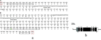
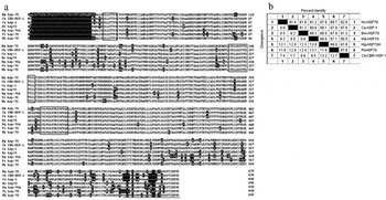
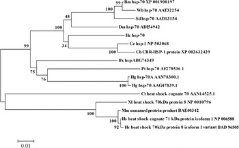
Genome walking generated a genomic DNA fragment of more than 5 kb containing the entire coding sequence of hsp70 of H. contortus (3925 bp). Combination of BLAST search, scaffold assembly and reverse transcript PCR revealed a 1941 bp hsp70 cDNA for H. contortus. A predicted sequence motif KANAVGIDLG, which is conserved among hsp70 family proteins, was seen at the N-terminus and matches well with Parastrongyloides trichosuri and Heterodera glycines between residues 3 and 10. The motifs GGMP and EEVD conserved in all hsp70 family proteins were present in the C-terminus (Fig. 1a). The hsp70 gene of H. contortus contains 7 exons (99 bp, 106 bp, 218 bp, 1005 bp, 111 bp, 135 bp and 267 bp) and 6 introns (55 bp, 65 bp, 87 bp, 69 bp, 78 bp and 74 bp) (Fig. 1b). The entire Hc-hsp70 ORF is 1941 bp encoding 646 aa, with a calculated molecular mass of 70·4 kDa and an isoelectric point of 5·50. The sequence includes 3 HSP70 signatures (9–16, 198–211 and 335–349) (Fig. 2a). The deduced amino acid sequence of H. contortus HSP70 had 92·0%, 91·4%, 91·8%, 91·2%, 88·7% and 87·8% identity with HSP70 protein of C. briggsae, C. elegans, Brugia malayi, Wuchereria bancrofti, Parastrongyloides trichosuri and Heterodera glycines, respectively (Fig. 2b). The hsp70-based phylogenetic analysis shows that H. contortus is closely related to Caenorhabditis. Thus, Hc-hsp70 is likely to be the orthologue of Ce-hsp-1, also known as hsp70A (Fig. 3).
(a) Sequence analysis of N-terminal and C-terminal of Hc-hsp70 gene by alignment. A. The N-terminal. B. The C-terminal. The conserved amino acids are shown in the box. The first and the last introns are underlined. The start codon and the stop codon are shown in the red box. The amino acid A shown in the green box represents the difference with other species. (b) Genomic structure of hsp70 from Haemonchus contortus. Exons are numbered and indicated as boxes (grey box 5′-UTR; blank box, 3′-UTR). The 5-pointed star indicates translation start codon ATG, and the 4-pointed star indicates translation stop codon TAA.

Alignment of the deduced amino acid sequence of Haemonchus contortus HSP70 with corresponding sequences of other parasitic nematodes. (a) Amino acid sequences are aligned using Clustal-W and BoxShade. Inconsistent amino acids are shaded black and the black boxes represent 3 HSP70 universal signatures. The GGMP tetrapeptide and the conserved sequence EEVD in the end of the protein are underlined. (b) The percentage identity was listed among nematodes. Ce, Caenorhabditis elegans; Hc, Haemonchus contortus; Bm, Brugia malayi; Wb, Wuchereria bancrofti; Sd, Setaria digitata; Pt, Parastrongyloides trichosuri; Hg, Heterodera glycines.

Neighbour-joining tree of amino acid sequences of HSP70 from a range of species. Abbreviation for species names are as follows: Ce, Caenorhabditis elegans; Hc, Haemonchus contortus; Bm, Brugia malayi; Wb, Wuchereria bancrofti; Sd, Setaria digitata; Dm, Dracunculus medinensis; Cb, Caenorhabditis briggsae; Pt, Parastrongyloides trichosuri; Hg, Heterodera glycines; Ct, Chironomus tentans; Xl, Xenopus laevis; Mm, Mus musculus; Hs, Homo sapiens; Bx, Bursaphelenchus xylophilus. Numbers above and below the branches represent the bootstrap values obtained using the neighbour-joining and maximum likelihood methods, respectively; and values ⩾ 70 are indicated.

Sequence analysis upstream of the H. contortus hsp70 gene
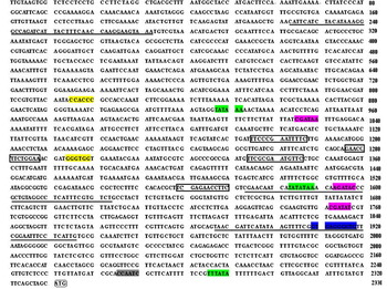
The 5′-flanking region of H. contortus hsp70 was analysed using promoter prediction algorithms based on the data obtained from human or Drosophila melanogaster genes (www.fruitfly.org/cgi-bin/seq-tools/promoter.pl). Three putative core promoter regions were identified at −395 to −444, −709 to −758 and −2059 to −2108, respectively (Fig. 4). The 5′-flanking region of the hsp70 gene was also analysed for putative transcription factor binding sites using the Transcription Element Search System (TESS). The region also contains the TATA box, CACCC binding factors, GATA binding sites, CAC binding sites and the CCAAT box. In addition, 4 binding sites for particular heat shock factors were found at −761 to −773, −984 to −997, −1043 to −1055 and −1144 to −1156 (Fig. 4).
Analysis of the upstream domain of the Haemonchus contortus HSP70 gene. The putative heat shock factor (HSF) binding sites (TTCNNGAANNTTC/GAANNTTCNNAA) are highlighted by boxes. The main predicted promoter elements are highlighted in different colours. TATA box (green); CACCC binding factor sites (yellow); GATA binding sites (pink); CAC binding sites (blue); CCAAT box (grey). The putative core promoter domains are underlined. The start codon ATG is highlighted by a rounded rectangle.

GFP expression in C. elegans transformed with a reporter construct
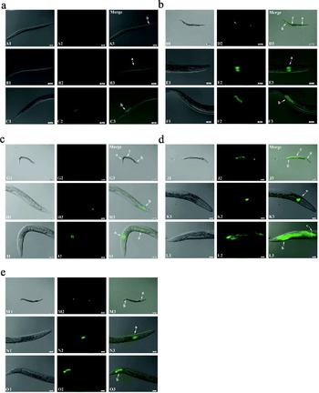
In order to analyse the promoter activity, we constructed a reporter plasmid with gfp fused to the putative promoter and used C. elegans as a heterologous expression system by microinjection. Transgenic lines showing the roller phenotype were selected. GFP was expressed in all larval stages and adults (Fig. 5a–e), although fluorescent intensity varied distinctly among individuals and with developmental stages (Fig. 5d and e). GFP was mainly distributed in the distal and anterior of the intestine in worms at all stages from larvae L1 to adult (arrows a, b in Fig. 5). However, some worms also had GFP in the posterior intestine (arrow c in Fig. 5).
Characterization of expression pattern of the 5′-flanking region GFP fusion. (a) Expression pattern of the promoter GFP fusion in L1 and L2 larvae of Caenorhabditis elegans. A1–A3: the whole view of L1 larvae; B1–B3: the anterior region of L2 larvae; and C1–C3, the posterior region of L2 larvae. (b) Expression pattern of the promoter GFP fusion in L3 larvae. D1–D3 represent the whole view of L3 larvae in lower power lens, E1–E3 represent the zoom head, and F1–F3 show the zoom tail of L3 larvae. (c) Expression pattern of the promoter GFP fusion in L4 larvae. G1–G3 represent the whole view of L4 larvae in lower power lens, H1–H3 demonstrate the zoom tail, and I1–I3 show the zoom head of L4 larvae. (d) Expression pattern of the promoter GFP fusion in Caenorhabditis elegans adults. J1–J3 represent the whole adult, K1–K3 represent the zoom tail, and L1–L3 indicate the zoom head of adult. (e) Characterization of another expression pattern of the promoter GFP fusion in Caenorhabditis elegans adults. M1–M3 represent the whole view of adult; N1–N3 represent the head zoom, and O1–O3 show the zoom tail of adult. The number 1 following the letter represents the worms were observed in bright field, 2 in GFP field, 3 was the merged picture of 1 and 2 by the computer. Arrows a, b, c, represent the front end, distal and back end of the intestine, respectively. The scale bar of D1–D3, G1–G3, J1–J3, M1–M3 = 100 μm, and A1–B3, E1–F3, H1–I3, K1–L3, N1–O3 = 25 μm.

Prokaryotic expression of HSP70
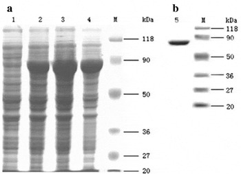
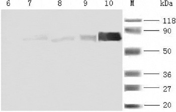
The recombinant Hc-hsp70 was highly expressed in E. coli BL21 harbouring the plasmid pET28b-hsp70 as shown by SDS-PAGE (Fig. 6a). Time-course analysis indicated that optimal expression occurred at 4 h post-induction. A prominent band was seen after purification on an immobilized metal affinity column (Fig. 6b). Western blot analysis showed that the purified protein could be recognized by His monoclonal antibody (Fig. 7 lane 10).
SDS-PAGE and purification of Haemonchus contortus HSP70 protein expressed in E. coli. (a) Lane 1: lysate of cells with plasmid pET28b-hsp70 before induced; lane 2: lysate of cells with plasmid pET28b-hsp70 induced 2 h; lane 3: lysate of cells with plasmid pET28b-hsp70 induced for 4 h; lane 4: lysate of cells with plasmid pET28b-hsp70 induced for 6 h. (b) Lane 5: purification of HSP70 protein by affinity column chromatography.

Western blot of Haemonchus contortus HSP70 expressed in E. coli and C. elegans. Lane 6 represents proteins from N2 worms with anti-6-His antibody; lanes 7–9 represent proteins from transgenic worms induced for 2, 4, 6 h with anti-6-His antibody, respectively; lane 10 represents purified pET28b-hsp70 protein with anti-6-His antibody.

Temporal expression of Hc-hsp70 throughout the life cycle of H. contortus
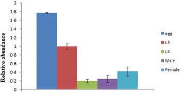
The transcription level of Hc-hsp70 was determined at different stages throughout the life cycle of H. contortus by qRT-PCR using the tubulin gene as internal control for normalization. Hc-hsp70 was expressed at readily detectable levels in eggs and at L3, L4, male and female stages. Highest expression was seen in eggs. Gender difference in the gene expression level of Hc-hsp70 was examined in adult worms. Since eggs expressed high levels of Hc-hsp70, the female worms without intrauterine eggs were selected. The results showed that there was no difference between male and female adults in the expression level (Fig. 8).
Transcriptional profile of of Hc-hsp70 in different developmental stages of Haemonchus contortus (eggs, L3 and L4) and genders, determined by qRT-PCR analysis. Data shown are mean values (±s.e.m.) derived from 3 replicates in repeat experiments. All gene expression levels were normalized to those of β-tublin gene. P-values (P < 0·01 are indicated by **, 0·01<P < 0·05 are indicated by *) were calculated by Dunnett's test.

Expression of Hc-hsp70 in transgenic worms
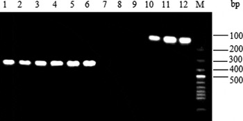
To examine the heat responsiveness of Hc-hsp70 in C. elegans, RT-PCR was used to detect the Hc-hsp70 transcript in C. elegans that were microinjected with pPD49·78/49·83-hsp70 and heat-stressed for 2, 4 or 6 h firstly. The expected 143 bp band was exclusively observed in transgenic worms (Fig. 9 lanes 10–12) while there was no product of Hc-hsp70 transcript in N2 worms (Fig. 9 lanes 7–9). Expression of Hc-HSP70 protein in heat-stressed transgenic C. elegans was further confirmed by Western blotting, as shown by specific recognition by anti-His monoclonal antibody. There was no Hc-hsp70 in N2 worms (Fig. 7).
Molecular analyses of the expression of Hc-hsp70-his in Caenorhabditis elegans after heat shock. Reverse transcriptase (RT)-PCR was performed on wild type C. elegans (N2) treated with heat shock 2, 4, 6 h (lanes 1–3, lanes 7–9), wild type worms transfected with Hc-hsp70 with his-tag (lanes 4–6, lanes 10–12) with the same treatment. Lanes 1–6 represent the positive control using the specific primers for C. elegans actin-1. Lanes 7–12 represent the PCR products using primers for Hc-hsp70. 1% agarose gel was chosen to resolve the PCR products. M, 100 bp ladder.

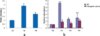
We also determined the expression pattern of Hc-hsp70 mRNA after heat shock in transgenic worms using qRT-PCR. As shown in Fig. 10a, expression of Hc-hsp70 was significantly up-regulated in the transgenic worms after 4 h and 6 h induction as compared with those at 2 h induction and the peak expression was observed after 4 h induction. These results suggest that Hc-hsp70 can be expressed in C. elegans in response to heat shock.
(a) Expression level of Hc-hsp70 transcript in C. elegans after heat shock was analysed by qRT-PCR. Data representing expression profiles are mean values derived from 3 independent experiments and each sample was carried out in triplicate. Relative expression values were normalized to actin-1 gene which was used as an endogenous control. P-values (P < 0·01 are indicated by **, 0·01<P < 0·05 are indicated by *) were calculated by Dunnett's test. (b) Relative mRNA level hsp-1 of C. elegans in wild type and transgenic worms after heat shock. Relative expression level of hsp-1 gene was determined by qRT-PCR to examine the changes between the wild type and transgenic worms before and after heat shock. Data representing expression profiles are mean values derived from 3 independent experiments and each sample was carried out in triplicate. Relative expression values were normalized to actin-1 gene which was used as an endogenous control. P-values (P < 0·01 are indicated by **, 0·01<P < 0·05 are indicated by *) were calculated by Dunnett's test.

Heat induction of Ce-hsp-1 in transgenic worms
To examine the effects of Hc-hsp70 on expression of hsp-1 of C. elegans, a fragment of hsp-1 mRNA between 171 bp and 579 bp (Accession no. Z80223.1) was amplified to examine the dynamics of hsp-1 expression upon heat shock in transgenic worms. The qRT-PCR showed that there was enhanced expression of hsp-1 in both the heat-shocked transgenic worms and the N2 worms. Expression of hsp-1 increased about 7-, 2·6- and 3-fold in N2 worms, respectively, after 2, 4 and 6 h heat shock (Fig. 10b). In transgenic worms, a moderate increase of expression of Ce-hsp-1 was also observed, about 1·8-, 1·3- and 2·1-fold higher after 2, 4 and 6 h heat shock than at 0 h. However, the overall increase of Ce-hsp-1 expression in transgenic worms was lower than that in N2 worms (Fig. 10b).
DISCUSSION
The present study revealed the entire encoding region of Hc-hsp70. The deduced amino acid sequence showed high similarity to the corresponding sequences of hsp70 of other organisms, especially nematodes. HSPs induced by heat shock may be involved in the process of parasite invasion (Watson, Reference Watson1990; Maresca and Carratfi, Reference Maresca and Carratú1992; Requena et al. Reference Requena, Jimenez-Ruiz, Soto, Assiego, Santaren, Lopez, Patarroyo and Alonso1992) since the nematodes must adapt to the temperature shift from the external environment to the mammalian host. Our results showed that free-living infective larvae also expressed HSP70, indicating involvement of HSP70 in larvae survival under adverse conditions.
So far, 3 subfamilies of the HSP70 family GRP170/LHSVP, HSP110 and Dnak have been identified. The HSP70 of H. contortus belongs to the Dnak subfamily characterized by a specific motif-DLGTT-S-V. Two repeats of putative conserved tetrapeptide GGMP (GGMP × 2) were located at the C-terminal region. Similar motifs have been identified in certain HSP70 s from various organisms, including Brugia (Selkirk et al. Reference Selkirk, Denham, Partono and Maizels1989), rat (O'Malley et al. Reference O'Malley, Mauron, Barchas and Kedes1985), human (Dworniczak and Mirault, Reference Dworniczak and Mirault1987), the nematodes (Parastrongyloides trichusuri (Tsuji et al. Reference Tsuji, Ohta and Fujisaki1997), Wuchereria bancrofti (Ravi et al. Reference Ravi, Kubofcik, Bandopathyaya, Geetha, Narayanan, Nutman and Kaliraja2004)). Some of these repetitive peptides have been proven to be antigenic (Engman et al. Reference Engman, Dragon and Donelson1990). The HSP70 of H. contortus also contained the conserved sequence EEVD at the C-terminus which is recognizable by the TPR domains of Hop, an adapter protein involved in the ordered assembly of HSP70-HSP90 multichaperone complexes (Scheufler et al. Reference Scheufler, Brinker, Bourenkov, Pegoraro, Moroder, Bartunik, Hartl and Moarefi2000). In C. elegans, Hc-hsp70 was expressed throughout the nematode's life cycle, suggesting that it could be indispensable for cell maintenance at all stages of development.
Hc-hsp70 has 91·2% similarity to the gene encoding HSP70 protein in C. elegans that is the orthologue of the protein encoded by the hsp-1 (syn, hsp-70A) gene (Snutch et al. Reference Snutch, Heschl and Baillie1988). The Hc-hsp70 is constitutively expressed in free-living and parasitic stages of the life cycle of H. contortus and hsp-1 gene is expressed throughout the lifespan of C. elgans. In this study, we showed that the transgenic C. elegans expressed Hc-hsp70 transcript in response to heat shock. We found that the hsp-1 transcript was up-regulated significantly less after heat shock in transgenic worms as compared with that of N2 worms. Taken together, our results suggest that the two genes are likely to have a similar function. Down-regulation of hsp-1 via RNA interference resulted in a small reduction in lifespan of an age-1 mutant (Morley and Morimoto, Reference Morley and Morimoto2004), indicating that hsp-1 plays some role in regulating longevity. Similarly, we speculate that the hsp70 might also be involved in the development of H. contortus.
On the other hand, growing evidence supports the idea that HSP70s are antigen candidates against parasites. Antibodies to HSP70 of Trichinella britovi were detected in Trichinella-infected rabbit serum (Vayssier et al. Reference Vayssier, Le-Guerhier, Fabien, Philippe, Vallet, Ortega-Pierres, Soule, Perret, Liu, Vega-Lopez and Boireau1999). Mice vaccinated with Trichinella spiralis HSP70 exhibited a decreased number of muscle larvae when they were challenged with T. spiralis larvae (Wang et al. Reference Wang, Zhu, Yang, Yang, Gu, Wei, Hao, Boireau and Cui2009). It was also found that the recombinant Leishmania infantum HSP70 induced Th1 type immune response in canines against L. infantum infection when vaccinated along with recombinant paraflagellar rod protein (PFR)-2 and kinetoplastida membrane protein (KMP)–II (Carrillo et al. Reference Carrillo, Crusat, Nieto, Chicharro, Thomas, Martínez, Valladares, Cañavate, Requena, López, Alvar and Moreno2008).
The ∼2329 bp of 5′-flanking sequence of hsp70 gene contained the TATA box sequence, CCAAT box sequence, Heat Shock Factor (HSF) binding region and GATA factor binding sites. B. malayi hsp-70 retained roughly half its activity when the TATA box at position −210 to −206 or core promoter at −227 to −171 was deleted (Shu et al. Reference Shu, Katholi, Higazi and Unnasch2003). In C. elegans, GATA region in the gut-specific esterase gene (ges-1) has been shown to act as both a gut-specific enhancer and the site of the pharynx/tail repression. The GATA region of the ges-1 gene can switch and coordinate expression between gut and pharynx (Egan et al. Reference Egan, Chung, Allen, Heschl, Van Buskirk and McGhee1995; Marshall and McGhee, Reference Marshall and McGhee2001). The HSF binding region recognizes TTCNNGAANNTTC elements, which can mediate the expression of heat-shock genes in response to elevated temperatures (Sorger and Pelham, Reference Sorger and Pelham1988; Wiederrecht et al. Reference Wiederrecht, Seto and Parker1988) or to other conditions (Tamai et al. Reference Tamai, Liu, Silar, Sosinowski and Thiele1994). Morgan et al. (Reference Morgan, Williams, Morimoto, Greene, Kingston and Tjian1987) demonstrated that the CCAAT-box-binding transcription factor and heat-shock transcription factor interact with a human hsp70 gene promoter. Moreover, the CCAAT-binding complex has been verified to coordinate the oxidative stress response in eukaryotes (Thon et al. Reference Thon, Abdallah, Hortschansky, Scharf, Eisendle, Haas and Brakhage2010). However, we did not find the transcription regulatory element Heat Shock Associated Site (GGGTGTC, HSAS) in H. contortus, which was identified in the promoters of heat-shock genes in C. elegans (Guha Thakurta et al. Reference Guha Thakurta, Palomar and Stormo2002).
We observed the expression of GFP in the intestine of transgenic C. elegans, mainly in the distal and front-end intestine, in all larval stages and adult worms. However, hsp-1 of C. elegans is expressed in the pharynx, anal depressor muscle, body wall muscle excretory cell and in unidentified cells besides the intestine (www.wormbase.org) in both larvae and adults. The different expression pattern may be explained by several aspects. One possibility is that GFP expression is prevented by repressors present in other parts of the worm body (Clark and Chiu, Reference Clark and Chiu2003). Another possibility is that some promoter regulatory elements are needed to drive GFP expression in other locations or some regulatory elements exist in 3′-flanking of the parasite gene, such as promoter of H. contortus AC-2 (Britton et al. Reference Britton, Redmond, Knox, McKerrow and Barry1999). The third possibility is that the spatial expression of parasite promoters in C. elegans correlates with their expression in the parasites as shown by Britton et al. (Reference Britton, Redmond, Knox, McKerrow and Barry1999). The fourth possibility is that unc-54 3′UTR used in C. elegans is permissive for expression in all somatic cells and the 3′UTR can regulate protein expression (Merritt et al. Reference Merritt, Rasoloson, Ko and Seydoux2008). Further work is needed to clarify those possibilities.
Hc-hsp70 transcript could be produced apparently in the transgenic worms, although different levels were observed following heat shock. Moreover, hsp-1 mRNA of C. elegans is significantly depressed with increased expression of Hc-hsp70, suggesting that over-expression of Hc-hsp70 may be detrimental to that of hsp-1, or maybe a similar function was offered by its homologue.
In conclusion, the present study has identified the full genomic region and coding sequence of hsp70 in H. contortus and found that the 5′-flanking region contains the promoter sequence. Furthermore, hsp-1 of C. elegans was depressed as the ectopic expression of Hc-hsp70 was increased in C. elegans. However, the biological significance of hsp70 in H. contortus needs further investigation.
ACKNOWLEDGEMENTS
We would like to thank Dr Yang CL (Institute of Genetics and Developmental Biology, Chinese Academy of Sciences), Dr Wang XC (National Institute of Biological Science, Beijing) and Dr Ren CH (Academy of Military Medical Science, Beijing) for their assistance with the transgenic technique in C. elegans and gifts of the vectors we needed.
FINANCIAL SUPPORT
This project was supported by a grant from the National Natural Science Foundation of China (No. 30771618, 30972174), and the Fundamental Research Funds for the Central Universities.