Introduction
Despite advances in in vitro production (IVP), the efficiency of in vitro maturation (IVM) is still lower than in vivo maturation, which limits its application in assisted reproduction techniques (Gilchrist, Reference Gilchrist2011). Maturation includes all of the events that allow the oocyte to achieve maximum potential development following fertilization. In this sense, maturation is one of the most important phases of IVP embryos because it is during this period that the oocyte acquires the ability to proceed to the next events (Gottardi & Mingoti, Reference Gottardi and Mingoti2009).
Although immature mammalian oocytes resume meiosis after isolation from the antral follicles (Pincus & Enzmann, Reference Pincus and Enzmann1935; Edwards, Reference Edwards1965), cytoplasmic maturation can occur asynchronously with respect to nuclear maturation (Janssenswillen et al., Reference Janssenswillen, Nagy and Van, Steirteghem1995; Huang et al., Reference Huang, Chang, Tsai, Lin, Kung, Wu and Lu1999). The oocyte requires a longer cytoplasmic maturation for the acquisition of greater developmental competence in vitro (Wu et al., Reference Wu, Cheung, Wen and Li2006). Therefore, it was suggested that the inhibition or delay of spontaneous nuclear in vitro maturation might allow for more time to accumulate the molecules important for embryonic development (Bilodeau-Goeseels, Reference Bilodeau-Goeseels2012).
The delay of meiosis can be obtained by incubating cumulus–oophorus complexes (COCs) with inhibitors, and maturation is initiated immediately after the removal of the inhibitors (Gilchrist & Thompson, Reference Gilchrist and Thompson2007). Treatment with agents that increase cyclic adenosine monophosphate (cAMP) confirmed the role of this compound in blocking nuclear maturation (Bilodeau et al., Reference Bilodeau, Fortier and Sirard1993; Gilchrist & Thompson, Reference Gilchrist and Thompson2007). Therefore, forskolin is an effective inhibitor of oocyte nuclear maturation because it stimulates cAMP levels (Dekel et al. Reference Dekel, Aberdam and Sherizly1984; Racowsky, Reference Racowsky1984; Sirard & First, Reference Sirard and First1988; Sirard, Reference Sirard1990; Bilodeau et al. Reference Bilodeau, Fortier and Sirard1993).
In turn, cytoplasmic maturation is characterized by various changes in the shape and location of the cellular organelles. These are closely related to the acquisition of meiotic competence and the ability of the oocyte to be fertilized and undergo embryonic development (Hyttel et al., Reference Hyttel, Greeve and Callesen1989).
Among the organelles, the mitochondria play a key role in cytoplasmic maturation because they are of fundamental importance to oocyte metabolism and are responsible for supplying the energy that is consumed during maturation. Migration of the mitochondria to areas of high energy consumption is crucial for the oocyte and blastomeres of the embryo. So the activity, morphology and distribution of the mitochondria can be used as criteria for evaluating the quality and competence of oocytes (Nagai et al., Reference Nagai, Mabuchi, Hirata, Shoda, Kasai, Yokota, Shitara, Yonekawa and Hoshi2006; Egerszegi et al., Reference Egerszegi, Alm, Ratky, Heleil, Brüssow and Torner2010) as well as being key in the initiation of apoptosis. There is an increase in mitochondrial fluorescence intensity during maturation (Katska-Ksiazkiewicz et al., Reference Katska-Ksiazkiewicz, Alm, Torner, Heleil, Tuchscherer and Rynska2011), and respiration of mitochondria in oocytes is closely related to the competence of embryonic development (Stojkovic et al., Reference Stojkovic, Machado, Stojkovic, Zakhartchenko, Hutzler, Gonçalves and Wolf2001; Torner et al., Reference Torner, Brüssow, Alm, Ratky, Pöhland, Tuchscherer and Kanitz2004).
This study aimed to examine the oocyte meiosis block caused by the addition of different concentrations of forskolin to the IVM medium by evaluating mitochondrial activity and distribution, ultrastructural organization, embryo production, the total number of viable cells and the rate of apoptosis in blastocysts.
Materials and methods
All chemicals were purchased from Sigma (Sigma-Aldrich® Corporation, St. Louis, MO, USA), except for those specified in the text.
Collection of ovaries and in vitro maturation (IVM)
Slaughterhouse-derived bovine Zebu ovaries were collected and carried to the laboratory of the School of Veterinary Medicine and Animal Science, UNESP, Botucatu-SP, Brazil. All follicles measuring 2–8 mm were aspirated. Oocytes (n ~ 1638) with at least three intact layers of cumulus cells and homogenous cytoplasm were selected for IVM. The oocytes were transferred to drops of Tissue Culture Medium 199 (TCM 199®; Gibco 11150–059, Grand Island, NY, USA) supplemented with 10% (v/v) fetal calf serum (FCS; Gibco 12657–029, Grand Island, NY, USA), 0.011 g/ml sodium pyruvate, 1 μg/μl follicle stimulating hormone (FSH; Pluset®, Hertape Calier Saúde Animal, Juatuba, Brazil), 5 μg/μl luteinizing hormone (LH; Lutropin®, Bioniche Co., Belleville, Ontário, Canada) and 16.6 mg/μl amicacin.
The oocytes remained in IVM medium in three different concentrations of forskolin® (F-6886, 0.1 mM, 0.05 mM or 0.025 mM) for 6 h. Then, they were maturated for an additional 18 h in forskolin-free medium for 24 h of maturation. The first time period above was an attempt to block and the second to resume oocyte meiosis (Block/Res). The oocytes were incubated in a humidified atmosphere with 5% CO2 at 38.5°C in an air incubator.
The experimental groups were divided into two additional groups to examine each concentration of forskolin at two time periods: 6 and 24 h.
Evaluation of oocytes nuclear stage
In each experimental group, oocytes were randomly collected at 6 or 24 h of maturation (n = 634). The oocytes were transferred to a 5 μl drop of Hoechst 33342 (concentration: 0.01 mg/ml) on a slide and covered with a cover slip. After 20 min, the nuclear morphology was observed using an inverted microscope with fluorescent ultraviolet (Leica DMIRB), classifying the nucleus into germinal vesicle (GV), germinal vesicle breakdown (GVB), metaphase I (MI), metaphase II (MII) or degenerate or unidentified (D/U) groups (excitation wavelength: 350 nm and emission wavelength: 461 nm).
Evaluation of mitochondrial activity and distribution of oocytes
To assess mitochondria, oocytes (10 per group in eight replicas = 320 oocytes) were stained with MitoTracker® Orange CMTMRos (Invitrogen, Molecular Probes, M7510). The oocytes were washed three times in phosphate-buffered saline (PBS) with 3% bovine serum albumin (BSA) and incubated for 30 min in the same medium containing 280 nM MitoTracker® Orange CMTMRos at 38°C under 5% CO2. After incubation, the oocytes were washed three times with pre-heated PBS without BSA. Then, the oocytes were stained with 2.5 mg/ml Hoechst 33342 in 3:1 (v/v) glycerol/PBS. The evaluation was performed using an inverted Confocal Laser-Leica TCS SP five microscope (excitation wavelength: 554 nm and emission wavelength: 576 nm).
Evaluation of oocytes ultrastructure
We used 5–10 oocytes from each experimental group. The samples were fixed for at least 24 h in a solution of 2.5% (v/v) glutaraldehyde in 0.1 M phosphate buffer, pH = 7.4. After fixation, the oocytes were embedded in 2% (v/v) agar stained with methylene blue to facilitate visualization. After washing in the same buffer, the oocytes were subjected to post-fixation in osmium tetroxide 1% (v/v) for 3 h at 4°C. They were then washed three times with twice-distilled water and dehydrated in a series of acetone solutions (30, 50, 70, 90 and 100%) for 5 min each, repeating the 100% wash concentration three times. The polymerization was performed with 502 Araldite for 72 h at 60°C.
To identify the location of the oocytes and to select significant fields for ultrastructural studies, semi-thin sections (0.5 mM) made primarily with glass knife on an ultramicrotome were generated. Ultra-thin sections were obtained with a diamond knife, collected on copper grids, stained with uranyl acetate and lead citrate and examined using a Model Tecnai Spirit brand Fei Company transmission electron microscope.
In vitro fertilization (IVF)
Matured oocytes from all groups were in vitro fertilized (IVF = day 0) with frozen Nelore bull semen (Bos taurus indicus). The semen was prepared using the Percoll method (Parrish et al., Reference Parrish, Krogenaes and Susko-Parrish1995), and the final concentration was adjusted to 2 × 10⁶ sperm/ml. The oocytes were incubated for 24 h in an incubator with 5% CO2 at a temperature of 38.5°C and absolute humidity.
The oocytes were fertilized in human tubal fluid (HTF®, Irvine Scientific, Santa Ana, California, USA) supplemented with 5 mg/ml BSA (A-8806), 0.011 g/ml sodium pyruvate, 0.5 mg/ml caffeine, 0.003 g/ml heparin, 0.0003 g/ml penicillamine, 0.00011 g/ml hypotaurine, 0.00018 g/ml epinephrine and 16.6 μg/μl amicacin.
In vitro culture (IVC)
Presumptive zygotes (n = 584) were cultured in synthetic oviductal fluid (SOFaa; Holm et al. Reference Holm, Booth, Schmidt, Greve and Callesen1999) supplemented with 2.7 mM myo-inositol, 0.2 mM sodium pyruvate, 5 mg/ml BSA (A-8806), 2.5% (v/v) FCS and 16.6 mg/μl amicacin. The embryos were kept in an incubator with 5% CO2, 5% O2 and 90% N2 at 38.5°C and absolute humidity. On day 7 (day 0 = IVF) the rate of blastocyst formation was observed.
Evaluation of apoptotic index
We assessed the rate of apoptosis after 7 days of CIV (day 0 = IVF). The protocol used was that previously described by Sudano et al. (Reference Sudano, Paschoal, Rascado, Magalhães, Crocomo, Lima-Neto and Landim-Alvarenga2011).
We used 10–15 embryos in each experimental group for the terminal deoxynucleotide transferase uridine nick-end labelling (TUNEL) technique that identifies in situ internucleosomal DNA fragmentation, a result of apoptosis. This evaluation was conducted using the In Situ Cell Death Detection Kit, Fluorescein® (Roche 11 684 795 910, Mannheim, Germany).
The embryos were washed in 0.1% (v/v) polyvinylpyrrolidone (PVP)/PBS and fixed in a solution of 4% (v/v) paraformaldehyde for 1 h at room temperature. Subsequently, the embryos were transfer to permeabilization solution [0.1% (v/v) Triton X-100 and 0.1% (v/v) sodium citrate in PBS] for 1 h. The embryos were incubated in the positive and negative control solution with DNase (Promega, M 6101, Madison, USA) for 1 h at 37°C in the dark.
Next, the positive control and the samples were incubated in drops of 10 μl MIX TUNNEL slides for 1 h at 37°C in a dark and humid chamber. The negative control was incubated only with tracer solution without the enzyme solution to verify the signal.
After washing, the samples and controls were subjected to a contrast step to visualize the DNA with Hoechst 33342 solution (1 mg/ml) and were analyzed using a fluorescence inverted microscope. Using excitation 450–490 nm and emission 515 nm, green fluorescent nuclei [fluorescein isothiocyanate (FITC)] were considered TUNEL-positive cells with fragmented DNA. Using excitation 365 nm and emission 420 nm, blue signal (Hoechst 333342 stain) indicated the presence and location of the cell nucleus.
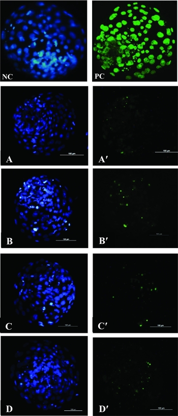
The number of apoptotic cells (stained in green) and the total number of cells (blue staining) were counted using Image J 1:41 software (Wayne Rasband National Institutes of Health, Bethesda, USA). Based on these data, we calculated the percentage of apoptotic occurrences (the number of apoptotic cells compared with the total number of cells in the embryo) (see Fig. 4).
Statistical analysis
Data were analyzed by analysis of variance (ANOVA) using the SAS PROC GLM procedure (SAS Inst. Inc., Cary, NC, USA). Sources of variation in the model, including treatment and replication, were respectively considered fixed and random effects. If ANOVA was significant, the comparisons means were performed using the least-squares difference. Data are presented as the mean and the standard error of least squares. For all analyses, we used a significance level of 5%.
Results
There was no difference in the stage of nuclear development between the control and treatments groups when the oocytes were blocked for 6 h with three concentrations of forskolin (Table 1). When these oocytes were blocked for 6 h with different concentrations of forskolin, and the block was reversed for 18 h, a significant difference was only observed in the MI stage between the groups (P < 0.05; Table 2).
Evaluation of nuclear maturation with Hoechst 33342 of oocytes matured for 6 h with different concentrations of forskolin. Total of eight replicas with 298 oocytes

D/U, Degenerate or unidentified; F, forskolin; GV, germinal vesicle; GVBD, germinal vesicle breakdown; MI, metaphase I; MII, metaphase II. Least-squares mean ± standard error mean. There was no statistical difference (P > 0.05).
Evaluation of nuclear maturation with Hoechst 33342 of oocytes matured for 6 h with different concentrations of forskolin followed by maturation agent free for 18 h. Total of eight replicas with 336 oocytes

D/U, Degenerate or unidentified; F, forskolin; GV, germinal vesicle; GVBD, germinal vesicle breakdown; MI, metaphase I; MII, metaphase II.
Least-squares mean ± standard error mean. Oocytes blocked followed by meiosis resumed (Block/Res).
a,b Values indicated by different superscript letters in the same column differ (P < 0.05).
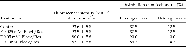
There was no difference in mitochondrial activity nor in the mitochondrial distribution throughout the cytoplasm between the groups, indicating that regardless of the forskolin dose, the mitochondrial activity remained stable (P > 0.05; Table 3).
Evaluation of cytoplasmic maturation from the fluorescence intensity and distribution of mitochondria with MitoTracker® Orange CMTMRos of the oocytes matured for 6 h with different concentrations of forskolin followed by maturation agent free for 18 h. Total of eight replicas with 320 oocytes
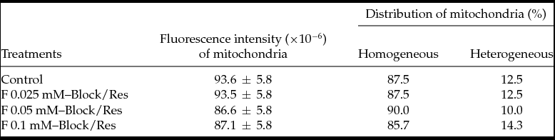
F, forskolin. Least-squares mean ± standard error mean. Oocytes blocked followed by meiosis resumed (Block/Res). There was no statistical difference (P > 0.05).
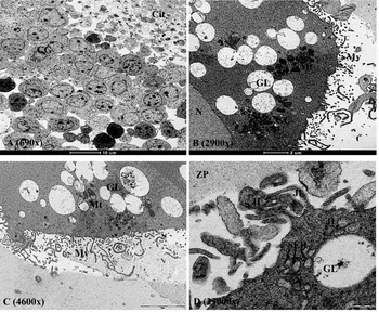
From the ultrastructural analysis, it was observed that the oocytes evaluated immediately after aspiration (immature group – 0 h) had more compact granulosa cells. The cortical granules were located in the central ooplasm-shaped cluster. Cumulus cell projections (CCPs) were present and ran through the zona pellucida (ZP). Furthermore, the immature group of oocytes was characterized by a larger perivitelline space than the mature group (Fig. 1).
Evaluation in transmission electron microscopy (TEM) of the immature oocytes group (0 h). (A) Compact granulosa cells (GC). (B) Presence of cluster of cortical granules (CG), mitochondria (Mt) and lipid granules (LG). (C) Dilated perivitelline space (PS). (D) Presence of microvilli (Mv) and junctions of granulosa cell like intermediate junction (IJ) and smooth endoplasmic reticulum (SER). Cyt: cytoplasm; N: nucleus; ZP: zona pellucida.

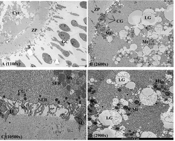
The oocytes were matured for 24 h (control group) and had expanded granulosa cells, with cortical granules in the periphery of the oocyte and many mitochondria adjacent to lipid droplets. CCPs with projections ending in the perivitelline space were also observed (Fig. 2).
Evaluation in transmission electron microscopy (TEM) of oocytes from the control group. (A) Characteristics of expanded granulosa cells (GC) after 24 h of maturation. (B) Associations of cytoplasm containing mitochondria (Mt) and lipid granules (LG). Note that the clusters of cortical granules (CG) are unmarked and directed to the periphery of the ooplasm. (C) Higher resolution ooplasma showing the association between mitochondria (Mt) and smooth endoplasmic reticulum (SER), the intermediate junctions (IJ) and peripheral cortical granules (CG). (D) Presence of lipid granules (LG) adjacent to mitochondria (Mt). Cyt, cytoplasm; ZP, zona pellucida.

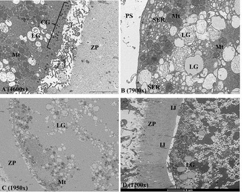
Oocytes from the F 0.05 mM group that were only blocked and that were blocked followed resumption of meiosis (F 0.05 mM–Block and F 0.05 mM–Block/Res groups) showed better preservation of the cytoplasm, with the cortical granules in the periphery of the oocyte and good microvilli quality. Moreover, the oocytes from the F 0.1 mM–Block and F 0.1 mM–Block/Res groups showed high lipid metabolism with a disruption of the cytoplasm and an absence of microvilli. In the F 0.1 mM–Block group, smooth endoplasmic reticulum was observed near the mitochondria, and in the F 0.1 mM–Block/Res group, the CCPs were limited to the perivitelline space. Regardless of the dose used, the expansion of granulosa cells was only observed in the oocyte groups in which the meiotic block was reversed (Fig. 3).
Evaluation by transmission electron microscopy (TEM) of oocytes that were maturated in presence of forskolin (A, B); and that were maturated in presence of forskolin and then in agent-free medium (C, D). (A) F 0.05 mM–Block (oocytes that were maturated in presence 0.05 mM of forskolin for 6 h) showed better preservation of the cytoplasm. (B) F 0.1 mM–Block (oocytes that were maturated in presence 0.1 mM of forskolin for 6 h) had a high metabolism of lipid granules (LG), demonstrated by the great disruption of the cytoplasm (Cyt). (C) F 0.05 mM–Block/Rev (oocytes that were maturated in presence 0.05 mM of forskolin for 6 h and then in agent-free medium) also showed better preservation of its structures. (D) F 0.1 mM–Block/Rev (oocytes that were maturated in presence 0.1 mM of forskolin for 6 h and then in agent-free medium) showed a disorganization of cytoplasmic structures. Mt: mitochondria. PS: perivitelline space. JI: junction intermediate. SER: smooth endoplasmic reticulum. ZP: zona pellucida.

The total number of blastocysts produced was 177 (number of oocytes = 584). Although there was no difference in blastocyst formation rates between the treatment and control groups (Table 4), the group of blastocysts produced from oocytes blocked for 6 h with the highest concentration of forskolin (0.1 mM) followed by reversal of the block for 18 h showed a higher rate of apoptotic cells (Table 5, Fig. 4; P < 0.05).
Evaluation of blastocyst rate in day 7 (day 0 = IVF) from oocytes matured for 6 h with different concentrations of forskolin followed by maturation agent free for 18 h. Total of eight replicas with 584 oocytes

Stage of embryo development evaluation on day 7: Eb, early blastocyst; B, blastocyst; Xb, expanded blastocyst; Hnb, blastocyst hatching; Hb, hatched blastocyst. Ooc, oocytes; Blast, blastocysts. F, forskolin. Least-squares mean ± standard error mean. Embryos produced after oocytes blocked and meiosis resumed (Block/Res). There was no statistical difference (P > 0.05).
Evaluation of the total number of cells and the apoptosis rate of day 7 blastocysts (day 0 = IVF) from oocytes matured for 6 h with different concentrations of forskolin followed by maturation agent free for 18 h. Total eight replicas with 10–15 embryos per group

F, forskolin. Least-squares mean ± standard error mean. Embryos produced after oocytes blocked and meiosis resumed (Block/Res).
a,b Values indicated by different superscript letters in the same column differ (P < 0.05).
Bovine embryos of different groups that were evaluated by TUNEL technique in inverted fluorescence microscope. (A, A′) Control group; (B, B′) F 0.025 mM group; (C, C′) F 0.05 mM group; (D, D′) F 0.1 mM group. NC, negative control; PC, positive control. Blue staining (Hoechst) represents the total number of viable cells. Green staining (FITC) indicates the fragmented DNA of apoptotic cells; ×400 magnification. Bar, 100 μm.

Discussion
In our experiment it was decided to use concentrations of forskolin lower than 0.1 mM (Dekel et al., Reference Dekel, Aberdam and Sherizly1984; Sirard, Reference Sirard1990; Bilodeau et al., Reference Bilodeau, Fortier and Sirard1993; Bilodeau-Goeseels, Reference Bilodeau-Goeseels2006) to not cause degeneration of the oocyte and the embryo. Therefore, we used three different concentrations to block or delay the resumption of meiosis over a period of 6 h (0.025 mM, 0.05 mM or 0.1 mM). Independent of the concentration used, most oocytes responded similarly and remained in GVBD or MI during the treatment period.
None of the group maintained its nucleus at the GV stage – which would have been required – regardless of the concentration of forskolin used. This negative effect might be a reflection of the short time that the oocytes were blocked because some authors block meiosis for 24 h (Bilodeau et al. Reference Bilodeau, Fortier and Sirard1993; Imai et al. Reference Imai, Kobayashi, Kaneyama, Kojima and Nagai2002). However, in our laboratory, we had tested blocking oocytes for 24 h in previous experiments but obtained high rates of nuclear degeneration (GV: 0 ± 1.6; GVBD: 8.7 ± 4.1; MI: 35.9 ± 7.4; MII: 1.9 ± 5.6; Deg/NI: 40.9 ± 5.9; P < 0.05).
The duration of the meiotic block was determined based on previous studies (Sirard & First, Reference Sirard and First1988; Sirard et al., Reference Sirard, Florman, Leibfried-Rutledge, Barnes, Sims and First1989), which showed that GVBD occurs at 6–9 h; therefore, approximately 8 h after the block, the oocyte resumes meiosis (Bilodeau et al. Reference Bilodeau, Fortier and Sirard1993).
Sirard (Reference Sirard1990) found a large number of oocytes in GVBD soon after aspiration. Likewise, our experiments also found a large number of oocytes with their nucleus in GVBD (data not shown). A fundamental explanation would be that in the time between oocyte collection at the slaughterhouse and arrival at the laboratory, the nucleus entered early maturation, perhaps due to the post-mortem processes of the animal.
There are many studies demonstrating meiotic reversal after blocking (Guixue et al., Reference Guixue, Luciano, Coenen, Gandolfi and Sirard2001; Bilodeau-Goeseels, Reference Bilodeau-Goeseels2003; Marques et al., Reference Marques, Nicacio, de Oliveira, Nascimento, Caetano, Mendes, Mello, Milazzotto, Assumpção and Visintin2007; Adona et al., Reference Adona, Pires, Quetglas, Schwarz and Leal2008). To determine if nuclear maturation is reversible, oocytes were cultured in medium that was free of blockers, and their nuclei were evaluated after blocking (Bilodeau-Goeseels, Reference Bilodeau-Goeseels2003).
However, until now, to our knowledge, any report of the effect of blocking bovine oocytes with forskolin followed by meiotic reversal was not present in the literature. For the first time, after blocking oocytes with three different concentrations of forskolin for 6 h (0.025 mM, 0.05 mM and 0.1 mM), meiotic reversal was tested after a period of 18 h.
There were a large number of oocytes in MI when forskolin was used at 0.1 mM, differing from the control group. A higher dosage of forskolin has a small effect on retarding MI, although it does not impair final maturation because at this concentration the contents of MII did not differ between groups.
In the present experiment, the reversal of the meiotic block was independent of the concentration of forskolin used because by the end of the maturation period the control group and all treatments were in MII. These results suggest improved oocyte viability compared with some studies, which found that most oocytes were still in GV (84.4%) and that none was in MII after IVM with forskolin (Katska-Ksiazkiewicz et al., Reference Katska-Ksiazkiewicz, Alm, Torner, Heleil, Tuchscherer and Rynska2011).
The MitoTracker Orange probe is readily sequestered only by actively respiring organelles depending on their oxidative activity (Torner et al., Reference Torner, Brüssow, Alm, Ratky, Pöhland, Tuchscherer and Kanitz2004; Ambruosi et al., Reference Ambruosi, Lacalandra, Iorga, Santis, Mugnier, Matarrese, Goudet and Dell’Aquila2009; Katska-Ksiazkiewicz et al., Reference Katska-Ksiazkiewicz, Alm, Torner, Heleil, Tuchscherer and Rynska2011). There was no difference in fluorescence between the groups, indicating that, regardless of the dose of forskolin, mitochondrial activity remained stable. Likewise, the distribution of mitochondria remained in the heterogeneous form, which according to the literature (Ambruosi et al., Reference Ambruosi, Lacalandra, Iorga, Santis, Mugnier, Matarrese, Goudet and Dell’Aquila2009) indicates mature cytoplasm. No differences were found between the groups in the distribution of this organelle, implying that even in the presence of forskolin, the ooplasm was able to develop to maturation.
From the evaluation of the ultrastructure by transmission electron microscopy (TEM), it was observed that the group of oocytes evaluated immediately after aspiration (immature group – 0 h) showed granulosa cells and compact cortical granules located in the central ooplasm in clusters. These features are marked signs of immature oocytes (Hyttel, Reference Hyttel, Tosti and Boni2011), which are expected in this experimental group. Furthermore, mitochondria and lipid droplets were observed in large numbers, as reported by Auclair et al. (Reference Auclair, Uzbekov, Elis, Sanchez, Kireev, Lardic, Dalbies-Tran and Uzbekova2013). This group of immature oocytes was characterized by a perivitelline space larger than the other groups. The cumulus cells have numerous projections, called CCPs, which pass through the ZP until they reach the oolemma (Hyttel et al., Reference Hyttel, Callesen and Greve1986).
Hyttel et al. (Reference Hyttel, Callesen and Greve1986) and Hyttel (Reference Hyttel, Tosti and Boni2011) reported that during oocyte development, mitochondria and lipid granules are directed towards the periphery of the oocyte; however, when the oocyte is in a mature state, these two structures are positioned in a more central region in the ooplasm, whereas the cortical granules go to the periphery (Hyttel, Reference Hyttel, Tosti and Boni2011). This was observed in our group of oocytes matured for 24 h. Although many differences were observed between the mature and immature groups, the arrangement of mitochondria and lipid droplets remained similar (Auclair et al., Reference Auclair, Uzbekov, Elis, Sanchez, Kireev, Lardic, Dalbies-Tran and Uzbekova2013).
In the control group, mitochondria, lipid droplets and smooth endoplasmic reticulum were observed in close association, forming a metabolic unit (Kruip et al., Reference Kruip, Cran, Van Beneden and Dieleman1983; Hyttel, Reference Hyttel, Tosti and Boni2011). The CCPs were still present in the form of intermediate junctions, although they were present through the ZP and did not connect over the oolemma, retracting their projections in the perivitelline space.
The oocytes in the F 0.05 mM–Block and F 0.05 mM–Block/Res groups showed a better preservation of the cytoplasm, with the cortical granules in the periphery of the oocyte and good microvilli quality. These characteristics are similar to the control group and mature oocytes in the literature (Hyttel et al., Reference Hyttel, Callesen and Greve1986; Hyttel, Reference Hyttel, Tosti and Boni2011).
Both groups of oocytes that were blocked with the highest dose of forskolin (F 0.1 mM–Block and F 0.1 mM–Block/Res) had high cytoplasmic lipid metabolism, indicated by the abundant myelin figures. Furthermore, disruption of cytoplasm was observed, indicating a possible metabolic alteration in these oocytes, with disruption of the cytoskeleton and an absence of microvilli in the perivitelline space.
The oocytes only blocked with forskolin (F 0.1 mM–Block and F 0.05 mM–Block) showed a much larger number of mitochondria when compared with their respective group followed by meiotic reversal. This event might indicate the ideal time of oocyte metabolism, which appeared to occur approximately 6 h after the block and was further stimulated by forskolin, regardless of its concentration.
In general, the groups that underwent reversal after the meiotic block were similar to the control group for all examined structures. In their ooplasma, associations between mitochondria, lipid granules and smooth endoplasmic reticulum were observed, and CCPs were limited to the perivitelline space; there also was expansion of granulosa cells in all groups (Kruip et al., Reference Kruip, Cran, Van Beneden and Dieleman1983; Hyttel et al., Reference Hyttel, Callesen and Greve1986; Hyttel, Reference Hyttel, Tosti and Boni2011).
Regardless of the dose used, expansion of granulosa cells was only observed in the groups after reversal because the groups of oocytes that were only blocked showed no expansion of their cells and remained similar to immature oocytes.
The literature indicates that forskolin is an efficient nuclear maturation inhibitor in oocytes (Ekholm et al. Reference Ekholm, Hillensjö, Magnusson and Rosberg1984; Racowsky, Reference Racowsky1984; Sirard, Reference Sirard1990; Bilodeau et al. Reference Bilodeau, Fortier and Sirard1993), but no study had reported on in vitro embryos after blocking oocytes with this agent followed by meiotic reversal. Therefore, this experiment produced the first in vitro embryos from oocytes blocked with three different concentrations of forskolin (0.025 mM, 0.05 mM or 0.1 mM) for a period of 6 h and reverted in agent-free medium for 18 h. All concentrations were able to produce embryos in similar manner to the control group, i.e., the blastocyst rate was not affected by the treatment.
According to Adona et al. (Reference Adona, Pires, Quetglas, Schwarz and Leal2008) the cleavage and blastocyst rate and total number of cells are not affected by adding of agent blocker to IVM. In contrast, Hashimoto et al. (Reference Hashimoto, Minami, Takaura and Imai2002) showed an increase in blastocyst formation rate using butyrolactone as a blocker associated with different cultivation conditions, including a low serum concentration of 5%. In this case, we note that we used a concentration of 10% FCS in IVM, and perhaps this is an aggravating factor during the meiotic block. Kubelka et al. (Reference Kubelka, Motlík, Schultz and Pavlok2000) reported that the use of FCS in the maturation medium results in partial loss of blocking ability.
When Lonergan et al. (Reference Lonergan, Dinnyes, Fair, Yang and Boland2000); Mermillod et al. (Reference Mermillod, Tomanek, Marchal and Meijer2000); Imai et al. (Reference Imai, Kobayashi, Kaneyama, Kojima and Nagai2002); Adona et al. (Reference Adona, Pires, Quetglas, Schwarz and Leal2008) used a blocker agent they did not find a difference in the total number of cells. These results are in agreement with this experiment, in which the total number of cells remained similar to the control group, independent of the forskolin concentration.
However, the best way to evaluate IVP embryo quality is to estimate apoptosis because a high incidence of apoptotic activity is associated with reduced embryonic viability (Byrne et al. Reference Byrne, Southgate, Brison and Leese1999). One way to quantitate apoptosis is the TUNEL assay, which allows the in situ detection of apoptotic cells by labelling oligonucleosomal DNA fragments generated by the endogenous activity of DNases during the process of apoptosis (Gjørret et al., Reference Gjørret, Knijn, Dieleman, Avery, Larsson and Maddox-Hyttel2003).
Most studies show that the optimal concentration of forskolin for meiotic block is 0.1 mM (Sirard, Reference Sirard1990; Bilodeau et al. Reference Bilodeau, Fortier and Sirard1993). However, although this dose of forskolin is effective at blocking meiosis and generates no difference in the rate of blastocyst formation or the total number of viable cells between the treatment and control groups, there was a higher rate of apoptotic cells in the group of blastocysts treated with this dose for 6 h and reversed for 18 h in IVM. This demonstrates that the highest dose of this agent can induce embryonic degeneration.
This finding is very interesting because the concentration of 0.1 mM normally used by the authors (Dekel et al., Reference Dekel, Aberdam and Sherizly1984; Sirard, Reference Sirard1990; Bilodeau et al., Reference Bilodeau, Fortier and Sirard1993; Bilodeau-Goeseels, Reference Bilodeau-Goeseels2006) caused the most damaged to oocytes in this experiment, in agreement with the cytoplasmic ultrastructure. Furthermore, the lower doses of forskolin used to block meiosis caused the least damage to cellular DNA.
In conclusion, all of the experimental groups reached the MII stage after different concentrations of forskolin were added. The highest concentration of the agent caused a high index of apoptosis, as confirmed by ultrastructural analysis, but did not affect the in vitro production of embryos.
Financial support
This work was supported by the São Paulo Research Foundation, Brazil (Fundação de Amparo à Pesquisa do Estado de São Paulo – FAPESP) (Grant number: 10/50410–2 and 2011/08926–4).